. 2008 Aug 13:8:79.
doi: 10.1186/1471-213X-8-79.

Newly identified patterns of Pax2 expression in the developing mouse forebrain

Affiliations

Newly identified patterns of Pax2 expression in the developing mouse forebrain

Vassiliki Fotaki et al. BMC Dev Biol. .

Abstract

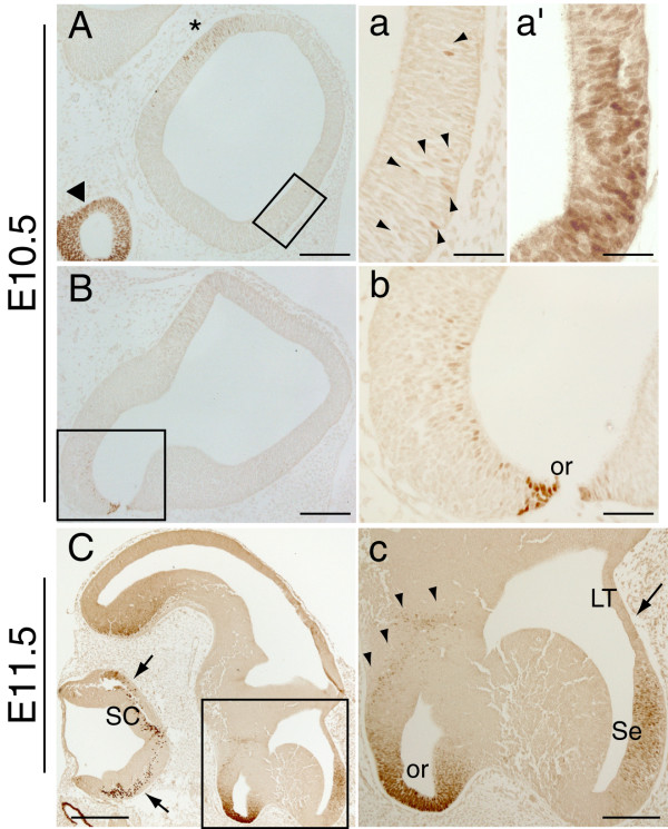
Background: The availability of specific markers expressed in different regions of the developing nervous system provides a useful tool for the study of mouse mutants. One such marker, the transcription factor Pax2, is expressed at the midbrain-hindbrain boundary and in the cerebellum, spinal cord, retina, optic stalk, and optic chiasm. We recently described a group of diencephalic cells that express Pax2 as early as embryonic day (E) 10.5, and become part of the eminentia thalami by E11.5. The discovery of this previously undescribed cell population prompted us to examine Pax2 protein expression in the developing mouse forebrain in more detail.

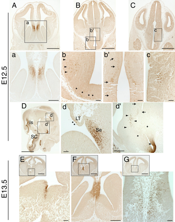
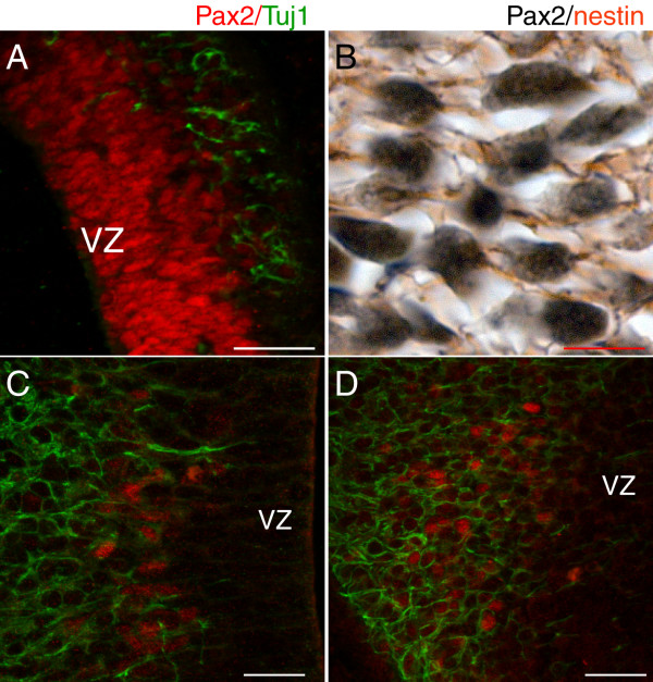
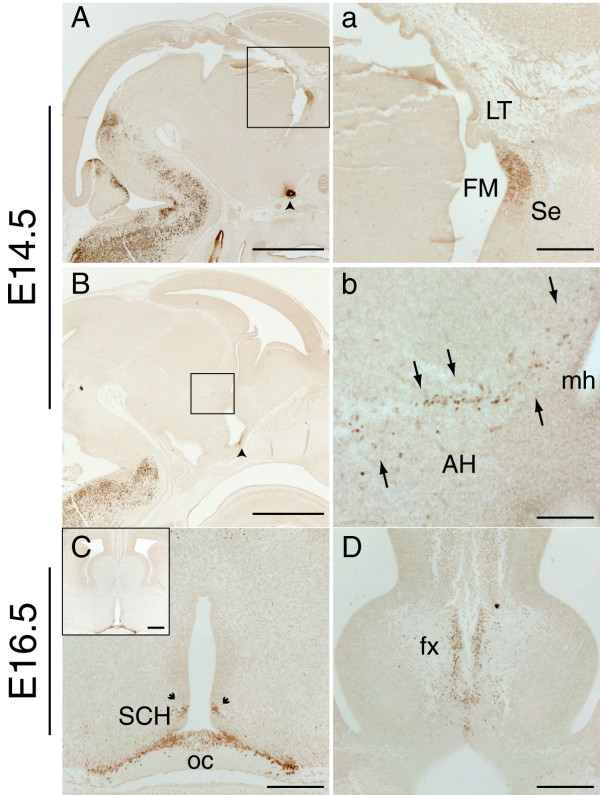
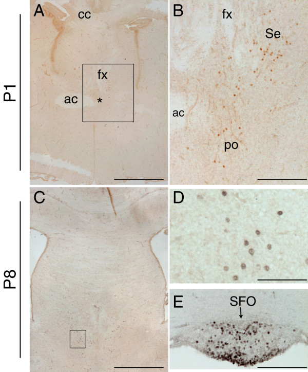
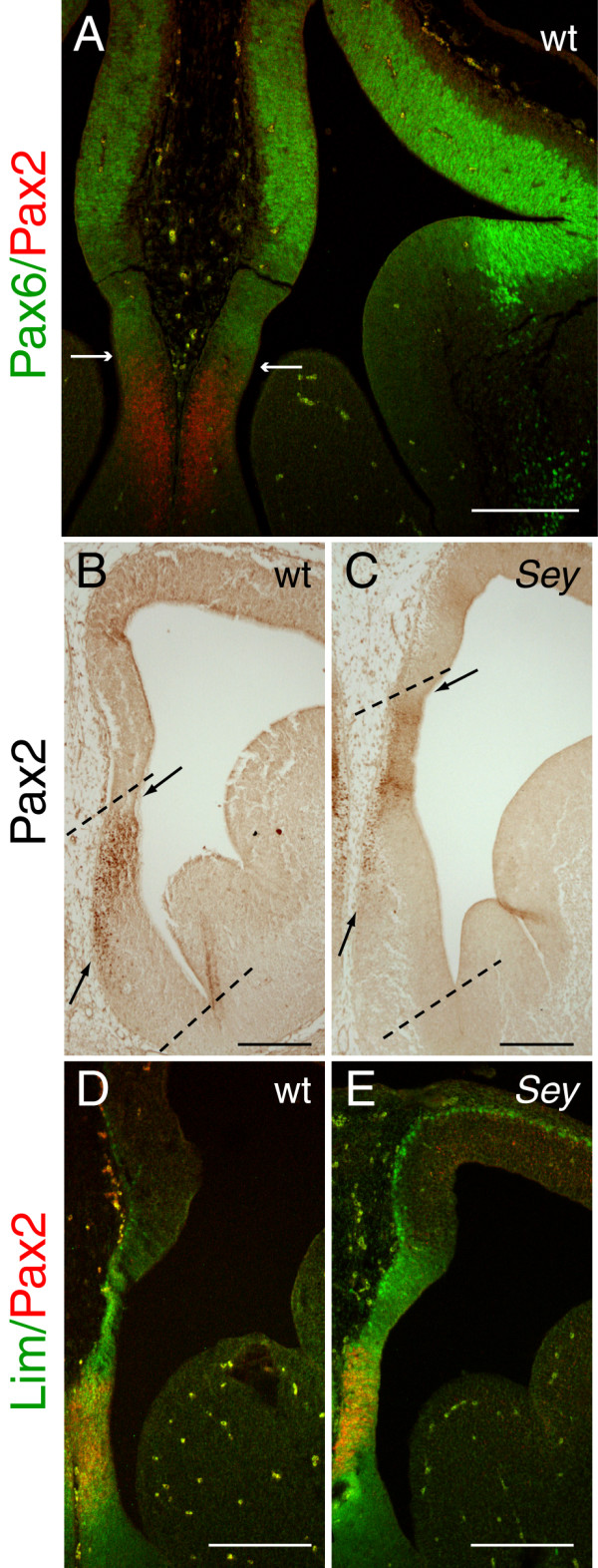
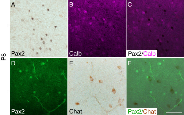
Results: We determined the expression pattern of Pax2 in the forebrain of wild type mouse embryos between E10.5 and postnatal day (P) 15. Pax2 expression was detected in the septum of the basal forebrain, hypothalamus, eminentia thalami and in the subfornical organ. To evaluate Pax2 as a marker for septal cells, we examined Pax2 expression in Pax6Sey/Sey mutants, which have an enlarged septum. We found that Pax2 clearly marks a population of septal cells equivalent to that seen in wild types, indicating its utility as a marker of septal identity. These cells did not express the GABAergic marker calbindin nor the cholinergic marker choline acetyltransferase and were not detectable after P15.

Conclusion: Pax2 is expressed in populations of cells within the developing septum, hypothalamus, and eminentia thalami. It seems especially useful as a marker of the telencephalic septum, because of its early, strong and characteristic expression in this structure. Further, its expression is maintained in the enlarged septum of Pax6Sey/Sey mutants.


Figures

Figure 1
Figure 1
Pax2 protein expression in the mouse forebrain at E10.5 and E11.5. A-B, a-b: E10.5 sagittal sections showing Pax2 expression in the developing forebrain. In the ventral telencephalon, a few Pax2-positive cells can be detected in lateral sections (arrowheads in a) and become more abundant in more medial sections (a'). A few Pax2-positive cells are detected in the hypothalamus, close to the strongly Pax2-positive optic recess (or) area (B, b). The previously described staining in the future eminentia thalami of the diencephalon (asterisk in A) and the optic cup (arrowhead in A) are also shown. C, c: E11.5 sagittal sections revealing strong Pax2 expression in the developing septum (Se). No Pax2 expression is detected in the neighbouring lamina terminalis (LT) (arrow in c) at this developmental stage. Pax2-immunopositive cells are also found in the anterior hypothalamus (arrowheads in c) and may originate from the ventral neuroepithelium of the optic recess area (or). Expression in the eminentia thalami is not shown. Note the Pax2 expression in the spinal cord (SC) (arrows in C). a, b and c are high power images of the boxed areas in A, B and C respectively. a' is a high power image of a sagittal section at a more medial level than that depicted in a. Scale bars: C, 400 μm; A, B, c, 200 μm; a, a', b, 50 μm.
Figure 2
Figure 2
Pax2 protein expression in the mouse forebrain at E12.5 and E13.5. A-C: Low power views of coronal E12.5 forebrain sections immunoreacted with Pax2; the boxed areas are shown at higher magnification in panels a-c. D: Low power view of a sagittal E12.5 section immunoreacted with Pax2. Note the strong expression of Pax2 in the isthmic region (Is) and the spinal cord (SC), in accordance with previous reports. d, d' higher magnification of the boxed areas in D. A, a, d: Strong Pax2 expression is detected in the septum (Se) of the basal forebrain, mainly in the septal neuroepithelium. A few Pax2-positive cells are detected in the lamina terminalis (LT) (arrows in d). b-c, d': Pax2 is detected in groups of cells found in the lateral hypothalamic area (arrowheads in b and d') and in the anterior hypothalamic neuroepithelium (arrows in b' and d', c). These cell populations might originate from cells located at the base of the hypothalamus (small arrows in b and d'), Pax2 expression can also be seen in the ventral neuroepithelium of the optic recess (asterisk in d'), as previously described. E-G: Low and high power images of E13.5 coronal sections reveal strong Pax2 expression in the septum, mainly in the neuroepithelium (F). A few immunopositive cells are found in the differentiating layer of the septum (G). The boxed areas in the inset panels in E-G indicate the areas shown in the respective high power images. A-C and E-G are sections from the same specimens respectively. Scale bars: D, E-G-insets, 1000 μm; A-C, 500 μm; a, d', F-high power, 200 μm; b-c, d, E, G-high power, 50 μm.
Figure 3
Figure 3
Pax2 is primarily expressed in neural progenitors in the septum and in early differentiated neurons in the hypothalamus. A: Double immunofluorescence with Pax2 (red) and β-tubulin III (Tuj1) (green) on a sagittal E12.5 telencephalic section shows that only a minority of Pax2-positive cells in the septum also express Tuj1, an early marker of differentiated neurons. This is further confirmed with double immunohistochemistry with Pax2 (black) and nestin (brown) (B), revealing that these Pax2-positive cells have nestin-positive filaments. Panels (A) and (B) show correspond to high power images taken within the region depicted in Fig. 2D, d. In the hypothalamus (C, D), most Pax2-positive cells express the early neural marker β-tubulin III (green), showing that they correspond to early generated neurons. Pax2-positive cells in (C) correspond to those shown in Fig. 2b', and those in (D) correspond to the cells indicated by arrowheads in Fig. 2b. The position of the ventricular zone (VZ) is indicated in sections A, C and D. Scale bars: A, 20 μm; B-D, 5 μm.
Figure 4
Figure 4
Pax2 protein expression in the mouse forebrain at E14.5 and E16.5. A, a: Low and high power images of an E14.5 sagittal section showing strong Pax2 staining in the neuroepithelium of the septum (Se) adjacent to the foramen of Monro (FM) in an E14.5 sagittal section. This intense Pax2 staining is observed at the level where the lamina terminalis (LT) joins the septum. a shows a higher magnification of the boxed area in panel A. B, b: In more lateral sagittal sections Pax2 expression is found in a cell population (arrows) within the anterior hypothalamus (AH) reaching the medial horn (mh) of the lateral ventricle. b shows a higher magnification of the boxed area in panel B. C: E16.5 coronal section showing Pax2 expression around the optic chiasm (oc) region, in the optic stalk epithelium and in a few cells of the hypothalamic neuroepithelium next to the suprachiasmatic nucleus (SCH) (arrows). D: E16.5 coronal section showing Pax2 expression in the differentiating layer of the medial septum surrounded by the axonal bundles of the fornix (fx). Scale bars: A, B, 1000 μm; a, C, D, 200 μm; C-inset, 250 μm; b, 100 μm.
Figure 5
Figure 5
Pax2 protein expression in the mouse forebrain during early postnatal development. A-E: Coronal forebrain sections immunostained for Pax2 at P1 (A-B) and at P8 (C-E), showing the presence of few disperse Pax2-positive cells in the septal area (Se) (A-C, high power in D), the medial preoptic nucleus (po) (asterisk in A and B), and the subfornical organ (SFO) (arrow in E). The fibre tracts are indicated in A and B for orientation purposes (ac, anterior commissure; cc, corpus callosum; fx, fornix). The boxed areas in A and C delineate the high power images depicted in B and D respectively. Scale bars: A, C, 100 μm; B, E, 25 μm; D, 12.5 μm.
Figure 6
Figure 6
The Pax2 expression domain in the telencephalic septum is located in a more dorsal position in the Pax6Sey/Sey mutant than in wild type. Pax2 expression in the telencephalic septum in E12.5 wild type (wt) (A, B, D) and Pax6Sey/Sey mutant (Sey) (C, E) coronal sections. A: Double immunofluorescence with Pax2 (red) and Pax6 (green) reveals that the two proteins are expressed in non-overlapping, mutually exclusive domains. The arrows in A indicate the ventral and dorsal limits of Pax6 and Pax2 expression respectively. B-C: Pax2 expression in wt and Sey embryos, showing that the Pax2 expression domain is shifted dorsally in the Sey mutant (C) compared to wt (B). The septum and the Pax2 expression domain are indicated by dashed lines and arrows respectively. D-E: Double immunofluorescence with Pax2 (red) and Lim1/2 (green) in the septum shows that Pax2 expression is found within the Lim-positive domain in both wild types (D) and Sey mutants (E), suggesting that the shifted Pax2 expression domain in the Sey mutant is still within the limits of the ventral telencephalon. Note that panels B and C correspond to slightly more rostral sections than those shown in D and E respectively. Scale bars: A, B, C, 200 μm; D, E, 100 μm.
Figure 7
Figure 7
Pax2 expression in the medial septum does not co-localise with that of calbindin and choline acetyltransferase (Chat). A-C: Double immunohistochemistry with Pax2 (black) (A) and calbindin (magenta) (B) on P8 coronal sections reveal the presence of both calbindin-positive and Pax2-positive neurons in the septal area, but not co-expression of these proteins (C). Similarly (D-F), septal neurons that express Pax2 (green) (D) and Chat (brown) (E), do not co-express these proteins (F). The panels are high powers of the level depicted in Fig. 5C. Scale bar for all panels, 10 μm.

Similar articles

Cited by

References

    1. Dressler GR, Deutsch U, Chowdhury K, Nornes HO, Gruss P. Pax2, a new murine paired-box-containing gene and its expression in the developing excretory system. Development. 1990;109:787–795. - PubMed
    1. Dressler GR, Douglass EC. Pax-2 is a DNA-binding protein expressed in embryonic kidney and Wilms tumor. Proc Natl Acad Sci USA. 1992;89:1179–1183. - PMC - PubMed
    1. Mansouri A, Hallonet M, Gruss P. Pax genes and their roles in cell differentiation and development. Curr Opin Cell Biol. 1996;8:851–857. - PubMed
    1. Stuart ET, Kioussi C, Gruss P. Mammalian Pax genes. Annu Rev Genet. 1994;28:219–236. - PubMed
    1. Nornes HO, Dressler GR, Knapik EW, Deutsch U, Gruss P. Spatially and temporally restricted expression of Pax2 during murine neurogenesis. Development. 1990;109:797–809. - PubMed

Publication types

LinkOut - more resources