

Differential wedging of vertebral body and intervertebral disc in thoracic and lumbar spine in adolescent idiopathic scoliosis - A cross sectional study in 150 patients
- PMID: 18700985
- PMCID: PMC2527554
- DOI: 10.1186/1748-7161-3-11
Differential wedging of vertebral body and intervertebral disc in thoracic and lumbar spine in adolescent idiopathic scoliosis - A cross sectional study in 150 patients
Abstract
Background: Hueter-Volkmann's law regarding growth modulation suggests that increased pressure on the end plate of bone retards the growth (Hueter) and conversely, reduced pressure accelerates the growth (Volkmann). Literature described the same principle in Rat-tail model. Human spine and its deformity i.e. scoliosis has also same kind of pattern during the growth period which causes wedging in disc or vertebral body.
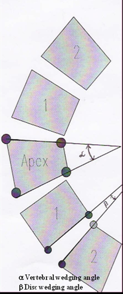
Methods: This cross sectional study in 150 patients of adolescent idiopathic scoliosis was done to evaluate vertebral body and disc wedging in scoliosis and to compare the extent of differential wedging of body and disc, in thoracic and lumbar area. We measured wedging of vertebral bodies and discs, along with two adjacent vertebrae and disc, above and below the apex and evaluated them according to severity of curve (curve < 30 degrees and curve > 30 degrees ) to find the relationship of vertebral body or disc wedging with scoliosis in thoracic and lumbar spine. We also compared the wedging and rotations of vertebrae.
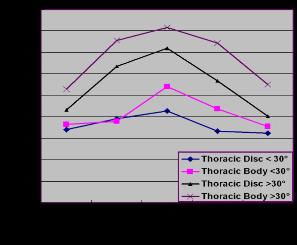
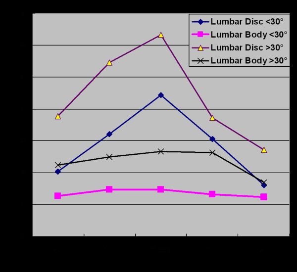
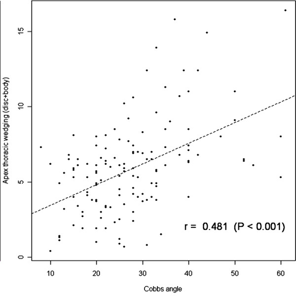
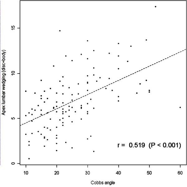
Results: In both thoracic and lumbar curves, we found that greater the degree of scoliosis, greater the wedging in both disc and body and the degree of wedging was more at apex supporting the theory of growth retardation in stress concentration area. However, the degree of wedging in vertebral body is more than the disc in thoracic spine while the wedging was more in disc than body in lumbar spine. On comparing the wedging with the rotation, we did not find any significant relationship suggesting that it has no relation with rotation.
Conclusion: From our study, we can conclude that wedging in disc and body are increasing with progression on scoliosis and maximum at apex; however there is differential wedging of body and disc, in thoracic and lumbar area, that is vertebral body wedging is more profound in thoracic area while disc wedging is more profound in lumbar area which possibly form 'vicious cycle' by asymmetric loading to spine for the progression of curve.
Figures
References
-
- Taylor TK, Ghosh P, Bushell GR. The contribution of the intervertebral disk to the scoliotic deformity. Clin Orthop. 1981;156:79–90. - PubMed
-
- Braun JT, Ogilvie JW, Akyuz E, Brodke DS, Bachus KN, Stefko RM. Experimental scoliosis in an immature goat model: a method that creates idiopathic-type deformity with minimal violation of the spinal elements along the curve. Spine. 2003;28:2198–203. doi: 10.1097/01.BRS.0000085095.37311.46. - DOI - PubMed
-
- Perdriolle R, Becchetti S, Vidal J, Lopez P. Mechanical process and growth cartilages. Essential factors in the progression of scoliosis. Spine. 1993;18:343–9. - PubMed
-
- Mehlman CT, Araghi A, Roy DR. Hyphenated history: the Hueter-Volkmann law. Am J Orthop. 1997;26:798–800. - PubMed
LinkOut - more resources
Full Text Sources
Miscellaneous
