. 2008 Aug 14;59(3):392-8.
doi: 10.1016/j.neuron.2008.06.009.

TRPC3 channels are required for synaptic transmission and motor coordination

Affiliations

TRPC3 channels are required for synaptic transmission and motor coordination

Jana Hartmann et al. Neuron. .

Abstract

In the mammalian central nervous system, slow synaptic excitation involves the activation of metabotropic glutamate receptors (mGluRs). It has been proposed that C1-type transient receptor potential (TRPC1) channels underlie this synaptic excitation, but our analysis of TRPC1-deficient mice does not support this hypothesis. Here, we show unambiguously that it is TRPC3 that is needed for mGluR-dependent synaptic signaling in mouse cerebellar Purkinje cells. TRPC3 is the most abundantly expressed TRPC subunit in Purkinje cells. In mutant mice lacking TRPC3, both slow synaptic potentials and mGluR-mediated inward currents are completely absent, while the synaptically mediated Ca2+ release signals from intracellular stores are unchanged. Importantly, TRPC3 knockout mice exhibit an impaired walking behavior. Taken together, our results establish TRPC3 as a new type of postsynaptic channel that mediates mGluR-dependent synaptic transmission in cerebellar Purkinje cells and is crucial for motor coordination.


Figures
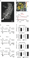
Figure 1
Figure 1. mGluR-Mediated Purkinje Cell Signaling in the Absence of TRPC1 and TRPC4 Subunits
(A) Confocal image of a patch-clamped Purkinje cell in a cerebellar slice from a wild-type (WT) mouse. The site of electrical stimulation (stim) is shown at higher magnification in (B). (B) Pseudocolor image of a synaptic Ca2+ signal evoked by parallel fiber (PF) stimulation. (C) Black traces: PF-evoked (five pulses, 100 Hz, in 10 μM CNQX) synaptic response consisting of an early rapid and a slow EPSC (bottom) and a Ca2+ transient (top). Red traces: Block of slow components by CPCCOEt (200 μM). (D) (Left) Slow EPSC (ten pulses, 100 Hz in 40 μM CNQX) in a WT mouse (lower trace) and the corresponding local Ca2+ response (upper trace). (Right) Pressure ejection of DHPG (200 μM for 100 ms) evoked a slow inward current (lower trace) and a local Ca2+ transient (upper trace). Similar experiments performed in TRPC1-knockout (E) and in TRPC1/TRPC4 DKO mice (F) using PF stimulation (left panels; 15 pulses, 50 Hz) and local DHPG pressure application (right panels; 1 mM, 100 ms), respectively. (G and H) Summary of the results obtained in WT and TRPC1-/- mice (mean ± SEM; [G]: n = 16 and n = 22 for synaptic transmission, n = 38 and n = 30 for DHPG applications in WT and TRPC1-/-, respectively) and in TRPC1/4 DKO mice ([H]: n = 12 and n = 21 for synaptic transmission, n = 13 and n = 8 for DHPG applications in WT and TRPC1/4 DKO, respectively). Responses in mutants were not significantly different from those in WT (Student’s t test).
Figure 2
Figure 2. Expression Pattern of TRPC Channel Subunits in the Mouse Brain and Single Purkinje Cells
(A) Copy numbers of TRPC subunit mRNA detected in 1 ng total RNA of mouse whole brain (left) and cerebellum (right; mean ± SEM). (B) Agarose gel electrophoresis of the TRPC1 amplicons in the neuropil material surrounding Purkinje cell somata. The gene-specific standard (1000 copies) was used as a positive control. (C) Analogous agarose gel electrophoresis of TRPC3 amplicons obtained from single Purkinje cells. (D) (Left) Real-time monitoring of the fluorescence emission of SYBR Green I during the PCR amplification of TRPC3 cDNA from single Purkinje cells (n = 34 cells). (Right) Copy numbers of transcripts of TRPC subunits in single Purkinje cells (mean ± SEM). (E) A dual-channel confocal scan of an immunohistochemical staining in an acute cerebellar slice. Calbindin-D28k immunoreactivity is shown in green (left) and that for TRPC3 is shown in red (middle). (Right) merged images.
Figure 3
Figure 3. Generation and Characterization of a TRPC3 Knockout Mouse
(A) (Top) Diagram of the intron-exon organization of the Mus musculus TRPC3 gene. Open boxes, untranslated exonic sequence; closed boxes, translated open reading frame; *, stop codon; F1, F2, R1, and R2: PCR primers. The lengths of the amplicons, including primers, are depicted. (Bottom) Diagram of the expected disruption after excision of exon 7 by the action of the Cre recombinase. (B) Image of the electrophoretic migration in an agarose gel of the amplicons obtained using the indicated primers. Plus, plus-RT reaction; minus, minus-RT controls. (C) Fluorescence image of an Alexa 594-filled Purkinje cell in a cerebellar slice from a TRPC3-/- mouse. (D) (Left) Blockade of PF-evoked fast EPSC in a TRPC3-deficient mouse by CNQX (40 μM). (Right) Summary graph of EPSC blockade by CNQX (n = 5). (E) (Top) Paired PF EPSCs evoked (interval of 100 ms) in a WT and in a TRPC3-/- mouse. (Bottom) Time course of facilitation in WT (n = 8 cells) and in TRPC3-/- (n = 11 cells) mice. (F) Footprint patterns in WT and TRPC3-/- mice. The images show mice walking on a glass plate. Dotted lines depict their transverse body diameter. The right part of the panel summarizes the superimposed paw positions for 24 steps of 8 WT and TRPC3-/- mice. (G) Percentage of hindpaw positions outside of the body diameter in both genotypes (summary of the results shown in (F). (H) Percentage of hindpaw slips relative to the total number of steps on a horizontal ladder with an irregular spacing (n = 9 WT and 12 TRPC3-/- mice). (I) Similar analysis of hindpaw slips during runs on an elevated beam (Ø = 1 cm; n=8WTand n=10 TRPC3-/- mice). Asterisks in (G)—(I) indicate high significance (p < 0.01, χ2 test [G], Mann-Whitney U test [H and I]). Error bars indicate SEM.
Figure 4
Figure 4. mGluR-Mediated Purkinje Cell Signaling in the Absence of TRPC3
(A) Slow EPSC in a WT mouse (lower trace) and the corresponding local dendritic Ca2+ response (upper trace). (B) Similar recording in a TRPC3-/- mouse. (C) Summary graphs for normalized (to stimulation strength) sEPCS (n = 41 in WT and n = 25 in TRPC3-/- mice) and Ca2+ transients (ΔF/F, n = 15 in WT and n = 17 in TRPC3-/- mice). (D and E) Synaptically evoked local dendritic Ca2+ transients in TRPC3-/- mice before (control) and after the addition of CPCCOEt (D) and CPA (E). Bar graphs: Summary of the results (n = 4 cells for CPCCOEt and 6 cells for CPA). (F) DHPG evoked a slow inward current (lower trace) and local Ca2+ transient (upper trace) in a WT mouse. (G) Similar recording in a TRPC3-/- mouse. (H) Summary of DHPG-evoked current (right) and Ca2+ (left) responses (n = 26 in WT and n = 30 in TRPC3-/- mice). (I) Dendritic Ca2+ transients evoked by synaptic simulation in the two genotypes as indicated. (J) Mean amplitudes of Ca2+ transients normalized to the first amplitude for ten subsequent stimuli in WT (n = 4 cells) and in TRPC3-/- mice (n = 6 cells). Error bars indicate SEM.

References

    1. Aiba A, Kano M, Chen C, Stanton ME, Fox GD, Herrup K, Zwingman TA, Tonegawa S. Deficient cerebellar long-term depression and impaired motor learning in mGluR1 mutant mice. Cell. 1994;79:377–388. - PubMed
    1. Amaral MD, Pozzo-Miller L. TRPC3 channels are necessary for brain-derived neurotrophic factor to activate a nonselective cationic current and to induce dendritic spine formation. J. Neurosci. 2007;27:5179–5189. - PMC - PubMed
    1. Barski JJ, Hartmann J, Rose CR, Hoebeek F, Mörl K, Noll-Hussong M, De Zeeuw CI, Konnerth A, Meyer M. Calbindin in cerebellar Purkinje cells is a critical determinant of the precision of motor coordination. J. Neurosci. 2003;23:3469–3477. - PMC - PubMed
    1. Batchelor AM, Garthwaite J. Novel synaptic potentials in cerebellar Purkinje cells: probable mediation by metabotropic glutamate receptors. Neuropharmacology. 1993;32:11–20. - PubMed
    1. Canepari M, Ogden D. Kinetic, pharmacological and activity-dependent separation of two Ca2+ signalling pathways mediated by type 1 metabotropic glutamate receptors in rat Purkinje neurones. J. Physiol. 2006;573:65–82. - PMC - PubMed

Publication types

MeSH terms