. 2008 Aug 15;29(2):205-16.
doi: 10.1016/j.immuni.2008.06.008.

The nuclear orphan receptor NR2F6 suppresses lymphocyte activation and T helper 17-dependent autoimmunity

Affiliations

The nuclear orphan receptor NR2F6 suppresses lymphocyte activation and T helper 17-dependent autoimmunity

Natascha Hermann-Kleiter et al. Immunity. .

Abstract

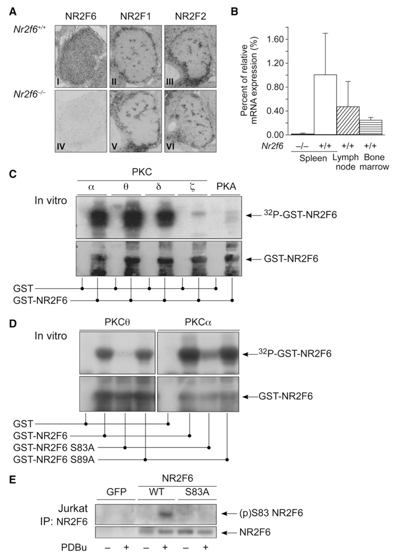
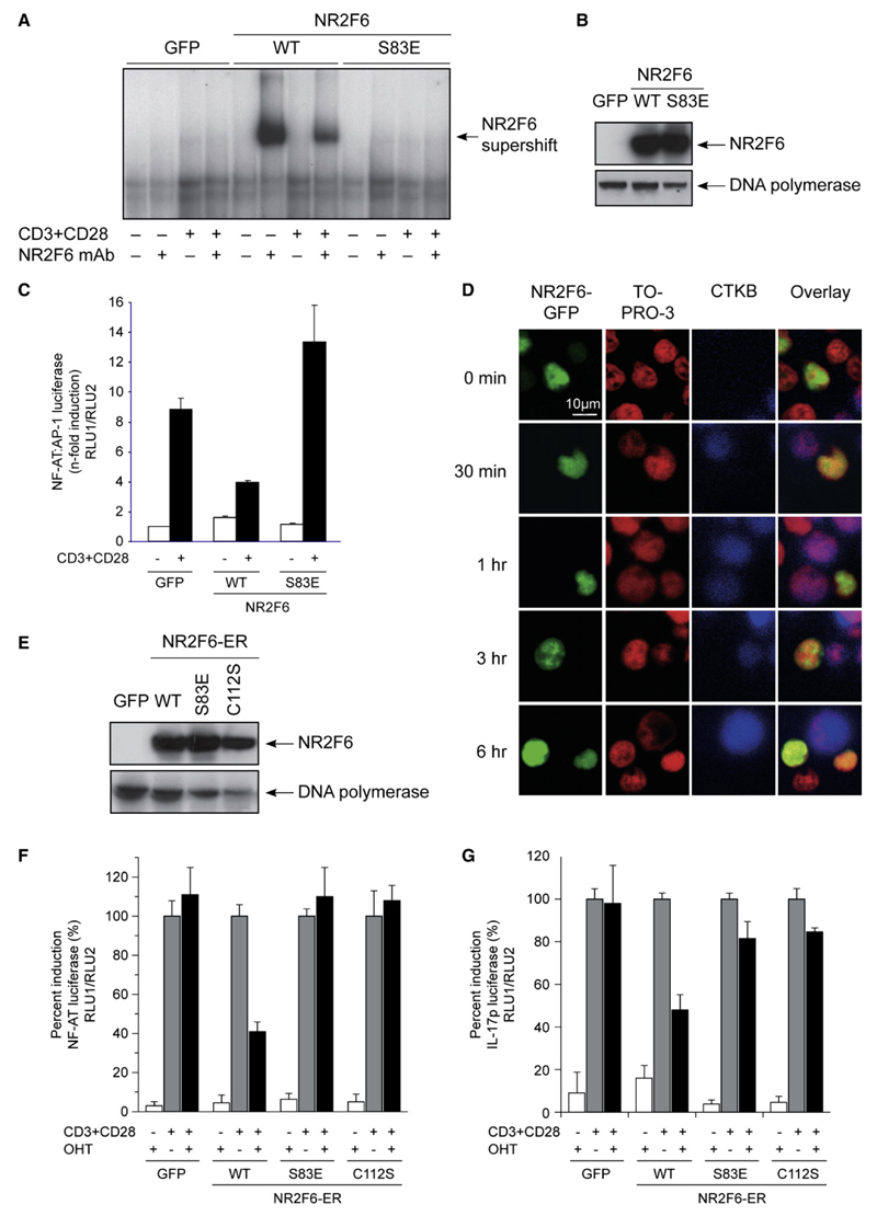
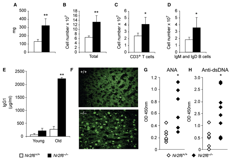
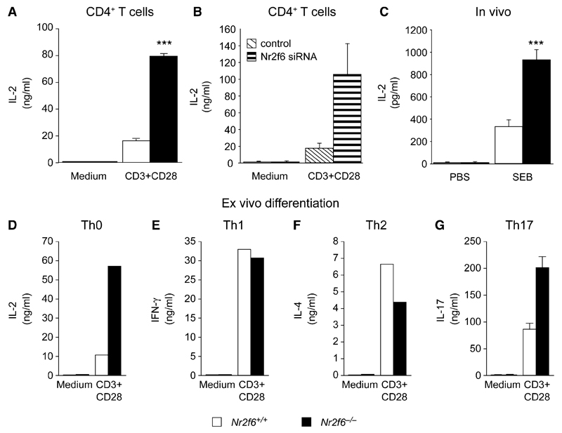
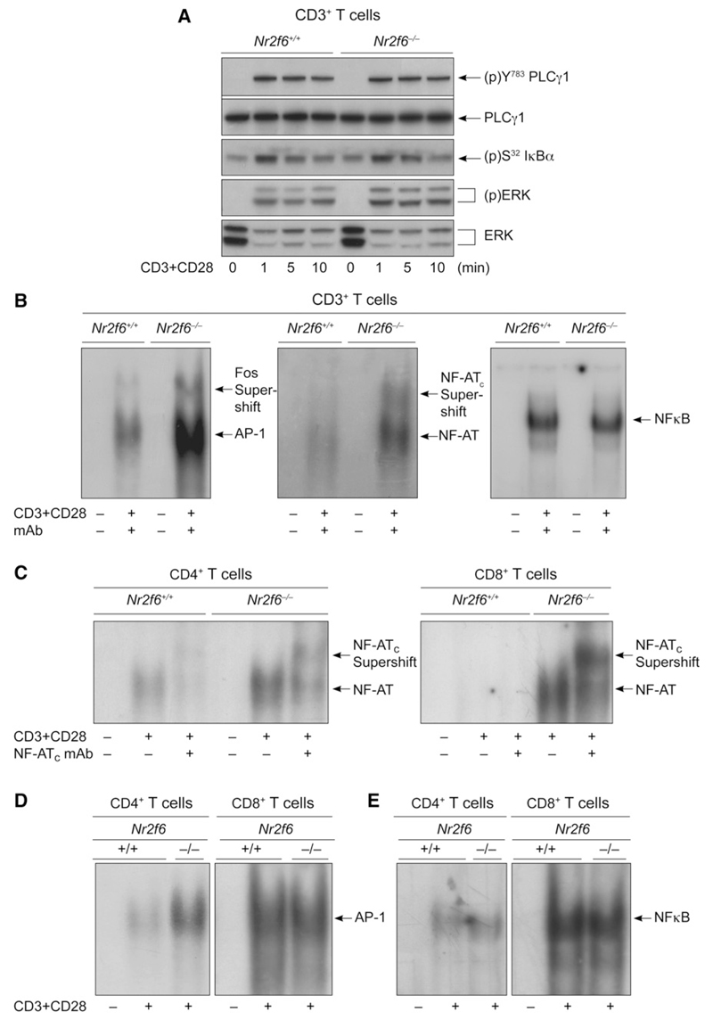
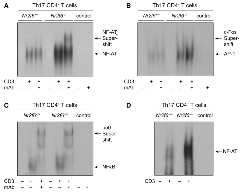
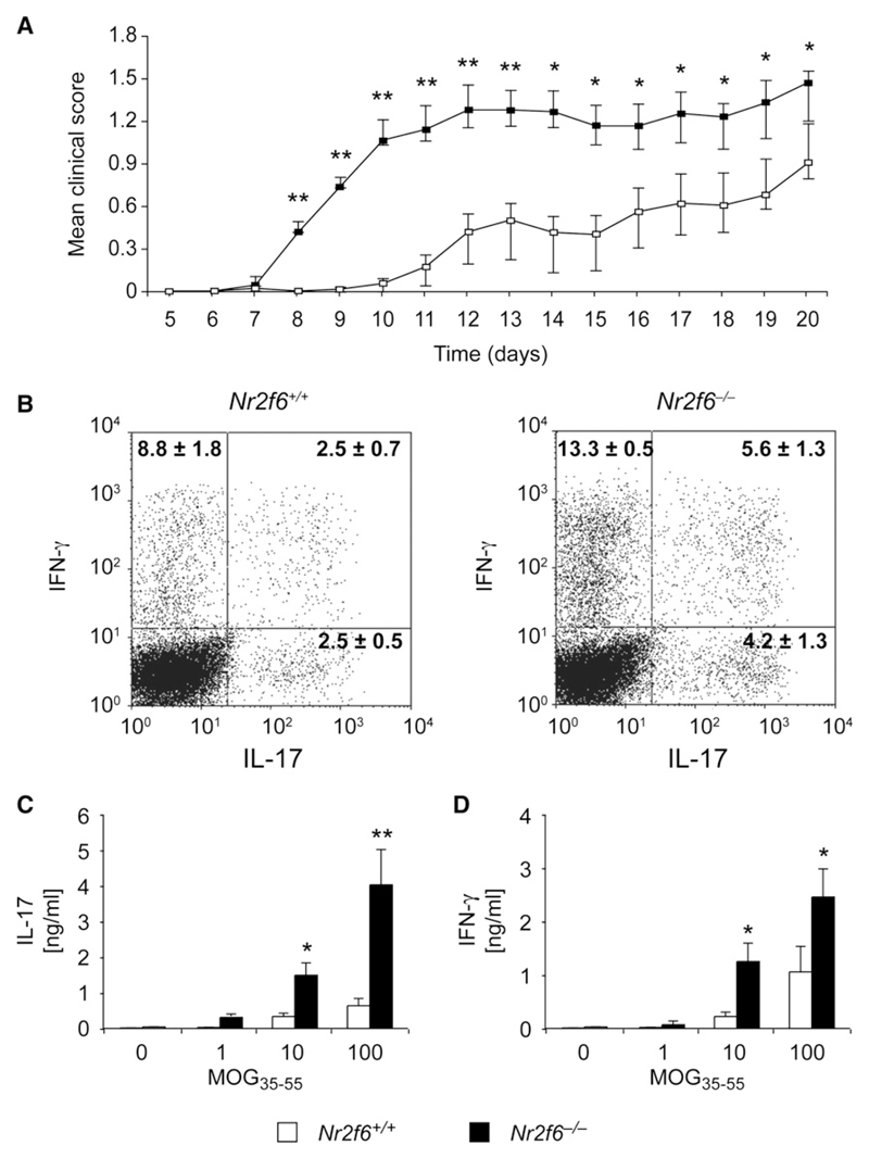
The protein kinase C (PKC) family of serine-threonine kinases plays a central role in T lymphocyte activation. Here, we identify NR2F6, a nuclear zinc-finger orphan receptor, as a critical PKC substrate and essential regulator of CD4(+) T cell activation responses. NR2F6 potently antagonized the ability of T helper 0 (Th0) and Th17 CD4(+) T cells to induce expression of key cytokine genes such as interleukin-2 (IL-2) and IL-17. Mechanistically, NR2F6 directly interfered with the DNA binding of nuclear factor of activated T cells (NF-AT):activator protein 1 (AP-1) but not nuclear factor kappaB (NF-kappa B) and, subsequently, transcriptional activity of the NF-AT-dependent IL-17A cytokine promoter. Consistent with our model, Nr2f6-deficient mice had hyperreactive lymphocytes, developed a late-onset immunopathology, and were hypersusceptible to Th17-dependent experimental autoimmune encephalomyelitis. Our study establishes NR2F6 as a transcriptional repressor of IL-17 expression in Th17-differentiated CD4(+) T cells in vitro and in vivo.


Figures

Figure 1
Figure 1. (p)Ser-83 on NR2F6 Is a PKC Phosphorylation Site
(A) Nr2f6 in situ hybridization was positive in thymus sections of wild-type (I) but not Nr2f6−/− (IV) E14.5 embryonic thymus sections. Neither the expression pattern nor the intensity of the other COUP-TF family members NR2F1 (II, V) and NR2F2 (II, VI) was altered in the Nr2f6−/− thymus. (B) qRT-PCR revealed Nr2f6 expression in spleen, lymph node, and bone marrow in the wild-type mouse. Nr2f6−/− samples served as specificity negative controls, data were normalized to GAPDH, and expression in the spleen was arbitrarily taken as 1. Mean of three independent experiments is shown; error bars represent standard error. (C and D) Kinase assays of full-length GST-NR2F6 (wild-type or S83A or S89A mutant) incubated with recombinant PKC family members (α, θ, δ, and ζ) or protein kinase A (PKA) as control. Specific phosphorylation of NR2F6 by the PKC isotypes α, θ, δ, and, to a much lower extent, PKC ζ and PKA could be observed. This specific PKC-mediated phosphorylation was mostly lost after mutation of the S83 site (but not Ser-89) into a S83A site. The anti-GST immunoblot (lower panel) confirmed equal loading. (E) Jurkat cells were transfected with GFP, NR2F6 wild-type, or S83A mutant, and NR2F6 was immunoprecipitated from resting cells (–) or cells stimulated for 20 min with 50 nM phorbol ester (PDBu) (+) and immunoblotted with the anti-(p)Ser-83 NR2F6-specific pAb.
Figure 2
Figure 2. (p)Ser-83 on NR2F6 Regulates NR2F6 DNA Binding in Jurkat T Cells
(A) NR2F6 Ser-83 phosphorylation directly influenced DNA binding in nuclear extracts as analyzed by EMSA. Jurkat T cells were transfected with NR2F6 wild-type, S83E mutant, or GFP control and were left unstimulated (–) or were CD3 plus CD28 stimulated (+). Supershift analysis was performed with a mAb for NR2F6. In contrast to the NR2F6 wild-type, the S83E mutant did not bind DNA. (B) Equal expression of NR2F6 wild-type and S83E mutant proteins in the nuclear fractions was confirmed by immunoblotting (DNA polymerase served as loading control). NR2F6 wild-type and mutant protein expression remained unaltered also during CD3 plus CD28 stimulation (not shown). (C) Transfected wild-type NR2F6 interfered with CD3 plus CD28-induced transcriptional activation of the NF-AT:AP-1 promoter luciferase reporter; this repressor activity was abolished by the S83E mutation in NR2F6. (D) Jurkat T cells transfected with NR2F6-GFP (green) were used to form conjugates with SEE-pulsed B cells stained with the cell tracker blue (CTKB). Cells were then fixed and nuclei were stained with TOPRO-3 (red). Conjugates were analyzed for NR2F6 localization at different time points. Similar results were obtained with an RGS-His6-tagged NR2F6 and an anti-RGSHis6 mAb staining (not shown). (E–G) Cotransfection experiments in Jurkat T cells showed that recombinant NR2F6-ER wild-type but not DNA-binding-defective mutants, S83E and C112S, induced repression of CD3 plus CD28-induced NF-AT-dependent reporter luciferase gene transcription. CD3 plus CD28-induced IL-17-dependent promoter luciferase reporter was similarly repressed by recombinant NR2F6-ER wild-type in transfected Jurkat T cells. Reporter induction rates were normalized for the transfected cells, and CD3 plus CD28-induced reporter activity without OHT treatment was arbitrarily set as 100%. Mean of at least two independent experiments analyzed in triplicates is shown. Error bars represent standard error. Equal expression of NR2F6 wild-type and S83E and C112S mutant proteins in the nuclear fractions was confirmed by immunoblotting (DNA polymerase served as loading control).
Figure 3
Figure 3. Nr2f6−/− Mice Develop a Late-Onset Immunopathology
(A–D) Twelve-month-old Nr2f6−/− mice displayed enlarged spleens with increased lymphocyte numbers in Nr2f6−/− mice. (A) Splenic weight, (B) total cellularity, and (C) T cell (CD3+) and (D) mature B cell numbers (IgM+ and IgD+) are shown (Nr2f6+/+, n = 11; Nr2f6−/−, n = 11; unpaired t test; (A) p = 0.009, (B) p = 0.006, (C) p = 0.033, (D) p = 0.046; means are shown with error bars). (E) Serum immunoglobulin levels of IgG1 young (6–10 weeks) and old (>12 months) Nr2f6−/− mice were determined via ELISA. Twelve-month-old Nr2f6−/− mice show significantly elevated IgG1 plasma titers (unpaired t test, Nr2f6+/+, n = 8; Nr2f6−/−, n = 8; p = 0.007). (F–H) Aged Nr2f6−/− mice generate autoantibodies against nuclear antigens (ANA; unpaired t test, Nr2f6+/+, n = 9; Nr2f6−\−, n = 9; p = 0.023) and double-stranded (ds) DNA (Nr2f6+/+ n = 9; Nr2f6−/− n = 9; p = 0.037) as determined by staining of rat liver sections with mouse serum (F) and ELISA (G and H). Means are shown; error bars represent standard error.
Figure 4
Figure 4. Nr2f6−/− T Cells Hyperrespond to Antigen-Receptor Stimulation
(A) CD3 plus CD28-induced IL-2 cytokine secretion responses of Nr2f6-deficient T cells (black bars) were significantly higher than wild-type controls (white bars). Data shown are the mean of three independent experiments performed in duplicate (split-plot ANOVA, p = 0.0007). (B) Similarly, siRNA-mediated Nr2f6 knockdown in CD4+ T cells resulted in enhanced IL-2 cytokine secretion upon CD3 plus CD28 stimulation, compared to siRNA nontargeting controls (n = 2). (C) IL-2 cytokine concentrations in plasma taken 2 hr after injection with SEB i.p. (10 μg/kg) were significantly higher in Nr2f6−/− mice (unpaired t test; Nr2f6+/+, n = 10; Nr2f6−/−, n = 10; p = 0.0004). (D–G) Naive CD4+ T cells were differentiated under neutral Th0 (D), (E) Th1, (F) Th2, and (G) Th17 conditions (conditions are defined in the Experimental Procedures), and relevant cytokines were measured from the supernatant after 4–5 days. One of two independent experiments with consistent results is shown for Th0, Th1, and Th2. IL-17 cytokine secretion in Nr2f6-deficient Th17 cells (black bars) was significantly higher than wild-type controls (white bars) (split-plot ANOVA Nr2f6+/+, n = 5; Nr2f6−/−, n = 5; p = 0.004162) Error bars represent standard error.
Figure 5
Figure 5. NR2F6 Acts as Repressor of NF-AT:AP-1 DNA-Binding Capability
(A) CD3+ T cells (5 × 10⁶ per lane) from Nr2f6+/+ and Nr2f6−/− mice were stimulated with CD3 plus CD28 for the indicated time periods, and the phosphorylation status of proteins was detected by immunoblotting, as indicated. One representative experiment of two is shown. (B–E) Analysis of DNA-binding activity in nuclear extracts of Nr2f6−/− CD4+ and Nr2f6−/− CD8+ T cells showed that NF-AT DNA binding is higher in both CD4+ and CD8+ T cells, whereas AP-1 DNA binding is higher only in CD4+ T cells. No change was detected in NF-κB binding. Supershift analysis was performed with antibodies against c-fos, NF-ATc, and p50, as indicated. One representative experiment out of four is shown.
Figure 6
Figure 6. NR2F6 Suppresses NF-AT:AP-1 DNA Binding Specifically in CD4+ Th17 Effector-Memory T Cells
(A–C) EMSA analysis of nuclear extracts prepared from Th17-differentiated and αCD3-antibody-re-stimulated cells (IL-23, TGF-β, IL-6, αIL-4, αIFN-γ). NF-AT:AP-1 DNA binding was higher in Nr2f6-deficient extract when the NF-AT:AP-1 derived from the minimal IL-2 promoter was used, whereas NF-κB remained unchanged. (D) To distinguish between NF-AT:AP-1 and NF-AT-only binding, we used the NF-AT-specific probe #3 derived from the IL17A minimal promoter region (Liu et al., 2004); this again revealed a higher NF-AT binding in the Nr2f6-deficient nuclear Th17 cell extracts when compared to the wild-type control. Supershift analysis was performed with antibodies against c-fos, NF-ATc, and p50 as indicated. Controls are the radiolabeled probe with or without the supershifting Ab. One representative experiment out of two is shown.
Figure 7
Figure 7. Nr2f6−/− Th17 Effector Cells Are Hyperreactive Ex Vivo and in Experimental Autoimmune Encephalomyelitis
(A) EAE disease course in age-matched female mice (Nr2f6+/+, n = 20; Nr2f6−/−, n = 20) is significantly higher in the Nr2f6−/− mice. Mean disease scores were calculated by comparison of the mean values of wild-type and Nr2f6−/− scores at the onset (day 7–day 13) and at disease progression (day 14–day 20) via a Welch two-sample t test. A significant difference was found in both periods (day 7–day 13, p = 0.0004; day 14–day 20, p = 0.025), although the difference was more profound during the disease onset. (B) Cyokine production by CD4+ T cells isolated from CNS mononuclear cells 14 days after disease induction. Cells were stimulated for 4 hr with PDBu plus ion-omycin in the presence of Golgi stop and analyzed for IFN-γ and IL-17 expression. A representative profile and mean ± SD of cytokine staining are shown. A significant increase of IL-17-IFN-γ double-positive cells (Nr2f6+/+, n = 7; Nr2f6−/−, n = 7; p = 0.0494) could be observed. (C and D) Production of (C) IL-17 and (D) IFN-γ in recall assays of splenocytes from MOG35-55-immunized mice was significantly enhanced in Nr2f6−/− T cells (split-plot ANOVA (C); Nr2f6+/+, n = 14; Nr2f6−/−, n = 14; p = 0.00019; (D) p = 0.0277).

Comment in

  • Orphans against autoimmunity.
    Bandukwala H, Sundrud MS, Rao A. Bandukwala H, et al. Immunity. 2008 Aug 15;29(2):167-8. doi: 10.1016/j.immuni.2008.07.008. Immunity. 2008. PMID: 18701076

Similar articles

Cited by

References

    1. Altman A, Kaminski S, Busuttil V, Droin N, Hu J, Hipskind RA, Villalba M. Positive feedback regulation of PLCγ1/Ca2+ signaling by PKCθ in restimulated T cells via a Tec kinase-dependent pathway. Eur J Immunol. 2004;34:2001–2011. - PubMed
    1. Anderson K, Fitzgerald M, Dupont M, Wang T, Paz N, Healy A, Xu Y, Ocain T, Schopf L, Jaffee B, et al. Mice deficient in PKCθ demonstrate impaired in vivo T cell activation and protection from T cell-mediated inflammatory diseases. Autoimmunity. 2006;39:469–478. - PubMed
    1. Baier G. The PKC gene module: Molecular biosystematics to resolve its T cell functions. Immunol Rev. 2003;192:64–79. - PubMed
    1. Bettelli E, Oukka M, Kuchroo VK. T(H)-17 cells in the circle of immunity and autoimmunity. Nat Immunol. 2007;8:345–350. - PubMed
    1. Chaudhary D, Kasaian M. PKCθ: A potential therapeutic target for T-cell-mediated diseases. Curr Opin Investig Drugs. 2006;7:432–437. - PubMed

Publication types

MeSH terms