. 2008 Sep;130(3):455-66.
doi: 10.1309/1QWTQFMF0Q9JEAGR.

Modification of solid phase red cell adherence assay for the detection of platelet antibodies in patients with thrombocytopenia

Affiliations

Modification of solid phase red cell adherence assay for the detection of platelet antibodies in patients with thrombocytopenia

Preeyanat Vongchan et al. Am J Clin Pathol. 2008 Sep.

Abstract

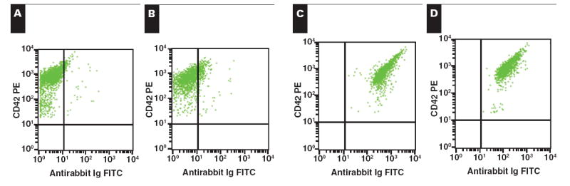
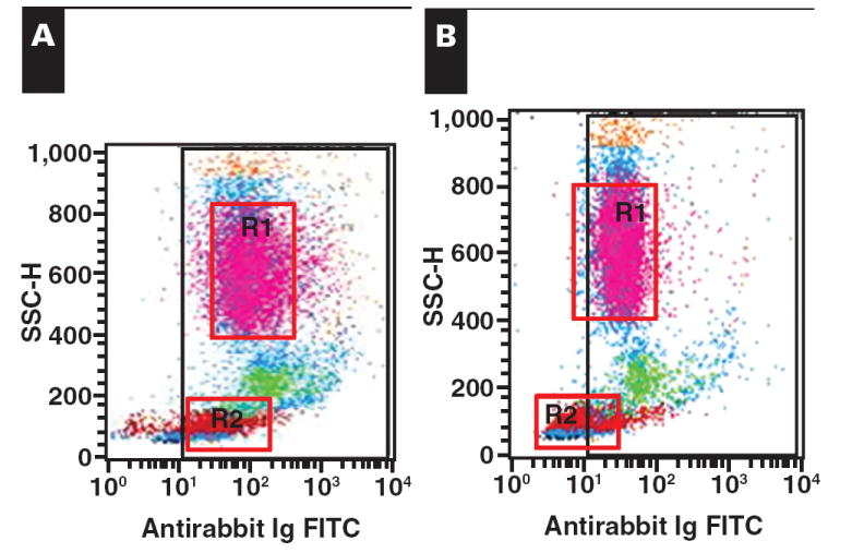
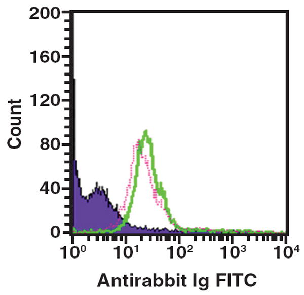
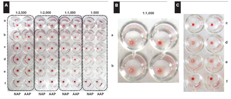
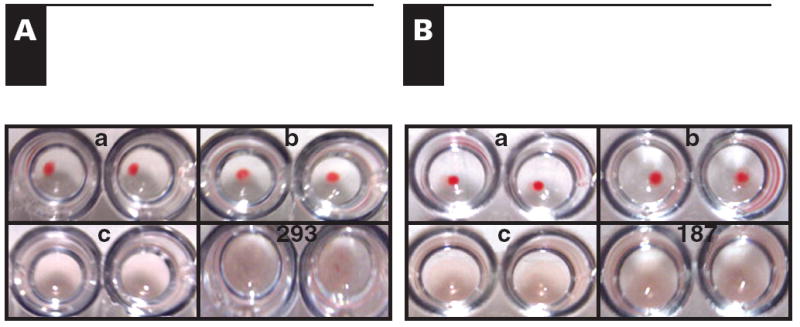
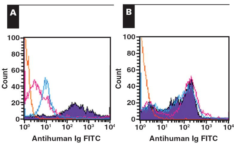
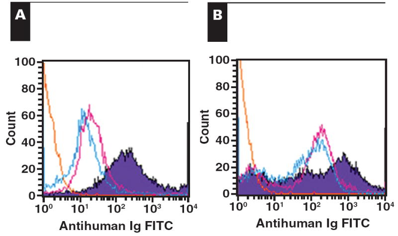
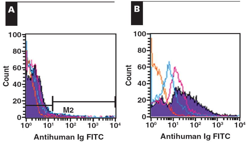
Platelet refractoriness is caused by HLA antibodies and platelet-specific antibodies. Current methods used to detect antiplatelet antibodies have limitations. Solid phase red cell adherence (SPRCA) lacks sensitivity and requires a second assay using chloroquine-treated intact platelets to specify the response due to anti-HLA. We modified SPRCA by using 2 types of antihuman platelet antibodies with different specificities toward platelet lysate and tested samples from 361 patients (69 with unexplained thrombocytopenia and 292 with poor response to platelet transfusions not explicable by alloimmunization or the clinical situation) and 50 from healthy volunteers. Our method compared favorably with platelet suspension direct immunofluorescence. All samples from healthy volunteers were negative; of the samples from the patient population, 240 were positive (147 samples had only antiplatelet and 3 samples had only anti-HLA antibodies). This modified technique had a sensitivity of 98% and a specificity of 91%.


Figures

Image 1
Image 1
Determination of rabbit antihuman platelet polyclonal antibody (NAP). Rabbits were immunized weekly, intramuscularly, with 5 pooled normal human platelets (1 × 10⁸ platelets/500 μL) for 8 weeks. Rabbit serum was collected before each booster and tested with pooled normal platelets by platelet suspension immunofluorescent technique. Fluorescein isothiocyanate (FITC)-conjugated goat antirabbit immunoglobulins (Igs) and CD42-phycoerythrin (PE) were used to detect the bound rabbit antihuman platelets and as platelet-specific marker, respectively. The reaction was analyzed by flow cytometry. A, Cell control. B, Conjugate control. C, Rabbit polyclonal antihuman platelet antibody 1:100. D, Rabbit polyclonal antihuman platelet antibody 1:50.
Image 2
Image 2
Adsorption (A, before; B, after) out of anti-HLA. Pooled normal human leukocytes were prepared and used to adsorb anti-HLA contaminated in rabbit antihuman platelet antibody as mentioned in the “Materials and Methods” section. The adsorbed rabbit antihuman platelet antibody (AAP) was proved to be free of anti-HLA by indirect immunofluorescent technique using 5 pooled normal human leukocytes. Washed human leukocytes (50 μL, 1 × 10⁷ cells/mL) were fixed with AB serum (final 10%) for 30 minutes on ice before adding AAP (50 μL). The incubation was performed on ice for another 30 minutes and washed 3 times with 1% bovine serum albumin phosphate-buffered saline, pH 7.2, containing 0.05% Tween-20. Fluorescein isothiocyanate (FITC)-conjugated goat antirabbit immunoglobulins (Igs; 1:50) were added and incubated for 30 minutes on ice before washing with the same buffer. Paraformaldehyde (0.5% in phosphate-buffered saline) was added, and the reaction was then analyzed by flow cytometry. R1, granulocyte population; R2, lymphocyte population; SSC, side scatter.
Image 3
Image 3
Detection of antiplatelet activity in adsorbed rabbit antihuman platelet antibody (AAP). The platelet suspension immunofluorescent technique was performed to investigate the remaining antiplatelet activity of AAP after anti-HLA was removed through adsorption. Five pooled normal human platelets were prepared and fixed with 1% paraformaldehyde. The paraformaldehyde-fixed platelets (50 μL, 1 × 10⁸ platelets/mL) were reacted with AAP (50 μL) or nonadsorbed antiplatelet antibody (NAP; 50 μL) for 30 minutes at room temperature before washing with EDTA–phosphate-buffered saline. Fluorescein isothiocyanate (FITC)-conjugated goat antirabbit immunoglobulins (Igs; 1:50) were added, and the reaction was performed for another 30 minutes at room temperature before washing. CD42-PE (1:50) was added and incubated under the same conditions. Finally, the reaction was washed, fixed with 0.5% paraformaldehyde, and analyzed by flow cytometry. Solid green line, NAP; dotted pink line, AAP.
Image 4
Image 4
Optimization of modified solid phase red cell adherence (SPRCA). Various concentrations of nonadsorbed antiplatelet antibody (NAP) or adsorbed rabbit antihuman platelet antibody (AAP) were coated (100 μL/well) onto a microtiter plate and incubated at 4°C overnight to optimize the modified SPRCA. After washing with bovine serum albumin (BSA)–phosphate-buffered saline (PBS), 5% BSA in PBS was added, 200 μL/well, and incubated for 1 hour at 37°C to block nonspecific binding sites. The microtiter plate was washed again 3 times with BSA-PBS. Various concentrations of platelet lysate (100 μL/well) were added after washing and incubated for another hour at 37°C. Glycine (1.9% in BSA-PBS) was then added followed with 50 μL/well of normal human serum or anti-HLA+ serum as negative and positive control samples, respectively. The reaction was allowed for 1 hour at 37°C and washed with the same buffer. Finally, 50 μL/well of various concentrations of Fc-specific rabbit antihuman IgG and 50 μL/well of 0.2% indicator cells were added sequentially. The microtiter plate was then centrifuged at 2,000 rpm for 2 minutes, and the agglutination of indicator cells was observed. A, Agglutination observed in various concentrations of rabbit antihuman IgG used (1:500-1:2,500). B and C, Enlarged picture of agglutination when rabbit antihuman IgG 1:1,000 was used. a, Platelet lysate, 2.5 μg/100 μL/well, anti-HLA positive serum, 1:10; b, Platelet lysate, 5.0 μg/100 μL/well, anti-HLA positive serum, 1:10; c, Platelet lysate, 2.5 μg/100 μL/well, Normal serum control sample; d, Platelet lysate, 2.5 μg/100 μL/well, anti-HLA positive serum, 1:100; e, Platelet lysate, 5.0 μg/100 μL/well, anti-HLA positive serum, 1:100; f, Cell control.
Image 5
Image 5
Various patterns of agglutination for interpretation of modified solid phase red cell adherence. In every investigation, the cell control (the only sensitized red cell in the well), negative control (no serum), and positive control (anti-HLA positive serum, 1:10) were performed in parallel. The agglutination pattern in the test well was interpreted following the pattern of control. a, Cell control; b, Negative control; c, Positive control; d, e, and f, Positive in both wells; g, h, and i, Negative (no antibody specific to platelet or HLA).
Image 6
Image 6
Positive in both wells (P-P pattern) of 2 samples with a difference in antibody combination. A, Sample 293. B, Sample 187. a, Cell control; b, Negative control; c, Positive control.
Image 7
Image 7
Positive in both wells (P-P pattern) of serum sample 293, which had only antiplatelet antibody. The serum sample was further investigated by the platelet suspension immunofluorescent technique (A) for antiplatelet antibodies and indirect immunofluorescence (B) using pooled normal human leukocytes for anti-HLA antibodies as mentioned in the “Materials and Methods” section. FITC, fluorescein isothiocyanate; Ig, immunoglobulin.
Image 8
Image 8
Positive in both wells (P-P pattern) of serum sample 187, which had antiplatelet and anti-HLA antibodies. The serum sample was further investigated by the platelet suspension immunofluorescent technique (A) for antiplatelet antibodies and indirect immunofluorescence (B) using pooled normal human leukocytes for anti-HLA antibodies as mentioned in the “Materials and Methods” section. FITC, fluorescein isothiocyanate; Ig, immunoglobulin.
Image 9
Image 9
Positive only in the nonadsorbed antiplatelet antibody well (P-N pattern) of 3 samples that had only anti-HLA antibody. a, Cell control. b, Negative control. c, Positive control.
Image 10
Image 10
Confirmation of anti-HLA alone of the P-N pattern (positive only in the nonadsorbed antiplatelet antibody well). The serum sample was further investigated by the platelet suspension immunofluorescent technique (PSIFT; A) for antiplatelet antibodies and indirect immunofluorescence (B) using pooled normal human leukocytes for anti-HLA antibodies as mentioned in “Materials and Methods” section. Weak positive was observed by PSIFT but strong positive for anti-HLA by indirect immunofluorescence. FITC, fluorescein isothiocyanate; Ig, immunoglobulin.

Similar articles

Cited by

References

    1. Slichter SJ. Platelet transfusions a constantly evolving therapy. Thromb Haemost. 1991;66:178–188. - PubMed
    1. Herman JH, Kamel HT. Platelet transfusion: current techniques, remaining problems, and future prospects. Am J Pediatr Hematol Oncol. 1987;9:272–286. - PubMed
    1. Kiefel V, Konig C, Kroll H, et al. Platelet alloantibodies in transfused patients. Transfusion. 2001;41:766–770. - PubMed
    1. Buetens O, Shirey RS, Goble-Lee M, et al. Prevalence of HLA antibodies in transfused patients with and without red cell antibodies. Transfusion. 2006;46:754–756. - PubMed
    1. Bajpai M, Kaura B, Marwaha N, et al. Platelet alloimmunization in multitransfused patients with haemato-oncological disorders. Natl Med J India. 2005;18:134–136. - PubMed

Publication types