. 2009 Jan;8(1):32-44.
doi: 10.1074/mcp.M800139-MCP200. Epub 2008 Aug 12.

Quantitative proteomics reveals GIMAP family proteins 1 and 4 to be differentially regulated during human T helper cell differentiation

Affiliations

Quantitative proteomics reveals GIMAP family proteins 1 and 4 to be differentially regulated during human T helper cell differentiation

Jan-Jonas Filén et al. Mol Cell Proteomics. 2009 Jan.

Abstract

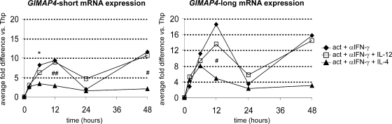
T helper (Th) cells differentiate into functionally distinct effector cell subsets of which Th1 and Th2 cells are best characterized. Besides T cell receptor signaling, IL-12-induced STAT4 and T-bet- and IL-4-induced STAT6 and GATA3 signaling pathways are the major players regulating the Th1 and Th2 differentiation process, respectively. However, there are likely to be other yet unknown factors or pathways involved. In this study we used quantitative proteomics exploiting cleavable ICAT labeling and LC-MS/MS to identify IL-4-regulated proteins from the microsomal fractions of CD4(+) cells extracted from umbilical cord blood. We were able to identify 557 proteins of which 304 were also quantified. This study resulted in the identification of the down-regulation of small GTPases GIMAP1 and GIMAP4 by IL-4 during Th2 differentiation. We also showed that both GIMAP1 and GIMAP4 genes are up-regulated by IL-12 and other Th1 differentiation-inducing cytokines in cells induced to differentiate toward Th1 lineage and down-regulated by IL-4 in cells induced to Th2. Our results indicate that the GIMAP (GTPase of the immunity-associated protein) family of proteins is differentially regulated during Th cell differentiation.


Figures

F<sc>ig</sc>. 1.
Fig. 1.
Sample preparation and data handling summary. Pb, plate-bound; pos., positive.
F<sc>ig</sc>. 2.
Fig. 2.
Correlation of the protein quantifications between ProICAT and XPRESS. The horizontal axis presents ProICAT quantifications, and XPRESS quantifications are on the vertical axis. Proteins, which were quantified only by another of the algorithms, are presented on the axis. All the proteins are present in A, and proteins showing >1.4-fold expression difference are in B. The correlation was determined also by Pearson's test resulting in a value of 0.77. A, proteins detected by both algorithms and up-regulated; B, proteins detected by ProICAT and up-regulated; C, proteins detected by SEQUEST/XPRESS and up-regulated; D, proteins detected by both algorithms and down-regulated; E, proteins detected by ProICAT or SEQUEST/XPRESS and down-regulated.
F<sc>ig</sc>. 3.
Fig. 3.
Western blot analysis of STAT1, MXA, and GIMAP4 confirmed the down-regulation by IL-4. Human cord blood CD4+ cells were activated by anti-CD3/anti-CD28 (Act.) and induced to differentiate toward Th2 cells with activation + IL-4. STAT1, MXA, and GIMAP4 were all down-regulated by IL-4.
F<sc>ig</sc>. 4.
Fig. 4.
MS and MS/MS data from protein GIMAP1. MS quantification of the peptide shows the down-regulation of GIMAP1 in the microsomal fraction of IL-4-treated cord blood CD4+ cells (A). Peptide fragmentation by MS/MS is shown in B, and Y- and B-ions present in the fragmentation spectrum are shown in C.
F<sc>ig</sc>. 5.
Fig. 5.
GIMAP1 and GIMAP4 are differentially regulated during Th1 and Th2 differentiation. A, human cord blood CD4+ T cells were activated with PHA and irradiated CD32/B7-transfected mouse L fibroblasts (act) and cultured in the presence of IL-12 or IL-4 to promote Th1 and Th2 differentiation, respectively, or in neutral Th0 conditions for 7 days. GIMAP1 and GIMAP4 mRNA expression was analyzed using quantitative real time RT-PCR. Each sample was analyzed in duplicate at least twice, and samples from at least three individuals were used for calculations. An asterisk (*) indicates a significant difference between Th1 and Th0, # indicates a significant difference between Th2 and Th0, and § indicates a significant difference between Th1 and Th2. *, p < 0.05; **, p < 0.01; ***, p < 0.001. B, human peripheral blood CD4+ cells were induced to the Th1 or Th2 direction by anti-CD3/anti-CD28 activation and cytokines IL-12 and IL-4, respectively. GIMAP4 expression was detected by Western blotting 2 and 6 days after culture. d, days; avg, average.
F<sc>ig</sc>. 6.
Fig. 6.
The regulation of differentially spliced variants of GIMAP4 mRNA during early Th1 and Th2 differentiation follows similar kinetics of gene expression. Human cord blood CD4+ T cells were activated by anti-CD3/anti-CD28 (act) and induced to differentiate toward Th1 and Th2 lineages by IL-12 and IL-4, respectively, or activated only. Anti-IFN-γ was added to culture medium to block the autocrine IFN-γ signaling. Samples from three independent cultures were harvested and analyzed using quantitative real time RT-PCR. Differences in gene expression were calculated as -fold differences versus naïve Thp cells, and the significance of difference between samples was determined by Student's t test as in Fig. 5.
F<sc>ig</sc>. 7.
Fig. 7.
GIMAP4 is up-regulated by Th1-inducing cytokines IFN-α and IL-18. Human umbilical cord blood CD4+ cells were cultured under the indicated conditions for 24 and 48 h, and the level of GIMAP4 protein expression was analyzed by Western blotting. A, besides IL-12, other Th1-inducing cytokines, namely IFN-α and IL-18, induced up-regulation of GIMAP4 expression. Cytokine-induced regulation of GIMAP4 was detectable only after 48 h of culture. GIMAP4/β-actin ratios were calculated from the Western blot presented. B, relative quantitation of GIMAP4 protein level after 48 h of culture showing average values from four individual cultures. Error bars, S.D.; p values were obtained using Student's t test. act, activation.
F<sc>ig</sc>. 8.
Fig. 8.
GIMAP4 gene expression is regulated by the STAT6 signaling pathway. Human CD4+ cells were transiently transfected with shRNA/siRNA targeting STAT6 or with control (scramble) and induced to differentiate toward the Th2 lineage using anti-CD3/anti-CD28 activation (act) and IL-4. Western blotting showed an effective knock down of STAT6 by STAT6 siRNA and high dependence for GIMAP4 expression on STAT6. The figure is a representative of three individual experiments showing a similar pattern of expression.

Similar articles

Cited by

References

    1. Agnello, D., Lankford, C. S., Bream, J., Morinobu, A., Gadina, M., O'Shea, J. J., and Frucht, D. M. ( 2003) Cytokines and transcription factors that regulate T helper cell differentiation: new players and new insights. J. Clin. Immunol. 23, 147–161 - PubMed
    1. Constant, S. L., and Bottomly, K. ( 1997) Induction of Th1 and Th2 CD4+ T cell responses: the alternative approaches. Annu. Rev. Immunol. 15, 297–322 - PubMed
    1. Lee, G. R., Kim, S. T., Spilianakis, C. G., Fields, P. E., and Flavell, R. A. ( 2006) T helper cell differentiation: regulation by cis elements and epigenetics. Immunity 24, 369–379 - PubMed
    1. Mosmann, T. R., Cherwinski, H., Bond, M. W., Giedlin, M. A., and Coffman, R. L. ( 1986) Two types of murine helper T cell clone. I. Definition according to profiles of lymphokine activities and secreted proteins. J. Immunol. 136, 2348–2357 - PubMed
    1. Mosmann, T. R., and Coffman, R. L. ( 1989) Heterogeneity of cytokine secretion patterns and functions of helper T cells. Adv. Immunol. 46, 111–147 - PubMed

Publication types

MeSH terms