. 2008 Oct 31;283(44):30101-11.
doi: 10.1074/jbc.M804613200. Epub 2008 Aug 13.

CDKL5 expression is modulated during neuronal development and its subcellular distribution is tightly regulated by the C-terminal tail

Affiliations

CDKL5 expression is modulated during neuronal development and its subcellular distribution is tightly regulated by the C-terminal tail

Laura Rusconi et al. J Biol Chem. .

Abstract

Mutations in the human X-linked cyclin-dependent kinase-like 5 (CDKL5) gene have been identified in patients with Rett syndrome (RTT), West syndrome, and X-linked infantile spasms, sharing the common feature of mental retardation and early seizures. CDKL5 is a rather uncharacterized kinase, but its involvement in RTT seems to be explained by the fact that it works upstream of MeCP2, the main cause of Rett syndrome. To understand the role of this kinase for nervous system functions and to address if molecular mechanisms are involved in regulating its distribution and activity, we studied the ontogeny of CDKL5 expression in developing mouse brains by immunostaining and Western blotting. The expression profile of CDKL5 was compared with that of MeCP2. The two proteins share a general expression profile in the adult mouse brain, but CDKL5 levels appear to be highly modulated at the regional level. Its expression is strongly induced in early postnatal stages, and in the adult brain CDKL5 is present in mature neurons, but not in astroglia. Interestingly, the presence of CDKL5 in the cell nucleus varies at the regional level of the adult brain and is developmentally regulated. CDKL5 shuttles between the cytoplasm and the nucleus and the C-terminal tail is involved in localizing the protein to the cytoplasm in a mechanism depending on active nuclear export. Accordingly, Rett derivatives containing disease-causing truncations of the C terminus are constitutively nuclear, suggesting that they might act as gain of function mutations in this cellular compartment.


Figures
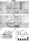
FIGURE 1.
FIGURE 1.
CDKL5 levels vary between the individual brain areas of P120 mice. A–P, immunohistochemistry experiments showing CDKL5 and MeCP2 expression in serial sections of an adult mouse brain (P120). The different panels show: cortex (A, E, I, M), hippocampus (B, F, J, N), cerebellum (C, G, K, O), and pons (D, H, L, P) of the adult mouse brain (P120). Panels E–H and M–P are magnifications of A–D and I–L, respectively. I-VI (panels A and I) indicate the different cortical layers; Dg, dentate gyrus; H, hilus; GL, granule cell layer; PL, Purkinje cell layer; MC, molecular cell layer. Q, Western blot showing CDKL5 and MeCP2 protein levels (upper and middle panels, respectively) in whole cell lysates prepared from dissected areas of a P120 brain. GAPDH was used as loading control (lower panel). R, graphic illustration showing the relative CDKL5 and MeCP2 levels (upper and lower graph, respectively) in hippocampus (Hip.), cortex (Ctx.), hypothalamus (Hyp.), thalamus (Th.), striatum (Str.), and cerebellum (Cer.) of the P120 mouse brain. CDKL5 and MeCP2 levels in the Western blot (panel Q) were normalized to GAPDH setting cortical levels arbitrarily to 1. Scale bars: A, I, D, L = 300 μm; B, J = 500 μm; C, K = 800 μm; E, H, M, p = 30 μm; G, O = 50 μm.
FIGURE 2.
FIGURE 2.
CDKL5 expression is highly induced at early postnatal stages of brain development. A–G, immunohistochemistry experiments showing CDKL5 expression in the cortex (A–D) and hippocampus (E–G) of mouse brains at the indicated embryonic or postnatal stages. H–N, MeCP2 expression in the cortex (H–K) and hippocampus (L--N) of mouse brains at different developmental stages. CP, cortical plate; IZ, intermediate zone; VZ, ventral zone; Hi, hippocampus; Dg, dentate gyrus; I-VI, different cortical layers. O and P, Western blot showing CDKL5 and MeCP2 protein levels (upper and middle panels, respectively) in total brain (O) or cerebellar (P) extracts at E18, P2, P5, P14, P28, and P120 stages. Actin was used as loading control (lower panel). Scale bars: A–D, H–K = 500 μm; E–G, L–N = 500 μm.
FIGURE 3.
FIGURE 3.
CDKL5 has a diffuse expression in most cells of the adult human brain. Immunohistochemistry experiments showing CDKL5 expression in cerebral cortex (A and B), corpus callosum (C and D), and cerebellum (E–H) of adult human brain. Arrows indicate some CDKL5-positive cells. PCL, Purkinje cell layer; GCL, granule cell layer; MCL, molecular cell layer. Scale bars: A, C, E, G = 30 μm; B, F = 10 μm; D, H = 20 μm.
FIGURE 4.
FIGURE 4.
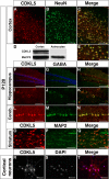
CDKL5 is a pan-neuronal protein in the adult mouse brain. A–C and E, Immunofluorescence experiments showing CDKL5 expression in cortical NeuN-positive neurons of a P120 mouse brain; D, Western blot showing CDKL5 and MeCP2 expression in whole cell extracts of cultured primary cortical neurons and astrocytes; F–N, CDKL5 expression in hippocampus (F–K) and cerebellum (L–N) is particularly high in GABAergic neurons (G, J, M); O–Q, CDKL5 colocalizes with MAP2-positive dendrites of neurons within the striatum. Panel E is a magnification of panel C. Panels C, E, H, K, N, and Q are merged images of CDKL5 (A, F, I, L, and O) and NeuN (B), GABA (G, J, M), and MAP2 (P), respectively. R and S, immunofluorescence of endogenous CDKL5 in primary cortical neurons (R; 10DIV). DAPI staining is shown in panel S and the merge in panel T. Scale bars: A–C, F–H = 150 μm; E, I–Q = 30 μm; R–T = 40 μm.
FIGURE 5.
FIGURE 5.
The subcellular distribution of CDKL5 varies between the individual areas of the adult mouse brain. A, fractionated cytoplasmic and nuclear extracts were prepared from carefully dissected brain areas of P120 mice and CDKL5 levels determined by Western blotting using MeCP2 and GAPDH as nuclear and cytoplasmic markers (upper, middle, and lower panels, respectively). B, graphic illustration showing the relative levels of cytoplasmic and nuclear CDKL5 (gray and black bars, respectively) in the different brain areas. Hip., hippocampus; Ctx, cortex; Hyp., hypothalamus; Th., thalamus; Str., striatum; Cer., cerebellum. The band intensities in A were quantified using a KODAK Image Station 2000R and the sum of cytoplasmic and nuclear CDKL5 set arbitrarily to 100%.
FIGURE 6.
FIGURE 6.
The nuclear localization of CDKL5 is induced at early postnatal stages of the developing mouse brain. A, fractionated cytoplasmic and nuclear extracts were prepared from total brains of E18, P2, P5, P14, P28, and P120 mice and relative levels of CDKL5 and MeCP2 analyzed by Western blotting (upper and middle panels, respectively). Actin was used as loading control (lower panels). B, graphic illustration showing the relative cytoplasmic and nuclear levels (gray and black bars, respectively) of CDKL5 (left) and MeCP2 (right). The band intensities in A were quantified with a KODAK Image Station 2000R and the sum of cytoplasmic and nuclear fractions set arbitrarily to 100%.
FIGURE 7.
FIGURE 7.
The C-terminal region mediates LMB-sensitive cytoplasmic localization of CDKL5. A, CDKL5, or the indicated derivatives, were fused to GFP, transiently overexpressed in human HeLa cells, and the subcellular localization analyzed 16 h after transfection. Right panels show the subcellular distribution in cells treated with 50 mm LMB for 3 h. DAPI was used to stain the nuclei of fixed cells. B, two CDKL5 derivatives carrying RTT causing truncating mutations were fused to GFP and their subcellular localization analyzed in HeLa cells. C, schematic illustration of the CDKL5 derivatives used in the above experiments. All derivatives carried GFP in the N terminus. Scale bar = 10 μm.

References

    1. Hagberg, B., Aicardi, J., Dias, K., and Ramos, O. (1983) Ann. Neurol. 14 471-479 - PubMed
    1. Hagberg, B., Goutieres, F., Hanefeld, F., Rett, A., and Wilson, J. (1985) Brain Dev. 7 372-373 - PubMed
    1. Armstrong, D. D. (2002) Ment. Retard. Dev. Disabil. Res. Rev. 8 72-76 - PubMed
    1. Hagberg, B. A., and Skjeldal, O. H. (1994) Pediatr. Neurol. 11 5-11 - PubMed
    1. Amir, R. E. (1999) Nat. Genet. 23 185-188 - PubMed

Publication types

Substances