. 2008 Aug 15;68(16):6762-9.
doi: 10.1158/0008-5472.CAN-08-0107.

The nuclear factor-kappaB pathway controls the progression of prostate cancer to androgen-independent growth

Affiliations

The nuclear factor-kappaB pathway controls the progression of prostate cancer to androgen-independent growth

Ren Jie Jin et al. Cancer Res. .

Abstract

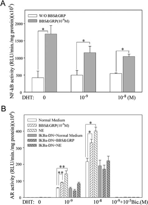
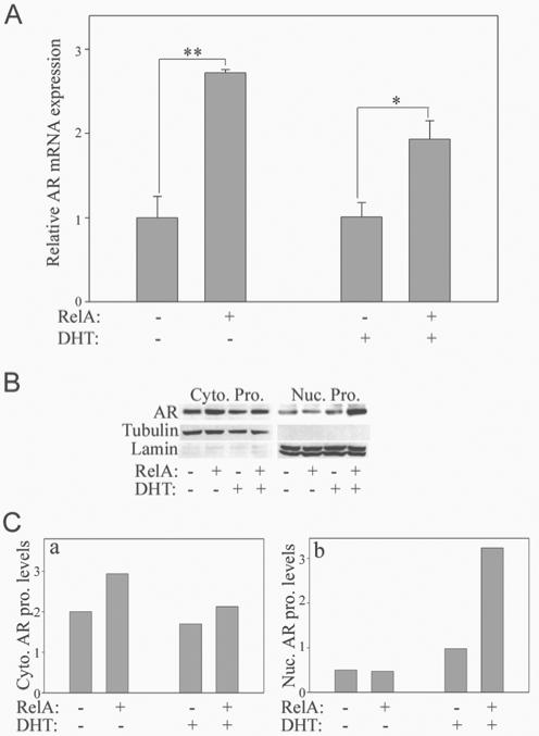
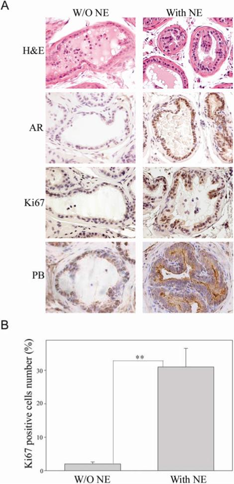
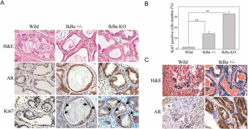
Typically, the initial response of a prostate cancer patient to androgen ablation therapy is regression of the disease. However, the tumor will progress to an "androgen-independent" stage that results in renewed growth and spread of the cancer. Both nuclear factor-kappaB (NF-kappaB) expression and neuroendocrine differentiation predict poor prognosis, but their precise contribution to prostate cancer progression is unknown. This report shows that secretory proteins from neuroendocrine cells will activate the NF-kappaB pathway in LNCaP cells, resulting in increased levels of active androgen receptor (AR). By blocking NF-kappaB signaling in vitro, AR activation is inhibited. In addition, the continuous activation of NF-kappaB signaling in vivo by the absence of the IkappaBalpha inhibitor prevents regression of the prostate after castration by sustaining high levels of nuclear AR and maintaining differentiated function and continued proliferation of the epithelium. Furthermore, the NF-kappaB pathway was activated in the ARR(2)PB-myc-PAI (Hi-myc) mouse prostate by cross-breeding into a IkappaBalpha(+/-) haploid insufficient line. After castration, the mouse prostate cancer continued to proliferate. These results indicate that activation of NF-kappaB is sufficient to maintain androgen-independent growth of prostate and prostate cancer by regulating AR action. Thus, the NF-kappaB pathway may be a potential target for therapy against androgen-independent prostate cancer.


Figures

Figure 1
Figure 1
NE peptides increase functional activation of NF-κB and AR in LNCaP cells. A. NE peptides (BBS and GRP) increase activation of NF-κB in LNCaP cells. The activity of NF-κB was determined by luciferase assay of protein extracts following transient transfection of NGL (NF-κB responsive reporter) vector. The values plotted represent the mean of at least three individual samples ± SEM. B. NE secreted factors increase activity of AR through the NF-κB pathway. The functional activity of AR was determined by luciferase assay of protein extracts following transient transfection of ARR2PB-Luc (AR responsive reporter) vector. Adenovirus expressing IκBα-DN was used to block NF-κB signaling. After transfection with ARR2PB-Luc and infection with IκBα-DN adenovirus (the empty adenovirus was used as control), LNCaP cells were treated with NE peptides (BBS and GRP, 10−8 M each) and NE secretions (conditioned medium containing NE extracts). The values plotted represent the mean of at least three individual samples ± SEM. Statistical significance was determined by student's t-test. *P <0.05; **P <0.01.
Figure 2
Figure 2
NF-κB activates transcription and/or stability of the AR in LNCaP cells. A. NF-κB (RelA) increases AR mRNA levels in LNCaP cells. The AR mRNA levels of LNCaP cells were quantified by real time RT-PCR after infection with RelA adenovirus. The amplification of AR was normalized to that of GAPDH. The values plotted represent the mean of at least three individual samples ± SEM. Statistical significance was determined by student's t-test. *P <0.05; **P <0.01. B. NF-κB (RelA) increases AR protein levels in LNCaP cells. Cytoplasmic (Cyto.) and nuclear (Nuc.) protein extracts were harvested from LNCaP cells after infection with RelA adenovirus. Western blot analysis was completed to detect AR protein levels. Lamin and Tubulin were used the control for nuclear and cytoplasmic protein, respectively. C. AR protein expression levels were quantified from the immunoblot by densitometry and the normalized expression of the AR protein (relative to Lamin or Tubulin) is represented by the bar graph adjacent to the immunoblot. a: Cytoplasmic AR protein; b: Nuclear AR protein.
Figure 3
Figure 3
NE-10 neuroendocrine tumor maintains prostate growth and AR expression after castration. A. Mice with or without NE tumor implantation were sacrificed at two weeks after castration. Immunohistochemical analysis was performed to determine AR, probasin (PB) expression and proliferation (Ki67) of the prostates. B. Cells positive for Ki67 were counted by monitoring at least 200 luminal epithelial cells from 3-5 different fields of each sample and plotted as a percentage of total counted. The results are reported as mean value (%); bars, ± SEM. ** P < 0.01 by Student's t test (t test).
Figure 4
Figure 4
Continuous activation of NF-κB signaling prevents regression of the mouse prostate after castration. A. Prostates from newborn mice (IκBα−/−, IκBα+/− and wild type) were grafted under the kidney capsule of male athymic nude mice and allowed to mature for 6 weeks in the male host. Then, host mice were castrated for two additional weeks. Immunohistochemical staining was performed to determine AR expression and proliferation (Ki67) of the prostates. Arrows indicate some of the Ki67 positive cells. B. The cells positive for Ki67 were counted by monitoring at least 200 luminal epithelial cells from 3-5 different fields of each sample and plotted as a percentage of total counted. The results are reported as mean value (%); bars, ± SEM. ** P < 0.01 by Student's t test (t test). C. Wild type and IκBα+/− transgenic mice were castrated at 7-8 weeks of age and the prostates were harvested six weeks after castration. H&E and immunohistochemical staining for AR were performed (pictures are showing dorsolateral lobes).
Figure 5
Figure 5
NF-κB signaling controls progression of prostate cancer to AI growth. A. Myc and Myc/IκBα+/− transgenic mice were castrated at 6 months of age and the prostates were harvested at two weeks after castration. Immunohistochemical staining was performed to determine AR expression and proliferation (Ki67) of the prostates (pictures are show lateral lobes). B. Cells positive for Ki67 were counted by monitoring at least 200 luminal epithelial cells from 3-5 different fields of each sample and plotted as a percentage of total counted. The results are reported as mean value (%); bars, ± SEM. ** P < 0.01 by Student's t test (t test).

Similar articles

Cited by

References

    1. Gregory CW, He B, Johnson RT, et al. A mechanism for androgen receptor-mediated prostate cancer recurrence after androgen deprivation therapy. Cancer Res. 2001;61:4315–4319. - PubMed
    1. Agoulnik IU, Weigel NL. Androgen receptor action in hormone-dependent and recurrent prostate cancer. J Cell Biochem. 2006;99:362–372. - PubMed
    1. Linja MJ, Savinainen KJ, Saramaki OR, Tammela TL, Vessella RL, Visakorpi T. Amplification and overexpression of androgen receptor gene in hormone-refractory prostate cancer. Cancer Res. 2001;61:3550–3555. - PubMed
    1. Jin RJ, Wang Y, Masumori N, et al. NE-10 neuroendocrine cancer promotes the LNCaP xenograft growth in castrated mice. Cancer Res. 2004;64:5489–5495. - PubMed
    1. Gregory CW, Johnson RT, Jr., Mohler JL, French FS, Wilson EM. Androgen receptor stabilization in recurrent prostate cancer is associated with hypersensitivity to low androgen. Cancer Res. 2001;61:2892–2898. - PubMed

Publication types

MeSH terms