. 2008 Oct;82(20):10247-61.
doi: 10.1128/JVI.01035-08. Epub 2008 Aug 13.

The vaccinia virus gene I2L encodes a membrane protein with an essential role in virion entry

Affiliations

The vaccinia virus gene I2L encodes a membrane protein with an essential role in virion entry

R Jeremy Nichols et al. J Virol. 2008 Oct.

Abstract

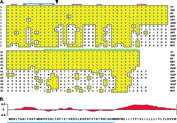
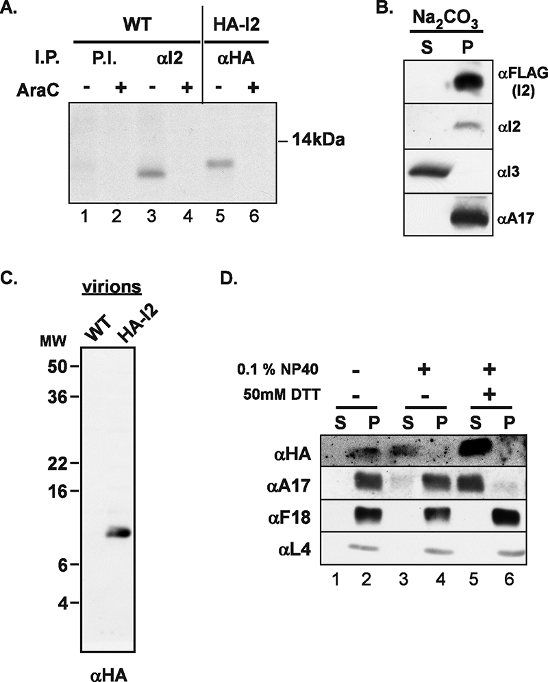
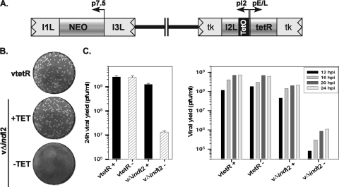
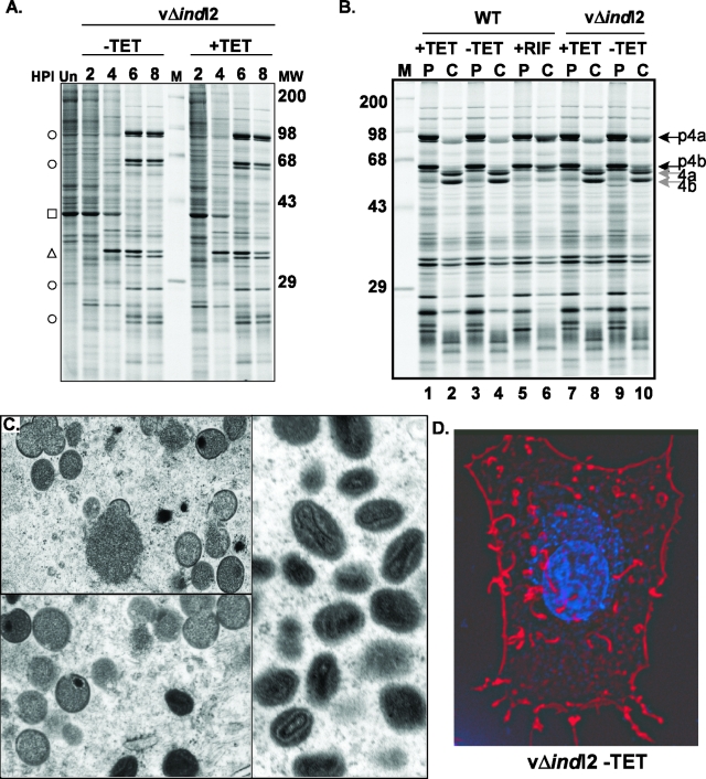
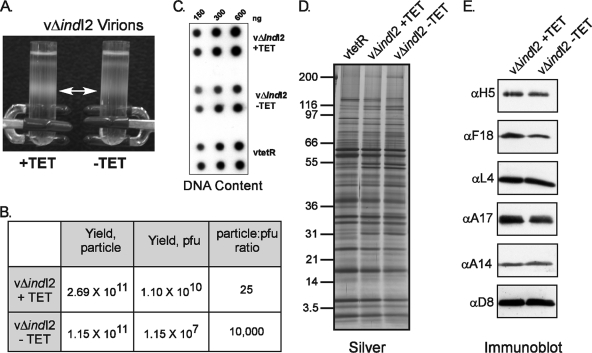
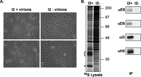
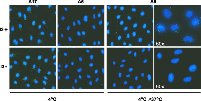
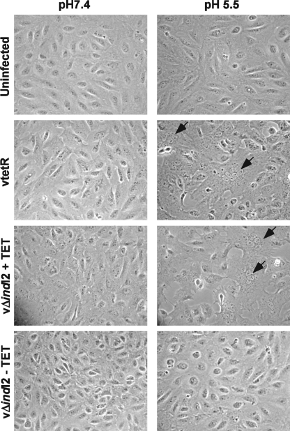
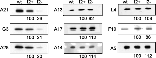
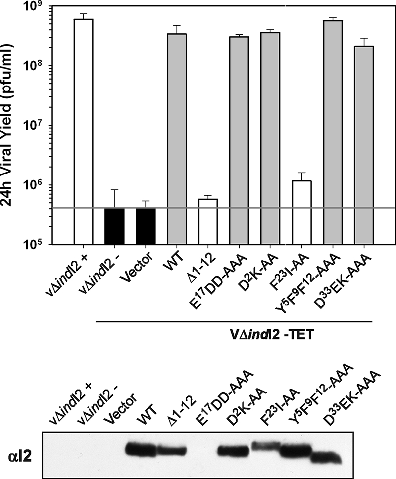
The previously unstudied vaccinia virus gene I2L is conserved in all orthopoxviruses. We show here that the 8-kDa I2 protein is expressed at late times of infection, is tightly associated with membranes, and is encapsidated in mature virions. We have generated a recombinant virus in which I2 expression is dependent upon the inclusion of tetracycline in the culture medium. In the absence of I2, the biochemical events of the viral life cycle progress normally, and virion morphogenesis culminates in the production of mature virions. However, these virions show an approximately 400-fold reduction in specific infectivity due to an inability to enter target cells. Several proteins that have been previously identified as components of an essential entry/fusion complex are present at reduced levels in I2-deficient virions, although other membrane proteins, core proteins, and DNA are encapsidated at normal levels. A preliminary structure/function analysis of I2 has been performed using a transient complementation assay: the C-terminal hydrophobic domain is essential for protein stability, and several regions within the N-terminal hydrophilic domain are essential for biological competency. I2 is thus yet another component of the poxvirus virion that is essential for the complex process of entry into target cells.


Figures

FIG. 1.
FIG. 1.
Conservation and structural organization of the I2 protein. (A) Sequence alignment of the poxviral I2 homologs. Shown is a sequence alignment of the I2 orthologs from vaccinia virus (WR strain) (VV [GenBank identification no. 29692238]), variola virus (VAR [GenBank no. 439035]), ectromelia virus (EV [GenBank no. 22164721]), monkeypox virus (MPV [GenBank no. 179750543]), cowpox virus (CPV [GenBank no. 20178507]), camelpox virus (CMLV [GenBank no. 18640364]), swinepox virus (SPV [GenBank no. 18640187]), Yaba-like disease virus (YLDV [GenBank no. 12085087]), swine fever virus (SFV [GenBank no. 18448493]), lumpy skin disease virus (LSDV [GenBank no. 151505037]), sheeppox virus (ShPV [GenBank no. 21492557]), myxoma virus (MYX [GenBank no. 18426922]), and molluscum contagiosum virus (MCV [GenBank no. 1492060]). (Sequences were obtained from www.poxvirus.org by using the POCs program.) Residues identical to vaccinia virus I2 are boxed and shaded in yellow; the green line marks a predicted membrane-anchoring domain, and the red and blue lines and arrowhead refer to residues chosen for mutagenesis. (B) Hydrophobicity plot of the I2 protein. The C terminus of I2 is predicted to contain a transmembrane domain (aa 47 to 73 [green line in panel A]). The region marked by the blue line was used to generate the anti-I2 serum.
FIG. 2.
FIG. 2.
I2 is a late protein that associates with membranes and is present within purified virions. (A) I2 is a late protein. Cells were infected with wt virus (lanes 1 to 4) or vHA-I2 (lanes 5 and 6) in the presence (lanes 2, 4, and 6) or absence (lanes 1, 3, and 5) of AraC and were metabolically labeled with [35S]Met from 3 to 8 hpi. Lysates were subject to immunoprecipitation (I.P.) analysis with preimmune (P.I. [lanes 1 and 2]), anti-I2 (αI2 [lanes 3 and 4]), or anti-HA (αHA [lanes 5 and 6]) antibodies. (B) I2 is a membrane protein. Cells infected with wt virus were transfected with pUC1246-I2, while cells infected with vTF7-3 were transfected with pTM13XFLAG-I2, so as to express wt I2 or 3× FLAG-tagged I2, respectively. Cells were harvested at 18 hpi and subjected to Na2CO3 fractionation; the supernatant (S) and pellet (P [membranes]) fractions were subjected to immunoblot analysis using anti-FLAG, anti-I2, anti-I3, or anti-A17 antibodies (αFLAG, αI2, αI3, and αA17, respectively), as indicated. (C) Encapsidation of I2 protein. Ten micrograms of purified wt and vHA-I2 virions was resolved by SDS-17% PAGE and subjected to immunoblot analysis with an anti-HA antibody. (D) Subvirion fractionation. To determine the localization of I2 within virions, 2 μg of purified HA-I2 virions was treated with buffer (lanes 1 and 2) or with NP-40, either alone (lanes 3 and 4) or in the presence of DTT (lanes 5 and 6); the soluble (S [lanes 1, 3, and 5]) and pellet (P [lanes 2, 4, and 6]) fractions were resolved by sedimentation and subjected to immunoblot analysis using anti-HA, anti-A17, anti-F18 (αF18), and anti-L4 (αL4) sera.
FIG. 3.
FIG. 3.
Repression of I2 expression causes a dramatic reduction in the yield of infectious virus and blocks plaque formation. (A) Schematic representation of the relevant regions of the vΔindI2 genome. An inducible copy of the I2 gene, under the regulation of its own promoter and the Tet operator, as well the Tet repressor gene, was inserted into the nonessential TK locus. The endogenous I2 allele was replaced by NEO. HPI, hours postinfection. (B) Plaque assay. Confluent monolayers of BSC40 cells were infected with the parental virus vTetR or vΔindI2 in the presence (+) or absence (−) of TET for 48 h; plaques were visualized after crystal violet staining. (C) Quantitation of viral yield. Cells were infected with vTetR or vΔindI2 in the presence (+) or absence (−) of TET for 24 h (left graph) or for 12, 16, 20, and 24 h (right graph). The yield of total cell-associated virus was determined by titration on BSC40 cells in the presence of TET.
FIG. 4.
FIG. 4.
I2 repression does not block viral gene expression or morphogenesis. (A) Temporal profile of viral protein synthesis. BSC40 cells were infected vΔindI2 (±TET) and pulse-labeled with [35S]methionine at sequential times after infection as indicated (2, 4, 6, and 8 hpi). Lysates were resolved by SDS-PAGE and visualized by autoradiography. The molecular masses of 14C protein standards (M) are indicated to the right (in kDa). Representative intermediate proteins are indicated by triangles and late proteins by circles; the cellular protein actin is indicated by a square. (B) Proteolytic processing of core proteins. BSC40 cells were infected with wt virus (lanes 1 to 6) or vΔindI2 (lanes 7 to 10) in the presence (+) or absence (−) of TET. RIF was included where indicated (lanes 5 and 6). At 8 hpi, cells were pulse-labeled with [35S]Met for 45 min before being harvested immediately (pulse [P]) or refed with complete media and incubated for an additional 15 h (chase [C]). Cell lysates were resolved by SDS-PAGE and visualized by autoradiography. 14C protein standards are shown to the left (M), with their molecular masses indicated (in kDa). The precursor forms of the core proteins 4a and 4b are indicated by black arrows, while the processed mature forms are indicated by gray arrows. (C) Electron microscopic analysis of vΔindI2 infections. BSC40 cells were infected with vΔindI2 in the presence (not shown) or absence of TET for 18 hpi and examined by transmission electron microscopy. Mature virions and the full range of morphogenesis intermediates were observed in both samples. (D) Visualization of actin tails. BSC40 cells were infected with vΔindI2 (MOI of 2) in the absence (or presence [not shown]) of TET; at 18 hpi, ceIls were fixed and stained with Texas Red-phalloidin to visualize actin tails (and DAPI to visualize cellular and viral DNA).
FIG. 5.
FIG. 5.
I2-deficient virions have a normal protein and DNA complement but are greatly reduced in specific infectivity. (A) Virion purification. BSC40 cells were infected with vTetR (not shown) or vΔindI2 (±TET) (MOI of 5), and virions were purified by sedimentation through a 36% sucrose cushion and a 25 to 40% sucrose gradient. The light-scattering bands of virions are indicated by the white arrow. (B) Quantitation of particle yield and infectious yield. The yields of virion particles and infectious virus were quantitated by measuring the OD260 and by plaque assay titration, respectively. (C) Determination of DNA content. One hundred fifty, 300, and 600 ng of purified vTetR, I2+ and I2 virions were subjected to Southern dot blot hybridization using a probe representing fragments of the vaccinia virus genome. (D and E) Equal amounts of vTetR, I2+, and I2 virions were resolved by SDS-PAGE and subjected to silver staining (D) or immunoblot (E) analysis with antisera specific for the H5, F18, L4, A17, A14, and D8 proteins (αH5 to αD8, respectively).
FIG. 6.
FIG. 6.
I2-deficient virions do not induce CPE or mediate early gene expression in target cells. (A) Visualization of CPE. BSC40 cells were infected with I2+ (left panels) or I2 (right panels) virions (MOI of 10) in the presence of [35S]Met. At 4 hpi, the cells were visualized by phase-contrast microscopy; representative images are shown. (B) Analysis of early gene expression. Lysates prepared from the infections described in panel A were either resolved electrophoretically and examined by autoradiography (left panel) or subjected to immunoprecipitation (IP) analysis with antibodies directed against the early viral proteins E9, D5, I3, and H5 (αE9 to αH5, respectively [right panel]) and then visualized by autoradiography. The brace symbol and dot indicate early viral proteins that are evident in the cells infected with I2+, but not I2, virions. The molecular masses of 14C protein standards (in kDa) are indicated to the right of the autoradiograph.
FIG. 7.
FIG. 7.
I2-deficient virions can bind to target cells but are deficient in entry. BSC40 cells were infected with 300 particles of I2+ or I2 virions (in duplicate) and maintained on ice for 1 h. One set of samples was fixed immediately (4°C), whereas a second set was incubated for at 37°C for 2 h in complete medium containing 300 μg/ml cycloheximide before being fixed (from 4°C to 37°C). Cells were permeabilized and stained with anti-A17 (left panels) or anti-A5 (right panels) sera. DAPI was added to visualize nuclei.
FIG. 8.
FIG. 8.
I2-deficient virions cannot mediate fusion from without. BSC40 cells were incubated with 10,000 particles per cell of purified vTetR, I2+, or I2 virions. After 1 h of absorption at 4°C, the cells were washed and subjected to a brief pulse with pH 5.5 or pH 7.4 medium. Cells were washed and incubated at 37°C for 3 h in the presence of cycloheximide. Phase-contrast microscopy images are presented. The arrows indicate syncytia formed by virion-mediated acid-induced fusion (right panels).
FIG. 9.
FIG. 9.
I2-deficient virions contain reduced levels of some components of the EFC. Three micrograms of purified wt, I2+, or I2 virions were resolved by SDS-PAGE and subjected to immunoblot analysis with antisera specific for components of the EFC (A21, G3, and A28), membrane proteins involved in virion morphogenesis (A13, A14, and A17), or core components (L4, F10, and A5). For each blot, the level of protein present in the I2+ virions was set at 100, and the relative level present in the I2 virions was determined.
FIG. 10.
FIG. 10.
The biological activities of various I2 alleles can be monitored via transient complemention of vΔindI2. BSC40 cells were infected with vΔindI2 (±TET); at 3 hpi, cells were transfected with empty vector or plasmids encoding wt or mutant alleles of I2 under the regulation of a viral promoter. Cells were harvested at 24 hpi, and plaque assays were performed to determine the viral yield. The data are shown graphically in the top panel, with the horizontal line representing the baseline yield obtained from uninduced infections. A portion of the harvested cells was also resolved by SDS-PAGE, and the expression of the plasmid-borne I2 protein was verified by immunoblot analysis with anti-I2 antibody (αI2). The I2 protein expressed from the viral genome is below the level of detection in this experiment.

Similar articles

Cited by

References

    1. Bayliss, C. D., and R. C. Condit. 1995. The vaccinia virus A18R gene product is a DNA-dependent ATPase. J. Biol. Chem. 2701550-1556. - PubMed
    1. Bisht, H., A. S. Weisberg, and B. Moss. 2008. Vaccinia virus L1 protein is required for cell entry and membrane fusion. J. Virol. [Epub ahead of print.] doi:10.1128/JVI.00852-08. - DOI - PMC - PubMed
    1. Brown, E., T. G. Senkevich, and B. Moss. 2006. Vaccinia virus F9 virion membrane protein is required for entry but not virus assembly, in contrast to the related L1 protein. J. Virol. 809455-9464. - PMC - PubMed
    1. Carter, G. C., M. Law, M. Hollinshead, and G. L. Smith. 2005. Entry of the vaccinia virus intracellular mature virion and its interactions with glycosaminoglycans. J. Gen. Virol. 861279-1290. - PubMed
    1. Chiu, W.-L., C.-L. Lin, M.-H. Yang, D.-L. M. Tzou, and W. Chang. 2007. Vaccinia virus 4c (A26L) protein on intracellular mature virus binds to the extracellular cellular matrix laminin. J. Virol. 812149-2157. - PMC - PubMed

Publication types

MeSH terms

LinkOut - more resources