Review
. 2008 Dec;295(6):F1601-12.
doi: 10.1152/ajprenal.00097.2008. Epub 2008 Aug 13.

A lipid-protein hybrid model for tight junction

Affiliations
Review

A lipid-protein hybrid model for tight junction

David B N Lee et al. Am J Physiol Renal Physiol. 2008 Dec.

Abstract

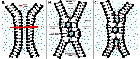
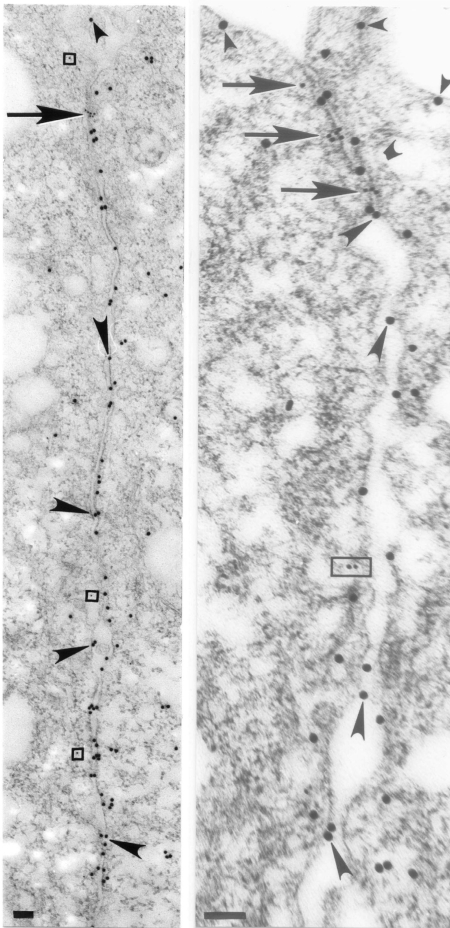
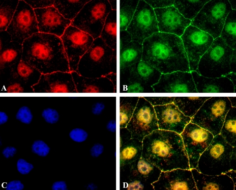
The epithelial tight junction (TJ) was first described ultrastructurally as a fusion of the outer lipid leaflets of the adjoining cell membrane bilayers (hemifusion). The discovery of an increasing number of integral TJ and TJ-associated proteins has eclipsed the original lipid-based model with the wide acceptance of a protein-centric model for the TJ. In this review, we stress the importance of lipids in TJ structure and function. A lipid-protein hybrid model accommodates a large body of information supporting the lipidic characteristics of the TJ, harmonizes with the accumulating evidence supporting the TJ as an assembly of lipid rafts, and focuses on an important, but relatively unexplored, field of lipid-protein interactions in the morphology, physiology, and pathophysiology of the TJ.


Figures

Fig. 1.
Fig. 1.
Tight junction (TJ) models. A: a protein model, in which extracellular components of integral TJ proteins (red) from adjacent cells fuse to form TJ barrier. B and C: speculative lipid-protein hybrid models formed by hemifusion of plasma membrane bilayers from 2 adjacent cells (cells 1 and 2) and formation of HII cylinders (TJ strands) or inverted micelles (TJ particles) or a combination of both. In B, protein channels or transporters (blue cylinders) insert into and populate this nonlamellar lipid infrastructure, forming a series of linked aqueous compartments across the TJ. In C, proteins (not shown) participate in trans-TJ transport through a process similar to “endosytosis-transcytosis-exocytosis.” In lipid-protein hybrid models, exoplasmic leaflet of 1 cell is linked to exoplasmic leaflet of the neighboring cell through a fusion region (F); in a protein-only model, no such linkage exists. Blue dots denote aqueous environment.
Fig. 2.
Fig. 2.
Lipid polymorphism. Schematic illustration of a micelle (A), bilayers (B), an inverted micelle (C), and a hexagonal (HI, D) or an inverted hexagonal (HII, E) cylinder. Each phospholipid molecule consists of a hydrophilic (water-loving) head group (blue spheres) facing an aqueous environment (blue background) and 1 or 2 hydrophobic (water-fearing) carbon tails (yellow) facing a lipid environment (yellow background).
Fig. 3.
Fig. 3.
Annexin A2 heterotetramer (AnxA2t)-mediated junction formation [modified from Lambert et al. (54)]. AnxA2-p11 complex (AnxA2t) forms a symmetrical bridge between 2 lipid bilayers (only 1 of the 2 participating lipid bilayers is shown at right). The 2 annexin A2 (AnxA2) subunits, each binding to a separate bilayer, are, in turn, linked by a p11 dimer. [Modified from Lee et al. (55).]
Fig. 4.
Fig. 4.
AnxA2t colocalizes with the TJ protein claudin-1. An immediately preconfluent Madin-Darby canine kidney (MDCK II) monolayer was triple-stained for AnxA2 subunit (A, red) of AnxA2t, TJ protein, claudin-1 (B, green), and the nucleus (C, blue). Nuclear staining was accomplished using 4,6-diamidino-2-phenylindole (DAPI; Molecular Probes, Eugene, OR). Merged image (D) shows TJ colocalization of AnxA2 and claudin-1 (yellow). [Triple staining of the nucleus (purple) is discussed in Lee et al. (56).] Magnification ×1,000. [From Lee et al. (56).]
Fig. 5.
Fig. 5.
Ultrastructural immunogold localization of AnxA2t and TJ proteins in MDCK II monolayers. AnxA2 subunit of AnxA2t (larger, 20-nm gold particles) is distributed in juxtaposition to the TJ proteins (smaller, 10-nm gold particles, arrows) zonula occludens-1 (ZO-1, left) and occludin (right). AnxA2 labels also extend beyond the TJ, along the length of the lateral plasma membrane and the apical, luminal membrane (small arrowheads). ZO-1 particles (arrow, left) aggregate in an area where apposing membranes seem to merge and appear mostly on the cytoplasmic side of the plasma membrane. Cytoplasmic ZO-1 labeling is sometimes observed outside the TJ (enclosed by squares). reflecting the general phenomenon of internalization of TJ proteins (115). Occludin particles (arrows, right) have a similar distribution, consistent with the use of antibody generated against a fusion protein consisting of the cytoplasmic COOH-terminal 150 amino acids of human occludin. Basolateral cytoplasmic labeling of occludin (enclosed by rectangle) is consistent with prior reports and represents the less phosphorylated form of this molecule, distributed in detergent-soluble lipid domains (81, 85). Although most AnxA2 labeling is distributed along the cytoplasmic side of the plasma membrane, it is also seen along the exoplasmic side of the plasma membrane and in the intercellular space (large arrowheads). Scale bars, 0.1 μm. [From Lee et al. (56).]
Fig. 6.
Fig. 6.
TJ protein claudin-2 colocalizes with lipid raft marker GM1. An immediately preconfluent MDCK II monolayer was triple-stained for claudin-2 (A, red), GM1 (B, green), and the nucleus (C, blue). Merged image (D) shows TJ colocalization of claudin-2 and GM1 (yellow). Differences in staining intensity of the 3 labels account for nuclear staining in merged image (D), which assumed a gold, rather than a purplish, hue, as in Fig. 4D. [See Lee et al. (56) for discussion of nuclear staining of claudin-2 and GM1.] Magnification ×1,500. [From Lee et al. (56).]

References

    1. Anderson JM Molecular structure of tight junctions and their role in epithelial transport. News Physiol Sci 16: 126–130, 2001. - PubMed
    1. Ayala-Sanmartin J, Henry JP, Pradel LA. Cholesterol regulates membrane binding and aggregation by annexin 2 at submicromolar Ca2+ concentration. Biochim Biophys Acta 1510: 18–28, 2001. - PubMed
    1. Balda MS, Whitney JA, Flores C, Gonzalez S, Cereijido M, Matter K. Functional dissociation of paracellular permeability and transepithelial electrical resistance and disruption of the apical-basolateral intramembrane diffusion barrier by expression of a mutant tight junction membrane protein. J Cell Biol 134: 1031–1049, 1996. - PMC - PubMed
    1. Blasig IE, Winkler L, Lassowski B, Mueller SL, Zuleger N, Krause E, Krause G, Gast K, Kolbe M, Piontek J. On the self-association potential of transmembrane tight junction proteins. Cell Mol Life Sci 63: 505–514, 2006. - PMC - PubMed
    1. Bruewer M, Hopkins AM, Hobert ME, Nusrat A, Madara JL. RhoA, Rac1, and Cdc42 exert distinct effects on epithelial barrier via selective structural and biochemical modulation of junctional proteins and F-actin. Am J Physiol Cell Physiol 287: C327–C335, 2004. - PubMed

Publication types

LinkOut - more resources