. 2008 Nov;295(5):C1071-82.
doi: 10.1152/ajpcell.00040.2008. Epub 2008 Aug 13.

Stimulation of glucose transport in response to activation of distinct AMPK signaling pathways

Affiliations

Stimulation of glucose transport in response to activation of distinct AMPK signaling pathways

Ming Jing et al. Am J Physiol Cell Physiol. 2008 Nov.

Abstract

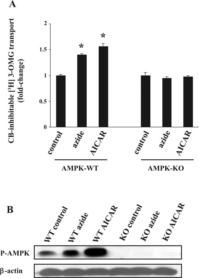
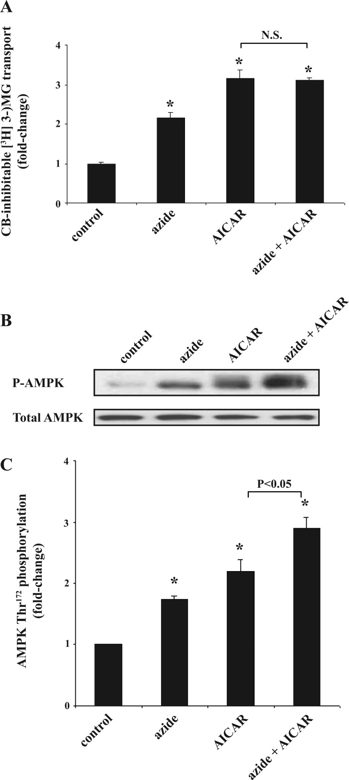
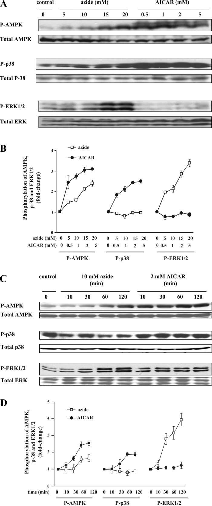
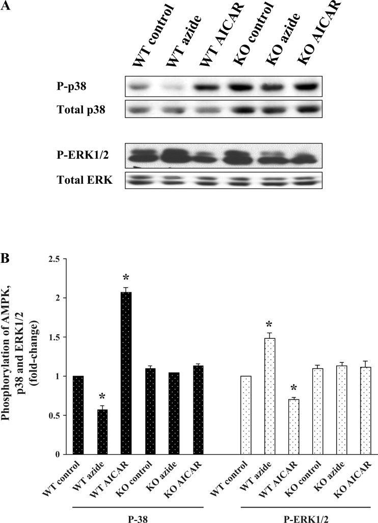
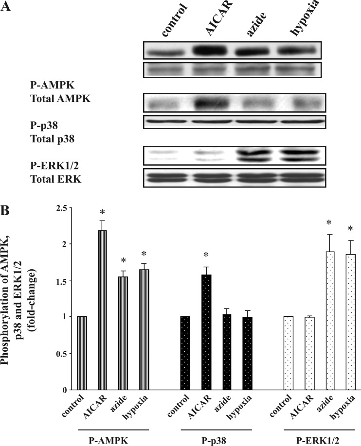
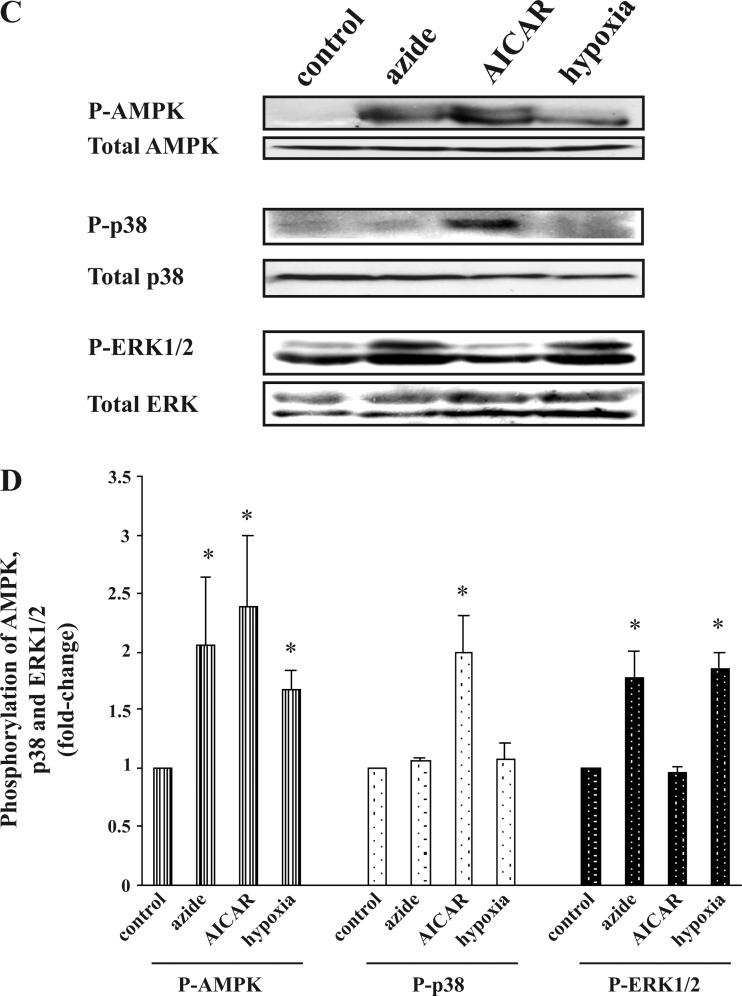
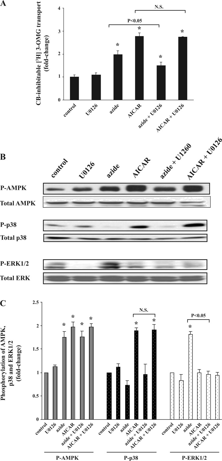
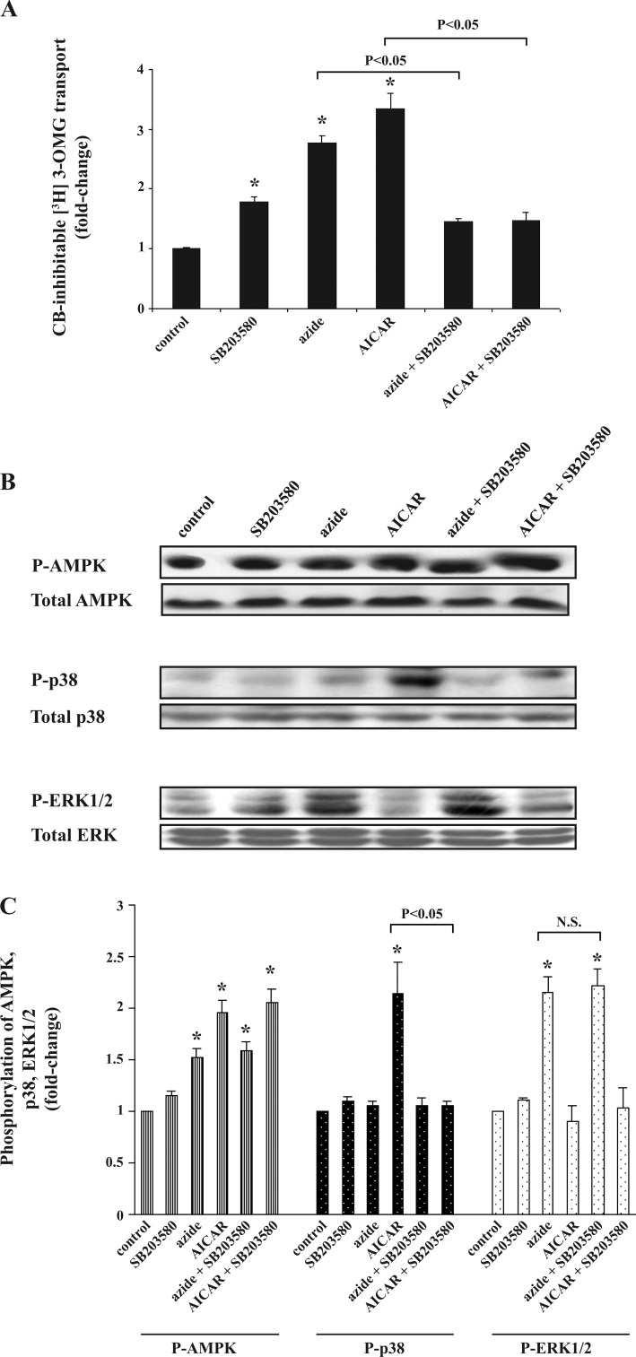
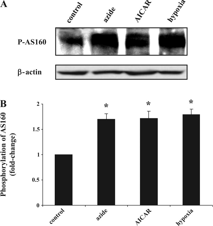
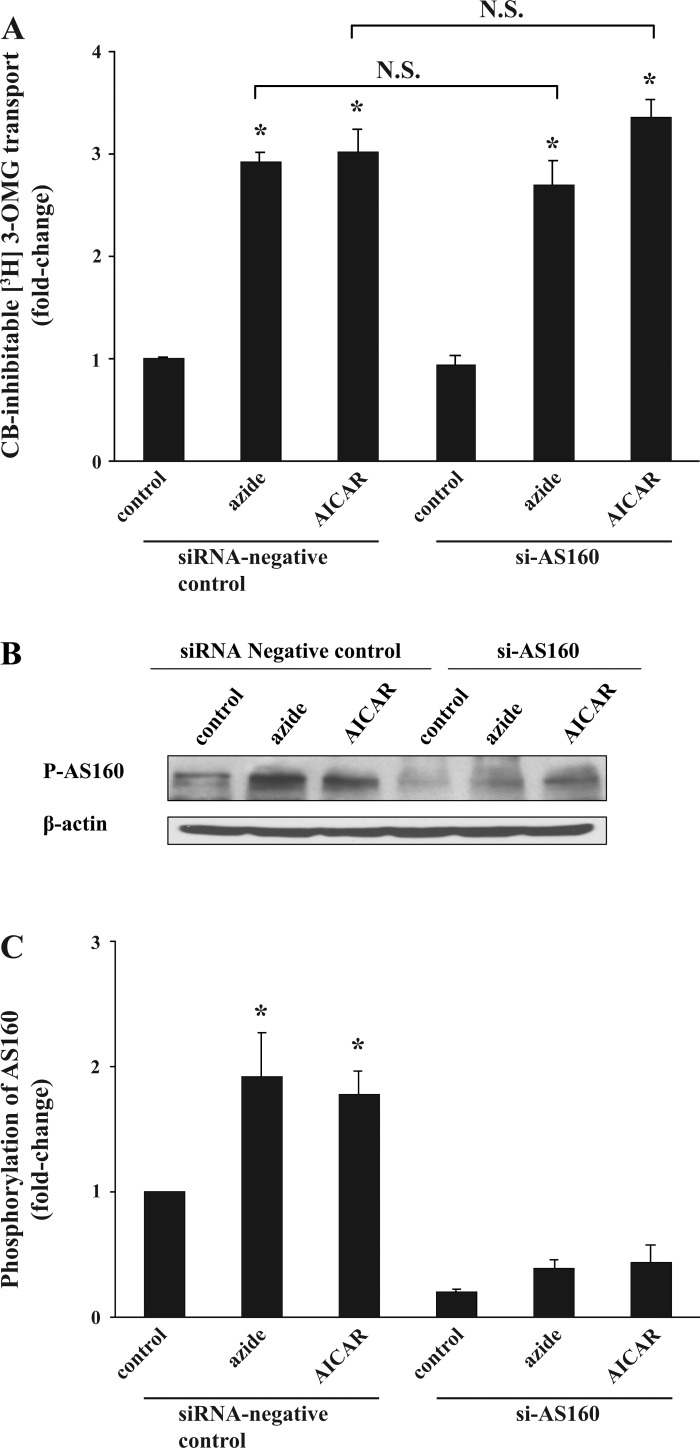
AMP-activated protein kinase (AMPK) plays a critical role in the stimulation of glucose transport in response to hypoxia and inhibition of oxidative phosphorylation. In the present study, we examined the signaling pathway(s) mediating the glucose transport response following activation of AMPK. Using mouse fibroblasts of AMPK wild type and AMPK knockout, we documented that the expression of AMPK is essential for the glucose transport response to both azide and 5-aminoimidazole-4-carboxamide-1-beta-d-ribofuranoside (AICAR). In Clone 9 cells, the stimulation of glucose transport by a combination of azide and AICAR was not additive, whereas there was an additive increase in the abundance of phosphorylated AMPK (p-AMPK). In Clone 9 cells, AMPK wild-type fibroblasts, and H9c2 heart cells, azide or hypoxia selectively increased p-ERK1/2, whereas, in contrast, AICAR selectively stimulated p-p38; phosphorylation of JNK was unaffected. Azide's effect on p-ERK1/2 abundance and glucose transport in Clone 9 cells was partially abolished by the MEK1/2 inhibitor U0126. SB 203580, an inhibitor of p38, prevented the phosphorylation of p38 and the glucose transport response to AICAR and, unexpectedly, to azide. Hypoxia, azide, and AICAR all led to increased phosphorylation of Akt substrate of 160 kDa (AS160) in Clone 9 cells. Employing small interference RNA directed against AS160 did not inhibit the glucose transport response to azide or AICAR, whereas the content of P-AS160 was reduced by approximately 80%. Finally, we found no evidence for coimmunoprecipitation of Glut1 and p-AS160. We conclude that although azide, hypoxia, and AICAR all activate AMPK, the downstream signaling pathways are distinct, with azide and hypoxia stimulating ERK1/2 and AICAR stimulating the p38 pathway.


Figures

Fig. 1.
Fig. 1.
Effect of azide and 5-aminoimidazole-4-carboxamide-1-β-d-ribofuranoside (AICAR) on glucose transport and on the abundance of phosphorylated AMP-activated protein kinase (p-AMPK) in wild-type (WT) and AMPK knockout (KO) fibroblasts. A: glucose transport. Confluent WT and AMPK KO fibroblasts in triplicate 6-cm culture dishes were serum-starved for 24 h. Cells were treated with diluent, 10 mM azide, or 2 mM AICAR for 1 h before measurement of cytochalasin B (CB)-inhibitable 3-O-methyl-d-[3H]glucose ([3H]3-OMG) transport in 3 independent experiments. Cells were lysed in 0.5 ml of H2O and used for radioactive counting. In each experiment, the rate of transport was normalized against that of untreated WT cells, and the results were averaged. The basal rate of glucose transport in WT and AMPK KO fibroblasts were not significantly different. *P < 0.05 compared with WT control (ANOVA). B: p-AMPK. Cells were treated as described in A, and lysates were subjected to SDS-PAGE. The abundance of p-AMPK was determined in the immunoblots. The abundance of p-AMPK increased 1.5 ± 0.1- and 2.3 ± 0.1-fold in WT cells treated with azide or AICAR, respectively. Blots were reprobed with antibodies against β-actin. Two additional independent experiments yielded similar results.
Fig. 2.
Fig. 2.
Effect of azide plus AICAR on glucose transport and p-AMPK abundance in Clone 9 cells. A: glucose transport. Confluent Clone 9 cells in triplicate 6-cm dishes were serum-starved for 24 h. Cells were treated with diluent, 10 mM azide, 2 mM AICAR, or both for 1 h before measurement of CB-inhibitable [3H]3-OMG. The experiment was repeated 3 times, and the results were averaged. Rates of transport were normalized against the control. *P < 0.05 compared with control. There was no significant difference (N.S.) between the rate of transport in AICAR- vs. AICAR + azide-treated cells (t-test; P > 0.05). B and C: p-AMPK and total AMPK abundance. Cell lysates treated in parallel with those described in A were subjected to immunoblotting to measure the relative content of cell p-AMPK and total AMPK. Results of the blots were analyzed using densitometry, and phosphorylation of AMPK is expressed as the degree of (fold) change compared with values in control cells after normalization against total AMPK. *P < 0.05 compared with control. p-AMPK in AICAR- vs. AICAR + azide-treated cells was significantly different. The content of total AMPK remained constant.
Fig. 3.
Fig. 3.
AICAR stimulates phosphorylation of p38 and azide activates phosphorylation of ERK1/2 in a dose- and time-dependent manner. A and B. dose response. Confluent Clone 9 cells were treated with diluent; 5, 10, 15, or 20 mM azide; or 0.5, 1.0, 2, or 5 mM AICAR for 1 h in 3 independent experiments. Cell lysates prepared in Nonidet P-40 (NP-40)-containing buffer were subjected to immunoblotting to measure the relative content of p-AMPK, AMPK, phosphorylated p38 (p-p38), total p38, phosphorylated ERK1/2 (p-ERK1/2), and total ERK. In each experiment, the densitometry value of each phosphoprotein in the different conditions was normalized against relative amounts of each protein. The results of 3 independent experiments were averaged and are expressed as fold change compared with the value of the control. C and D: time course. Confluent Clone 9 cells were treated with diluent, 10 mM azide, or 2 mM AICAR for 10, 30, 60, or 120 min. Cell lysates were subjected to immunoblotting to measure the relative content of cell p-AMPK, total AMPK, p-p38, total p38, p-ERK1/2, and total ERK. In each experiment, the densitometry value of each phosphoprotein was normalized against the relative abundance of each protein. The experiment was repeated 3 times, and the results were averaged and are expressed as fold change.
Fig. 4.
Fig. 4.
Effect of azide and AICAR on the abundance of p-p38 and p-ERK1/2 in mouse fibroblasts. A and B: confluent AMPK WT and KO mouse fibroblasts were treated with diluent, 10 mM azide, or 2 mM AICAR for 1 h. Cell lysates were subjected to immunoblotting to measure the relative content of the phospho- and total proteins. The experiment was repeated 3 times; a representative blot is shown. Results of the blots were analyzed using densitometry and normalized against total p38 and total ERK1/2. Phosphorylation of p38 and ERK1/2 is expressed as fold change compared with values in individual control cells. *P < 0.05 compared with individual control. P-p38 increased response to AICAR treatment, and p-ERK1/2 increased response to azide treatment in WT mouse fibroblasts, whereas there was no change in phosphoprotein in KO mouse fibroblast exposed to either azide or AICAR.
Fig. 5.
Fig. 5.
Effect of hypoxia on the abundance of p-AMPK, p-p38, and p-ERK1/2 in Clone 9 and H9C2 cells. A and B: Clone 9 cells. Confluent Clone 9 cells were exposed to hypoxia for 1 h, while cells treated with diluent, 10 mM azide, or 2 mM AICAR served as controls. C and D: H9C2 cells. Confluent H9C2 cells were exposed to hypoxia for 1 h, while cells treated with diluent, 10 mM azide, or 2 mM AICAR served as controls. The experiment was repeated 3 times; a representative blot is shown. Results of the blots were analyzed using densitometry and normalized against total AMPK, total p38, and total ERK1/2. Phosphorylation of AMPK, p38, and ERK1/2 is expressed as fold change compared with values in control cells. *P < 0.05 compared with individual control.
Fig. 5.
Fig. 5.
Effect of hypoxia on the abundance of p-AMPK, p-p38, and p-ERK1/2 in Clone 9 and H9C2 cells. A and B: Clone 9 cells. Confluent Clone 9 cells were exposed to hypoxia for 1 h, while cells treated with diluent, 10 mM azide, or 2 mM AICAR served as controls. C and D: H9C2 cells. Confluent H9C2 cells were exposed to hypoxia for 1 h, while cells treated with diluent, 10 mM azide, or 2 mM AICAR served as controls. The experiment was repeated 3 times; a representative blot is shown. Results of the blots were analyzed using densitometry and normalized against total AMPK, total p38, and total ERK1/2. Phosphorylation of AMPK, p38, and ERK1/2 is expressed as fold change compared with values in control cells. *P < 0.05 compared with individual control.
Fig. 6.
Fig. 6.
Effect of U0126 on azide- or AICAR-induced stimulation of glucose transport, p-AMPK, p-p38, and p-ERK1/2. A: glucose transport. Confluent Clone 9 cells in triplicate 60-mm dishes were pretreated with diluent or 10 μM U0126 for 20 min before being treated with diluent, 10 mM azide, or 2 mM AICAR for 1 h. Uptakes in control and treated cells were performed in parallel. The experiment was repeated 3 times using triplicate dishes for each condition, and the results were averaged. Values are expressed as fold change (±SE) compared with control. *P < 0.05 compared with control (ANOVA). Glucose transport was lower in cells treated with both U0126 and azide compared with azide alone (t-test; P < 0.05). U0126 had no effect on glucose transport in AICAR-treated cells. B and C: abundance of p-AMPK, p-p38, and p-ERK1/2. Cells were treated as described in A, lysed in NP-40-containing buffer, and used for immunoblotting. In each experiment, the densitometry value of phosphorylation of each protein was normalized against the control, and the results of 3 independent experiments were averaged. *P < 0.05 compared with control. Cells exposed to U0126 plus azide had decreased abundance of p-ERK1/2 compared with azide alone (P < 0.05). U0126 had no effect on p-p38 in AICAR-treated cells.
Fig. 7.
Fig. 7.
Effect of SB 203580 on azide- or AICAR-induced stimulation of glucose transport, p-AMPK, p-p38, and p-ERK1/2. A: glucose transport. Confluent Clone 9 cells in triplicate 60-mm dishes were serum-starved for 24 h before the experiment. Cells were pretreated with 10 μM SB 203580 or diluent for 60 min before being treated 10 mM azide or 2 mM AICAR for 1 h. Uptakes in control and all treated cells were performed in parallel. The experiment was repeated 2 times using triplicate dishes for each condition, and the results were averaged. Values are expressed as fold change (±SE) compared with basal values. *P < 0.05 compared with control (ANOVA). Glucose uptake was significantly different in cells treated with both SB 203580 and AICAR vs. AICAR alone and in cells treated with both SB 203580 and azide vs. azide alone (t-test; P < 0.05). Exposure to SB 203580 alone also stimulated the rate of glucose transport. B and C: abundance of p-AMPK, p-p38, and p-ERK1/2. Cell treated as described in A were lysed in NP-40-containing buffer and used for immunoblotting. In each experiment, the densitometry value of each phosphoprotein in each condition was normalized against the control, and the results of 3 independent experiments were averaged. *P < 0.05 compared with control. Cells preexposed to SB 203580 and AICAR had decreased abundance of p-p38 compared with cells treated with AICAR alone (P < 0.05). SB 203580 had no effect on the effect of azide on p-ERK1/2.
Fig. 8.
Fig. 8.
Effect of azide, hypoxia, and AICAR on the abundance of phosphorylated Akt substrate of 160 kDa (p-AS160). A: Western blot of p-AS160. Confluent Clone 9 cells were treated with diluent, 10 mM azide, hypoxia, or 2 mM AICAR for 1 h. Cell lysates were subjected to immunoblotting to measure the relative content of p-AS160; β-actin was used as a control for loading of the gel. B: quantification of p-AS160. Results of 3 independent experiments were averaged and are expressed as fold change compared with control. *P < 0.05 compared with control (n = 3).
Fig. 9.
Fig. 9.
Effect of small interfering RNA directed against AS160 (siAS160) on glucose transport in response to azide and AICAR and on p-AS160. A: glucose transport. Seventy percent confluent Clone 9 cells in triplicate dishes were transfected with 5 μl/ml Lipofectamine 2000 containing 100 pmol/ml siAS160 or scrambled siRNA. Forty-eight hours after transfection, cells were treated with diluent, 10 mM azide, or 2 mM AICAR for 1 h before measurement of CB-inhibitable 3-OMG transport. The experiment was repeated another time, and the results were averaged. *P < 0.05 compared with control siRNA. The stimulation of transport in response to azide or AICAR was not decreased by exposure to the siRNA (P > 0.05). B and C: p-AS160. Cells lysates were subjected to immunoblotting for p-AS160. Similar results were obtained in a second independent experiment. The content of β-actin remained constant. Results of the blots were analyzed using densitometry and normalized against the respective control. *P < 0.05 compared with control.

Similar articles

Cited by

References

    1. Abbud W, Habinowski S, Zhang JZ, Kendrew J, Elkairi FS, Kemp BE, Witters LA, Ismail-Beigi F. Stimulation of AMP-activated protein kinase (AMPK) is associated with enhancement of Glut1-mediated glucose transport. Arch Biochem Biophys 380: 347–352, 2000. - PubMed
    1. Antonescu CN, Huang C, Niu W, Liu Z, Eyers PA, Heidenreich KA, Bilan PJ, Klip A. Reduction of insulin-stimulated glucose uptake in L6 myotubes by the protein kinase inhibitor SB203580 is independent of p38MAPK activity. Endocrinology 146: 3773–3781, 2005. - PubMed
    1. Asahi Y, Hayashi H, Wang L, Ebina Y. Co-localization of the translocated GLUT4 with rearranged actin by insulin treatment in CHO cells and L6 myotubes. J Med Invest 46 :192–199, 1999. - PubMed
    1. Barnes K, Ingram JC, Porras OH, Barros LF, Hudson ER, Fryer Lee GD, Foufelle F, Carling D, Hardie DG, Baldwin SA. Activation of GLUT1 by metabolic, and osmotic stress: potential involvement of AMP-activated protein kinase (AMPK). J Cell Sci 115: 2433–2442, 2002. - PubMed
    1. Begonja AJ, Geiger J, Rukoyatkina N, Rauchfuss S, Gambaryan S, Walter U. Thrombin stimulation of p38 MAP kinase in human platelets is mediated by ADP, and thromboxane A2 and inhibited by cGMP/cGMP-dependent protein kinase. Blood 109: 616–618, 2007. - PubMed

Publication types

MeSH terms