Comparative Study
. 2008 Aug 13;28(33):8189-98.
doi: 10.1523/JNEUROSCI.2218-08.2008.

Depletion of 26S proteasomes in mouse brain neurons causes neurodegeneration and Lewy-like inclusions resembling human pale bodies

Affiliations
Comparative Study

Depletion of 26S proteasomes in mouse brain neurons causes neurodegeneration and Lewy-like inclusions resembling human pale bodies

Lynn Bedford et al. J Neurosci. .

Abstract

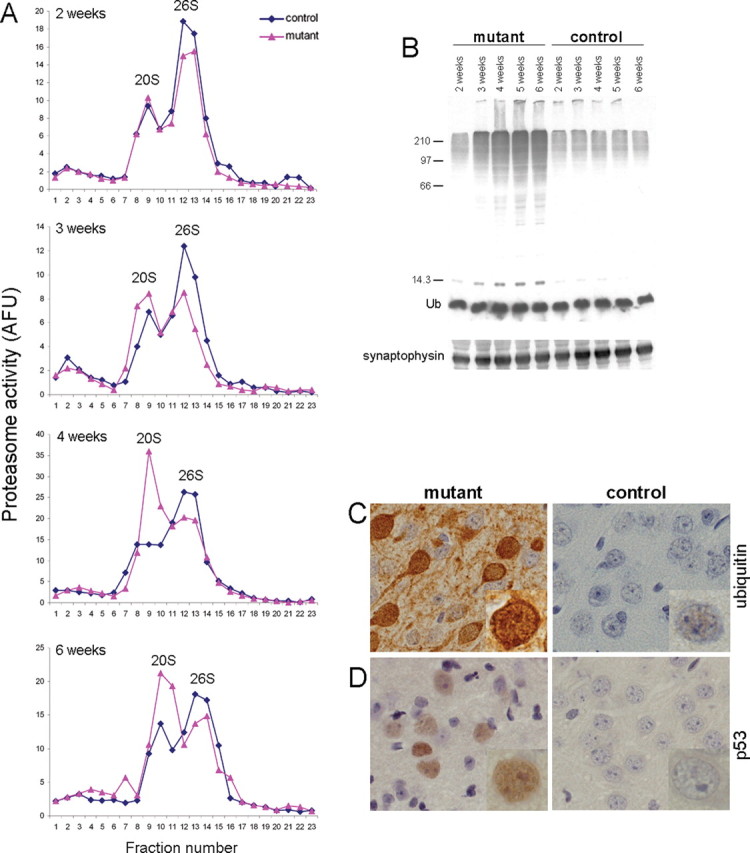
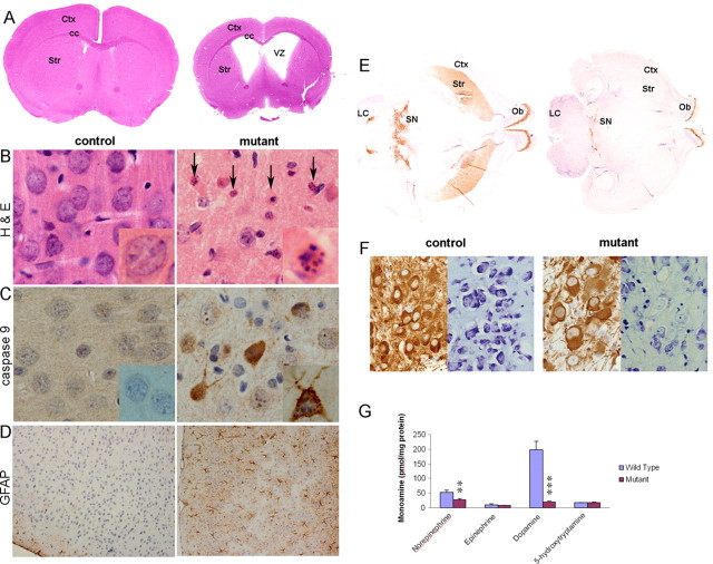
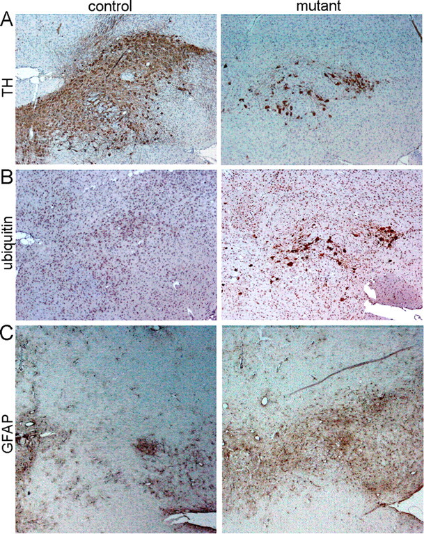
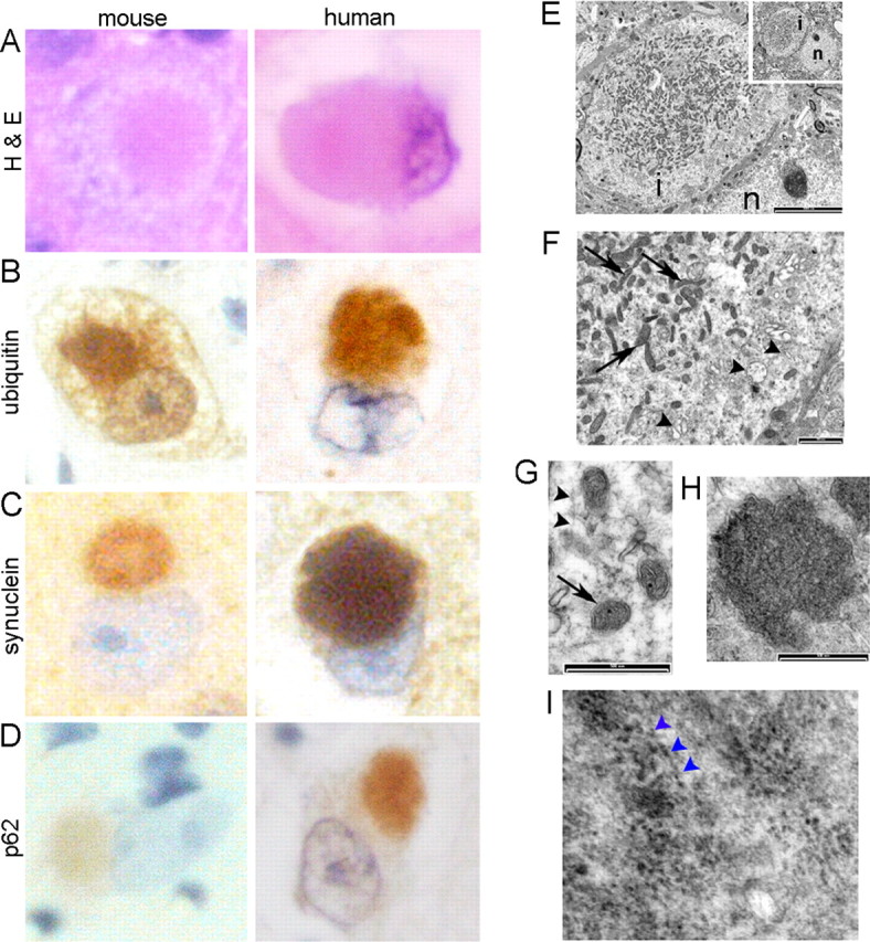
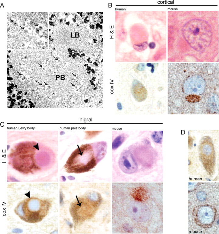
Ubiquitin-positive intraneuronal inclusions are a consistent feature of the major human neurodegenerative diseases, suggesting that dysfunction of the ubiquitin proteasome system is central to disease etiology. Research using inhibitors of the 20S proteasome to model Parkinson's disease is controversial. We report for the first time that specifically 26S proteasomal dysfunction is sufficient to trigger neurodegenerative disease. Here, we describe novel conditional genetic mouse models using the Cre/loxP system to spatially restrict inactivation of Psmc1 (Rpt2/S4) to neurons of either the substantia nigra or forebrain (e.g., cortex, hippocampus, and striatum). PSMC1 is an essential subunit of the 26S proteasome and Psmc1 conditional knock-out mice display 26S proteasome depletion in targeted neurons, in which the 20S proteasome is not affected. Impairment of specifically ubiquitin-mediated protein degradation caused intraneuronal Lewy-like inclusions and extensive neurodegeneration in the nigrostriatal pathway and forebrain regions. Ubiquitin and alpha-synuclein neuropathology was evident, similar to human Lewy bodies, but interestingly, inclusion bodies contained mitochondria. We support this observation by demonstrating mitochondria in an early form of Lewy body (pale body) from Parkinson's disease patients. The results directly confirm that 26S dysfunction in neurons is involved in the pathology of neurodegenerative disease. The model demonstrates that 26S proteasomes are necessary for normal neuronal homeostasis and that 20S proteasome activity is insufficient for neuronal survival. Finally, we are providing the first reproducible genetic platform for identifying new therapeutic targets to slow or prevent neurodegeneration.


Figures

Figure 1.
Figure 1.
Ablation of Psmc1 causes 26S proteasome depletion. A, Representative proteasome profiles from cortex of 2-, 3-, 4-, and 6-week-old control and Psmc1 fl/fl;CaMKIIα-Cre (mutant) mice after glycerol density gradient centrifugation. Fractions 1 and 23 correspond to the top (10%) and bottom (40%) of the gradient, respectively. 20S and 26S proteasomal distribution was revealed by succinyl-Leu-Leu-Val-Tyr-7-amido-4-methylcoumarin hydrolysis activity of gradient fractions. Individual experiments at different ages are not directly comparable because of, for example, the amount of protein loaded and slight variability in fraction collection. AFU, Arbitrary fluorescent unit. B, Accumulation of polyubiquitinated proteins, but not free ubiquitin (Ub), in the Psmc1 fl/fl;CaMKIIα-Cre (mutant) cortex with increasing age, shown by Western analysis of cortex homogenates with an anti-ubiquitin antibody. Anti-synaptophysin was used as a loading control. C, D, Immunohistological staining of cortex sections from 6-week-old control and Psmc1 fl/fl;CaMKIIα-Cre (mutant) mice using anti-ubiquitin (C) and anti-p53 (D) antibodies (40×). Higher-magnification images are inset into the representative sections.
Figure 2.
Figure 2.
26S proteasomal dysfunction causes neurodegeneration. A–D, Histological examination of coronal sections from control and Psmc1 fl/fl;CaMKIIα-Cre (mutant) mouse brains at 8 weeks of age. A, Hematoxylin and eosin (H&E)-stained whole-brain sections, shown at higher magnification in B (40×). The arrows point to pyknotic and fragmented nuclei in the mutant cortex. Immunohistochemistry with antibodies to cleaved caspase 9 (40×) (C) and GFAP (10×) (D) in cortex sections. Representative individual neurons are magnified and shown in the inset of appropriate figures. E, TH-stained horizontal sections of whole brain at the level of the nigrostriatal pathway from Psmc1 fl/fl;TH Cre (mutant) and control mice. F, TH and Nissl staining of sequential sections from the substantia nigra of control and Psmc1 fl/fl;TH Cre (mutant) mice (20×). G, Catecholamine and indolamine levels measured by HPLC with electrochemical detection in the striatum of Psmc1 fl/fl;TH Cre (mutant) and control mice. Graph represents mean ± SEM (controls, n = 5; mutants, n = 7). **p < 0.01 and ***p < 0.001 from control mice by unpaired Student's t test. Ctx, Cerebral cortex; cc, corpus callosum; Str, striatum; VZ, ventricular zone; Ob, olfactory bulb; SN, substantia nigra; LC, locus ceruleus.
Figure 3.
Figure 3.
Neurodegeneration of the substantia nigra in Psmc1 fl/fl;TH Cre mice. A–C, Immunohistochemistry using antibodies to TH (A), ubiquitin (B), and GFAP (C) (10×). A, TH immunostaining identifies nigral neurons in control and Psmc1 fl/fl;TH Cre (mutant) horizontal brain sections at 3 weeks of age. B, C, Increased ubiquitin (B) and astrocytic gliosis (C) in the substantia nigra of the mutant brain.
Figure 4.
Figure 4.
Lewy-like intraneuronal inclusions in 26S proteasome-depleted neurons. A–D, Representative Lewy-like inclusions in mouse cortical neurons at 6 weeks of age stained with H&E and ubiquitin, α-synuclein, and p62 antibodies, directly compared with Lewy bodies from the brains of patients with dementia with Lewy bodies (100×). No inclusions were observed in the control mouse brains (data not shown). E–I, Ultrastructure of mouse Lewy-like inclusions by EM. The paranuclear inclusion (E; inset; n, nucleus; i, inclusion) contains mitochondria (F, G, arrows), a background matrix of fine filaments (G, arrowheads) and granular material, with peripheral membrane-bound vesicles (F, arrowheads), including double-membraned autophagosome-like dense bodies (H). I, Filamentous α-synuclein is associated with 10 nm gold particles by immunoelectron microscopy. The arrowheads point to particles aligned along distinct filaments. Scale bars: E, 5000 nm; E, 1000 nm; G, H, 500 nm.
Figure 5.
Figure 5.
Mitochondria in human and mouse Lewy bodies. A, EM showing mitochondria (arrows) among filamentous material in a human nigral PB, which is adjacent to a classical LB that is devoid of mitochondria. Enlarged view of mitochondria within the pale body is shown in the inset. B, C, Shown is H&E and cox IV immunostaining of cortical Lewy bodies (B) and nigral Lewy and pale bodies (C) from human disease and Psmc1 fl/fl;CaMKIIα-Cre and Psmc1 fl/fl;TH Cre mice (100×). The arrows and arrowheads identify the pale and classical dense core Lewy bodies of human disease, respectively. D, The normal pattern of cox IV in human and mouse neurons (100×). The heavy brown pigment in H&E human nigral neurons is neuromelanin.

References

    1. Abou-Sleiman PM, Muqit MM, Wood NW. Expanding insights of mitochondrial dysfunction in Parkinson's disease. Nat Rev Neurosci. 2006;7:207–219. - PubMed
    1. Bayer SA, Wills KV, Triarhou LC, Ghetti B. Time of neuron origin and gradients of neurogenesis in midbrain dopaminergic neurons in the mouse. Exp Brain Res. 1995;105:191–199. - PubMed
    1. Bennett EJ, Shaler TA, Woodman B, Ryu KY, Zaitseva TS, Becker CH, Bates GP, Schulman H, Kopito RR. Global changes to the ubiquitin system in Huntington's disease. Nature. 2007;448:704–708. - PubMed
    1. Bové J, Prou D, Perier C, Przedborski S. Toxin-induced models of Parkinson's disease. NeuroRx. 2005;2:484–494. - PMC - PubMed
    1. Burgin KE, Waxham MN, Rickling S, Westgate SA, Mobley WC, Kelly PT. In situ hybridization histochemistry of Ca2+/calmodulin-dependent protein kinase in developing rat brain. J Neurosci. 1990;10:1788–1798. - PMC - PubMed

Publication types

MeSH terms

Substances