Comparative Study
. 2008 Aug 13;28(33):8199-207.
doi: 10.1523/JNEUROSCI.0979-08.2008.

Loss of PINK1 function affects development and results in neurodegeneration in zebrafish

Affiliations
Comparative Study

Loss of PINK1 function affects development and results in neurodegeneration in zebrafish

Oleg Anichtchik et al. J Neurosci. .

Abstract

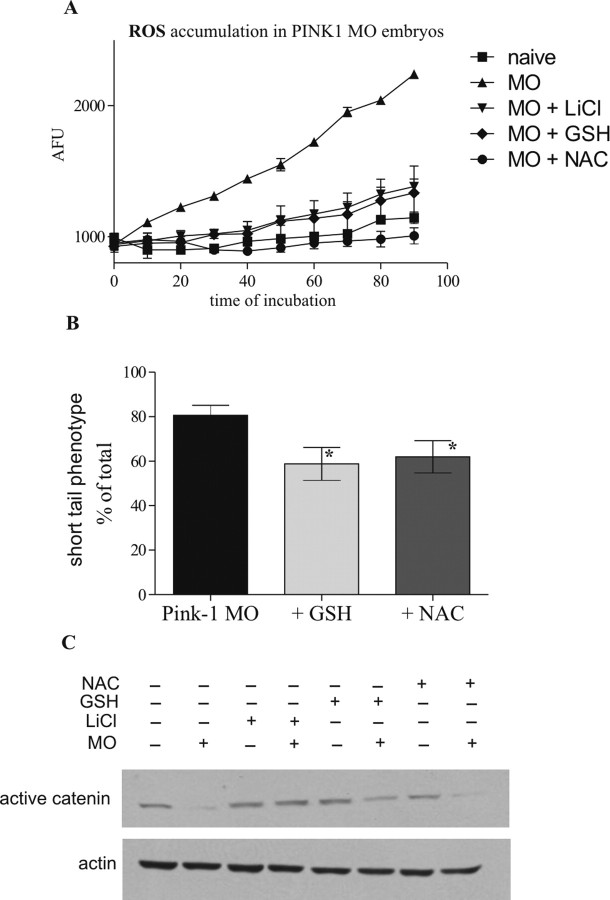
Parkinson's disease (PD) is the second most prevalent neurodegenerative disorder in the Western world. PTEN (phosphatase/tensin homolog on chromosome 10)-induced putative kinase 1 (PINK1), a putative kinase that is mutated in autosomal recessive forms of PD, is also implicated in sporadic cases of the disease. Although the mutations appear to result in a loss of function, the roles of this protein and the pathways involved in PINK1 PD are poorly understood. Here, we generated a vertebrate model of PINK1 insufficiency using morpholino oligonucleotide knockdown in zebrafish (Danio rerio). PINK1 knockdown results in a severe developmental phenotype that is rescued by wild-type human PINK1 mRNA. Morphants display a moderate decrease in the numbers of central dopaminergic neurons and alterations of mitochondrial function, including increases in caspase-3 activity and reactive oxygen species (ROS) levels. When the morphants were exposed to several drugs with antioxidant properties, ROS levels were normalized and the associated phenotype improved. In addition, GSK3beta-related mechanisms can account for some of the effects of PINK1 knockdown, as morphant fish show elevated GSK3beta activity and their phenotype is partially abrogated by GSK3beta inhibitors, such as LiCl and SB216763 [3-(2,4-dichlorophenyl)-4-(1-methyl-1H-indol-3-yl)1H-pyrrole-2,5-dione]. This provides new insights into the biology of PINK1 and a possible therapeutic avenue for further investigation.


Figures

Figure 1.
Figure 1.
Characterization of zebrafish PINK1. A, RT-PCR analysis of PINK1 mRNA expression at different hours after fertilization. B, In situ hybridization showing expression of zebrafish PINK1 mRNA in 24 and 48 hpf embryos. Right, Negative control staining with sense probe. C, Immunohistochemical analysis of PINK1 protein (green) and TH (red) in adult zebrafish brain (i–iii). PINK1 protein is localized ubiquitously with the tendency to be enriched along ventricular surfaces. In some areas (hypothalamus), PINK1 protein is colocalized with tyrosine hydroxylase. i, Posterior hypothalamus; ii, optic tectum; iii, areas around diencephalic ventricle; iv, posterior tuberculum (PINK1 protein, red; TH, green). D, The PINK1 antibody used (Cayman Chemicals, catalog #10006283) detects bands of ∼66 and ∼33 kDa in mouse liver mitochondria preparations. Similarly sized bands are seen in the wild-type zebrafish, and these disappear in PINK1 MO-treated fish. The trend in this gel is representative of at least five replicate experiments. E, PINK1 morphants at 24 hpf after injection of 7 ng of MOs show long-tail, short-tail, and no-tail phenotypes. Five-mismatch MO control zebrafish look the same as uninjected siblings. F, Phenotype of PINK1 morphants at 48 hpf after injection of 7 ng of MO. Enlarged brain ventricle, slim tail-yolk extension, and curved spines are indicated by arrows in mildly (middle) and severely (right) affected morphants. Scale bars: B, E, F, 100 μm; C, 50 μm.
Figure 2.
Figure 2.
Morphological characterization of PINK1-deficient fish. A, B, Acetylated tubulin immunostaining in MO-injected (A) and five-mispair MO control (B) embryos at 48 hpf. The decrease in staining in the tectal area is indicated by an arrow. C, D, TH and 5-HT staining in representative MO-injected (C) and five-mispair MO control (D) embryos at 48 dpf. The diencephalic cluster of dopaminergic cells is outlined. E, In situ hybridization of PINK1 MO-injected and control embryos at 48 hpf. The increase in fezl and ngn-1 expression in morphants is indicated by arrows. F, G, Immunohistochemistry with antibody 40.2D6 (islet-1) in the trunk of control (F) and MO-injected (G) fish, showing decreased staining in MO fish. H, I, Acetylated tubulin staining in the trunk of control (H) and MO-injected (I) fish. Changes in the morphology (e.g., decrease in neuron numbers) of spinal neurons are indicated by arrows. RB, Sensory Rohon-Beard neuron; MN, primary motoneuron. Scale bars, 100 μm.
Figure 3.
Figure 3.
Rescue of PINK1 MO phenotype with full-length human PINK1 mRNA. A, Rescue of the overall MO phenotype at 48 hpf by coinjected full-length human PINK1 mRNA. The phenotypes assessed (curved back, small tail-yolk extension) are indicated with arrows. Scale bar, 100 μm. B, Coinjection of MOs with human full-length mRNA partially restores PINK1 levels in 24 hpf embryos. C, Coinjection of mutant human mRNA did not produce a rescue of PINK1-deficient phenotype. The bars represent changes in the assessed phenotype relative to the MO-induced phenotype in three independent experiments. Error bars indicate SEM.
Figure 4.
Figure 4.
Rescue of PINK1 MO phenotype via modulation of AKT/GSK3β pathway. A, Decrease of Ser9-phosphorylated GSK3β in lysates of 24 hpf PINK1 MO embryos. Because this antibody detects both Ser9 GSK3β (47 kDa) and GSK3α (51 kDa), the lower band (GSK3β) was used for quantification. Blots obtained from three independent experiments were used for quantification and the levels of Ser9 GSK3β were normalized to the corresponding levels of the total GSK3β. Ser9 GSK3β phosphorylation was reduced by the morpholino to 27.6% (±16.56%, SEM) of the control level (set to 100%). p = 0.01 as analyzed by t test. B, Decrease of the active form of β-catenin in 24 hpf embryos injected with 7 ng of PINK1 MO. Active β-catenin bands were quantified as a function of the actin loading control in three independent experiments. Active β-catenin levels were reduced by the morpholino to 40.2% (±11.1%, SEM) of the control levels (set to 100%). p < 0.01 as analyzed by t test. C, Rescue of the short tail phenotype in 24 hpf PINK1 MO-injected embryos by nonspecific (50 mm of LiCl) and specific (10 μm of SB216763) GSK3β inhibitors. Bars represent three individual experiments, with 15–20 fish in each group per experiment. *p < 0.05, **p < 0.01, as analyzed by unconditional logistical regression analysis. D, Recovery of active β-catenin levels in PINK1 MO-injected embryos after treatment with 50 mm LiCl. The numbers under the blots are the ratios between the active β-catenin band intensities and actin loading control band intensities in relation to the control values. Independent replicate of the experiment could be seen in Figure 6C. E, The activity of AKT is decreased in PINK1 MO-injected embryos at 24 hpf. Ser473 AKT bands were quantified as a function of total AKT in three independent experiments. ***p < 0.01, as analyzed by t test. Error bars indicate SEM.
Figure 5.
Figure 5.
Increase of apoptosis in PINK1 MO-injected fish. A, Acridine orange staining of MO-injected embryos. Stained cells (fluorescent) represent dead cells. The arrows show increased acridine orange accumulation in brain and spine of short-tail PINK1 MO-injected embroys. B, Caspase-3 activity is increased in MO-injected fish at 24 hpf. Values represent means of three samples, each of which is the total lysate of 15 fish. C, LiCl (50 mm) rescues the increased levels of caspase-3 in 24 hpf PINK1 MO-injected embryos. Values represent means of three samples, each of which is the total lysate of 15 fish. *p < 0.05; **p < 0.01; ***p < 0.01, as analyzed by one-way ANOVA with Bonferroni post hoc test. D, The mitochondrial Δψ membrane potential, represented as the ratio 590/530 nm (orange/green) of JC-1, is decreased in PINK1 morphants. LiCl (50 mm) does not change decreased levels of JC-1 in MO embryos. Bars represent three independent experiments, each of which uses the total lysate of 80–100 24 hpf embryos. Scale bars, 100 μm. Error bars indicate SEM.
Figure 6.
Figure 6.
ROS levels are increased in PINK1 MO-injected fish. A, Accumulation of the CM-H2DCFDA dye, as the indicator of ROS levels, in live 24 hpf PINK1 MO-injected embryos. The increased levels of ROS are decreased using 100 μm GSH or 100 μm NAC, compounds with antioxidant qualities. Values represent means of three samples, each of which is the total lysate of 15 fish. B, Quantification of the short-tail phenotype at 24 hpf in embryos from A representing three independent experiments, each of which is the quantification of 15–20 24 hpf embryos. *p < 0.05, one-tailed t test. Error bars indicate SEM. C, Detection of active catenin in PINK1 MO-injected fish, treated with 50 mm LiCl, 100 μm GSH, and 100 μm NAC. Gel is representative of data from two independent experiments.

References

    1. Abdelilah S, Mountcastle-Shah E, Harvey M, Solnica-Krezel L, Schier AF, Stemple DL, Malicki J, Neuhauss SC, Zwartkruis F, Stainier DY, Rangini Z, Driever W. Mutations affecting neural survival in the zebrafish Danio rerio. Development. 1996;123:217–227. - PubMed
    1. Blader P, Fischer N, Gradwohl G, Guillemot F, Strähle U. The activity of neurogenin1 is controlled by local cues in the zebrafish embryo. Development. 1997;124:4557–4569. - PubMed
    1. Carmichael J, Sugars KL, Bao YP, Rubinsztein DC. Glycogen synthase kinase-3beta inhibitors prevent cellular polyglutamine toxicity caused by the Huntington's disease mutation. J Biol Chem. 2002;277:33791–33798. - PubMed
    1. Castelo-Branco G, Rawal N, Arenas E. GSK-3beta inhibition/beta-catenin stabilization in ventral midbrain precursors increases differentiation into dopamine neurons. J Cell Sci. 2004;117:5731–5737. - PubMed
    1. Clark IE, Dodson MW, Jiang C, Cao JH, Huh JR, Seol JH, Yoo SJ, Hay BA, Guo M. Drosophila pink1 is required for mitochondrial function and interacts genetically with parkin. Nature. 2006;441:1162–1166. - PubMed

Publication types

MeSH terms