. 2008 Oct;19(10):4319-27.
doi: 10.1091/mbc.e08-05-0470. Epub 2008 Aug 13.

A protein methylation pathway in Chlamydomonas flagella is active during flagellar resorption

Affiliations

A protein methylation pathway in Chlamydomonas flagella is active during flagellar resorption

Mark J Schneider et al. Mol Biol Cell. 2008 Oct.

Abstract

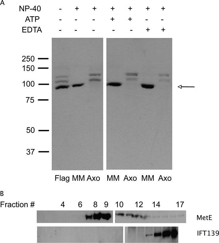
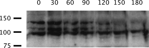
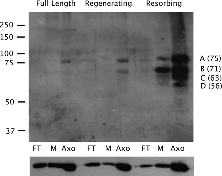
During intraflagellar transport (IFT), the regulation of motor proteins, the loading and unloading of cargo and the turnover of flagellar proteins all occur at the flagellar tip. To begin an analysis of the protein composition of the flagellar tip, we used difference gel electrophoresis to compare long versus short (i.e., regenerating) flagella. The concentration of tip proteins should be higher relative to that of tubulin (which is constant per unit length of the flagellum) in short compared with long flagella. One protein we have identified is the cobalamin-independent form of methionine synthase (MetE). Antibodies to MetE label flagella in a punctate pattern reminiscent of IFT particle staining, and immunoblot analysis reveals that the amount of MetE in flagella is low in full-length flagella, increased in regenerating flagella, and highest in resorbing flagella. Four methylated proteins have been identified in resorbing flagella, using antibodies specific for asymmetrically dimethylated arginine residues. These proteins are found almost exclusively in the axonemal fraction, and the methylated forms of these proteins are essentially absent in full-length and regenerating flagella. Because most cells resorb cilia/flagella before cell division, these data indicate a link between flagellar protein methylation and progression through the cell cycle.


Figures

Figure 1.
Figure 1.
Two-dimensional gel analysis of flagellar proteins. Proteins from long and short flagella samples were fluorescently labeled and then analyzed on the same 2D gel. (A) Cy3 image of a 2D gel showing the proteins from full-length flagella. (B) Cy5 image of the same 2D gel showing the proteins from short, regenerating flagella. The similar shape and intensity of the tubulin (T) region of each image indicates the same amount of total flagellar protein was loaded for each sample. Numbers 1–4 indicate proteins referred to in Results. The small circles with plus signs in the center (two per gel image) were used for image alignment.
Figure 2.
Figure 2.
Immunoblot analysis of flagellar samples. (A) Flagellar (Flag), membrane/matrix (MM), and axonemal (Axo) fractions were probed with affinity purified antibodies to MetE. In whole flagella, MetE (∼86.5 kDa) runs as a major band (arrow), with two more slowly migrating isoforms that may be phosphorylated forms of the enzyme. The images shown here are from two halves of the same immunoblot. (B) Sucrose gradient analysis of MetE from the MM fraction indicates MetE runs at ∼9 S and is hence not associated with IFT particles, which appear in fractions 14–16, i.e., at 15–16.5 S. The two sections of each gradient image indicate the samples were run on two different gels (fractions 1–9 and 10–17 for MetE and fractions 1–10 and 11–16 for IFT139).
Figure 3.
Figure 3.
Double label immunofluorescence microscopy. (A) Isolated flagella stained with antibodies to IFT139 (an IFT complex A component) and MetE. (B) Two color overlay showing IFT139 in green and MetE in red. (C) Collage of double labeled flagella to show more clearly both individual (green and red) and coincident (yellow) localization of IFT139 and MetE, respectively.
Figure 4.
Figure 4.
MetE requires an active anterograde IFT system for its presence in flagella. fla10 cells were shifted to the restrictive temperature (32°C) to inactive anterograde IFT; the flagella were isolated every 30 min after the temperature shift and analyzed for MetE via immunoblotting. Time (min) after the temperature shift is shown across the top, and the positions of molecular weight standards (×10−3) are shown to the left.
Figure 5.
Figure 5.
Analysis of MetE in samples of flagella at varying stages. Flagella were isolated from full-length, regenerating, and resorbing flagella. Each sample was then fractionated into freeze-thaw (FT), membrane (M), and axoneme (Axo) fractions and analyzed by immunoblotting with affinity purified antibodies to MetE. For a given flagellar fraction, immunoblotting with tubulin antibody was used to determine that the same amount of total protein was loaded for each sample. The results shown here are lanes from a single immunoblot, cut, and pasted into a collage, indicated by the spaces between the respective sections.
Figure 6.
Figure 6.
Protein methylation varies with the state of the flagella. Flagella were isolated from full-length, regenerating, and resorbing flagella, fractionated into freeze-thaw (FT), membrane (M), and axoneme (Axo) fractions, and analyzed by immunoblotting using antibodies to asymmetrically dimethylated arginine residues (top panel). The migration of standards is shown to the left (×10−3), and the masses (kDa) of proteins A–D, estimated from the migrations of the standards, are shown to the right. The blot was then stripped and probed with antibodies to α-tubulin to serve as a loading control (bottom panel).
Figure 7.
Figure 7.
Localization of proteins containing asymmetric dimethyl arginine residues in resorbing flagella. Cells were stained with Asym24 antibodies demonstrating two classes of label: enrichment of proteins containing dimethyl arginine at the FTC (top row) or numerous puncta along the length of the flagellum, similar to IFT particle staining.

Comment in

  • Mol Biol Cell. 19:4019.

References

    1. Baldwin G. S., Carnegie P. R. Isolation and partial characterization of methylated arginines from the encephalitogenic basic protein of myelin. Biochem. J. 1971;123:69–74. - PMC - PubMed
    1. Berman S. A., Wilson N. F., Haas N. A., Lefebvre P. A. A novel MAP kinase regulates flagellar length in Chlamydomonas. Curr. Biol. 2003;13:1145–1149. - PubMed
    1. Bloodgood R. A. Resorption of organelles containing microtubules. Cytobios. 1974;9:142–161. - PubMed
    1. Bloodgood R. A. Calcium-regulated phosphorylation of proteins in the membrane-matrix compartment of the Chlamydomonas flagellum. Exp. Cell Res. 1992;198:228–236. - PubMed
    1. Bloodgood R. A., Salomonsky N. L. The transmembrane signaling pathway involved in directed movements of Chlamydomonas flagellar membrane glycoproteins involves the dephosphorylation of a 60-kD phosphoprotein that binds to the major flagellar membrane glycoprotein. J. Cell Biol. 1994;127:803–811. - PMC - PubMed

Publication types

Substances

LinkOut - more resources