. 2008 Aug 19;105(33):11845-50.
doi: 10.1073/pnas.0804529105. Epub 2008 Aug 13.

Double-strand breaks associated with repetitive DNA can reshape the genome

Affiliations

Double-strand breaks associated with repetitive DNA can reshape the genome

Juan Lucas Argueso et al. Proc Natl Acad Sci U S A. .

Abstract

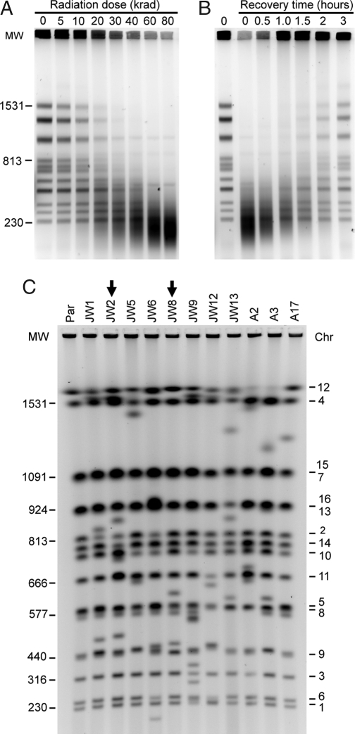
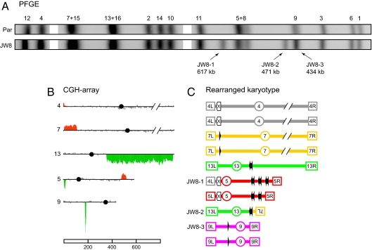
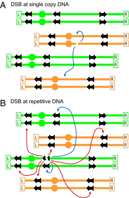
Ionizing radiation is an established source of chromosome aberrations (CAs). Although double-strand breaks (DSBs) are implicated in radiation-induced and other CAs, the underlying mechanisms are poorly understood. Here, we show that, although the vast majority of randomly induced DSBs in G(2) diploid yeast cells are repaired efficiently through homologous recombination (HR) between sister chromatids or homologous chromosomes, approximately 2% of all DSBs give rise to CAs. Complete molecular analysis of the genome revealed that nearly all of the CAs resulted from HR between nonallelic repetitive elements, primarily Ty retrotransposons. Nonhomologous end-joining (NHEJ) accounted for few, if any, of the CAs. We conclude that only those DSBs that fall at the 3-5% of the genome composed of repetitive DNA elements are efficient at generating rearrangements with dispersed small repeats across the genome, whereas DSBs in unique sequences are confined to recombinational repair between the large regions of homology contained in sister chromatids or homologous chromosomes. Because repeat-associated DSBs can efficiently lead to CAs and reshape the genome, they could be a rich source of evolutionary change.


Conflict of interest statement

The authors declare no conflict of interest.

Figures

Fig. 1.
Fig. 1.
DNA DSB induction, chromosomal restoration, and identification of rearrangements. (A) PFGE showing fragmentation of chromosomes in nocodazole-arrested (G2) diploid cells after the indicated dose of γ-radiation. (B) PFGE showing a time course of chromosomal restoration after exposure to 80 krad. (C) PFGE molecular karyotyping of the parental diploid strain (Par) and of the 11 radiation-survivor isolates that were investigated in detail. Molecular weight in kilobases is indicated to the left and specific chromosomes (numbers) to the right. Arrows emphasize the lanes with the JW8 and JW2 isolates.
Fig. 2.
Fig. 2.
Molecular dissection of CAs in the JW8 isolate. (A) Cropped alignment of the PFGE profiles from Fig. 1C. (Par) Parental diploid strain. JW8-1, -2, and -3 indicate the CAs characterized in JW8. (B) CGH-array data for chromosomes involved in CAs. Chromosome numbers are shown to the left of each plot and the horizontal lines correspond to the genomic position of microarray probes from the left to the right telomeres; black circles indicate the position of centromeres. Vertical bars correspond to the average signal of seven consecutive probes. Coloring indicates gene dosage as follows: gray. no significant change; red, gene amplifications; green, gene deletions. (C) Schematic representation of CAs and parental chromosomes with the respective genomic sites involved in rearrangements. Terminal boxes with internal labeling represent the left (L) and right (R) telomeres, and labeled circles represent centromeres. Each chromosome is drawn in a different color. Solid black arrows represent full-length Ty elements with their respective LTRs; arrowheads represent solitary LTR insertions. Empty box arrows with an internal “X” label represent the HXT loci. Chromosomes in B and C were scaled according to the reference bar in kilobases, except for Chr 4 and 7, which are truncated.
Fig. 3.
Fig. 3.
Molecular dissection of CAs in the JW2 isolate. All numbers and drawings are presented according to the legend in Fig. 2. (A) Cropped alignment of the PFGE profiles. (B) CGH-array data for chromosomes involved in CAs. (C) Band-array data for CAs. The plots for the specific chromosomes involved in the CAs are shown, with red rising bars, indicating the genomic segments enriched in each band. Background signal from comigrating parental chromosomes are not shown. (D) Schematic representation of the CAs and of the parental chromosomes with the respective genomic sites involved in rearrangements. The JW2-1, -3, and -4 CAs resulted from tripartite recombination and were structured as follows: JW2–1 was composed of a region of Chr 13 from the left telomere, passing through the centromere (CEN13) up to YMRCTy1-4, and a region of Chr 14 from YNLWTy1-2 to the left telomere; JW2-3 was a translocation including Chr 5 sequences from the right telomere to YERCTy1-2, and Chr 2 sequences from YBLWTy1-1, passing through CEN2 and including the entire right arm; finally JW2-4 was a translocation involving Chr 2 sequences from the left telomere to YBLWTy1-1 and Chr 14 DNA from YNLWTy1-2 passing through CEN14 to include the entire right arm. The remaining CA, JW2-2, was a complex nonreciprocal translocation involving the Chr 8 sequences from the left telomere, passing through CEN8 and including most of the right arm up to YHRCTy1-1, combined with sequences from Chr 5 represented by an interstitial duplication between YERCTy1-1 and YERCTy1-2 and a single copy of the distal region up to the right telomere.
Fig. 4.
Fig. 4.
Model for generation of CAs through the repair of repeat-associated DSBs. Given the random distribution of induced DSBs, most are expected to appear in single-copy DNA sequences as indicated in A, where efficient recombinational repair can occur between a sister chromatid or homolog (blue arrows). In contrast, DSBs that occur within the repetitive DNA sequences shown in B also have numerous opportunities for the ectopic recombination (red arrows), generating the CAs. The two ends formed by a single DSB can act independently in these interactions. The ectopic repair of DSBs in repetitive elements is in competition with the repair involving the sister chromatid or the homologue.

Comment in

  • From the shards of a shattered genome, diversity.
    Scheifele LZ, Boeke JD. Scheifele LZ, et al. Proc Natl Acad Sci U S A. 2008 Aug 19;105(33):11593-4. doi: 10.1073/pnas.0805812105. Epub 2008 Aug 13. Proc Natl Acad Sci U S A. 2008. PMID: 18701721 Free PMC article. No abstract available.

References

    1. Muller HJ. Artificial transmutation of the gene. Science. 1927;66:84–87. - PubMed
    1. McClintock B. Cytological observations of deficiencies involving known genes, translocations and inversions in Zea mays. MO Agric Exp Station Res Bull. 1931;163:1–30.
    1. Kupiec M. Damage-induced recombination in the yeast Saccharomyces cerevisiae. Mutat Res. 2000;451:91–105. - PubMed
    1. Fasullo M, Dave P, Rothstein R. DNA-damaging agents stimulate the formation of directed reciprocal translocations in Saccharomyces cerevisiae. Mutat Res. 1994;314:121–133. - PubMed
    1. Myung K, Kolodner RD. Induction of genome instability by DNA damage in Saccharomyces cerevisiae. DNA Repair (Amst) 2003;2:243–258. - PubMed

Publication types

Associated data