Termination zones of functionally characterized spinothalamic tract neurons within the primate posterior thalamus
- PMID: 18701750
- PMCID: PMC2576190
- DOI: 10.1152/jn.90810.2008
Termination zones of functionally characterized spinothalamic tract neurons within the primate posterior thalamus
Abstract
The primate posterior thalamus has been proposed to contribute to pain sensation, but its precise role is unclear. This is in part because spinothalamic tract (STT) neurons that project to the posterior thalamus have received little attention. In this study, antidromic mapping was used to identify individual STT neurons with axons that projected specifically to the posterior thalamus in Macaca fascicularis. Each axon was located by antidromic activation at low stimulus amplitudes (<30 microA) and was then surrounded distally by a grid of stimulating points in which 500-microA stimuli were unable to activate the axon antidromically, thereby indicating the termination zone. Several nuclei within the posterior thalamus were targets of STT neurons: the posterior nucleus, suprageniculate nucleus, magnocellular part of the medial geniculate nucleus, and limitans nucleus. STT neurons projecting to the ventral posterior inferior nucleus were also studied. Twenty-five posterior thalamus-projecting STT neurons recorded in lumbar spinal cord were characterized by their responses to mechanical, thermal, and chemical stimuli. Sixteen of 25 neurons were recorded in the marginal zone and the balance was located within the deep dorsal horn. Thirteen neurons were classified as wide dynamic range and 12 as high threshold. One-third of STT neurons projecting to posterior thalamus responded to noxious heat (50 degrees C). Two-thirds of those tested responded to cooling. Seventy-one percent responded to an intradermal injection of capsaicin. These data indicate that the primate STT transmits noxious and innocuous mechanical, thermal, and chemical information to multiple posterior thalamic nuclei.
Figures

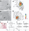
 ) indicating each point at which antidromic stimulation was tested. Contour plot indicates the current amplitude required for antidromic activation. B1: the lesion marking the LTP in Sg (→) of the same axon. B2: illustration as in A2. C: recording point in the marginal zone. D: antidromic activation from VPL had a longer latency than from Sg. E: antidromic activation from VPL. Top: 3 antidromic action potentials recorded in the dorsal horn. Middle: collision of an orthodromic action potential (▾) with an antidromic action potential (▿). Bottom: train of 333-Hz stimuli at the VPL LTP (12 μA). F: antidromic activation from Sg as in E. Stimulus artifacts reduced for clarity. CL, center lateral; CM, center median; eml, external medullary lamina; L, limitans; LD, lateral dorsal; LP lateral posterior; MG, medial geniculate; MGmc, magnocellular part of medial geniculate; MD, medial dorsal; Pla, anterior pulvinar; Pli, inferior pulvinar; Pll, lateral pulvinar; Po, posterior nucleus; VMb, basal ventral medial; VPI, ventral posterior inferior; VPM, ventral posterior medial.
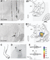
 ). B1: photomicrograph of a lesion marking the LTP in the Po (→). B2: illustration of B1 with each stimulation test site indicated and a contour plot showing the amplitude of current required to activate the neuron antidromically. C: receptive field. D: the neuron was recorded in nucleus proprius (→). E: antidromic activation from Po: Top: series of 3 antidromic action potentials time locked to the 3 stimuli. Middle: collision between an orthodromic (▾) and blocked antidromic action potential (▿). Bottom: 333-Hz, 8-μA stimulus train.
 , electrode penetrations that were unable to evoke antidromic action potentials with ≥500 μA. Bottom: coronal view corresponding to the most caudal plane above. B: photomicrograph of the coronal plane shown in A with a lesion marking the LTP in Po. Scale bar = 1.0 mm. C: characterization of the response to mechanical stimuli. B, brush; Pr, pressure; Pi, pinch; S, squeeze. This cell was classified as a high-threshold (HT) neuron. D: receptive field. E: recording point in nucleus proprius.


Similar articles
-
Spinothalamic and spinohypothalamic tract neurons in the sacral spinal cord of rats. I. Locations of antidromically identified axons in the cervical cord and diencephalon.J Neurophysiol. 1996 Jun;75(6):2581-605. doi: 10.1152/jn.1996.75.6.2581. J Neurophysiol. 1996. PMID: 8793765
-
Pruriceptive spinothalamic tract neurons: physiological properties and projection targets in the primate.J Neurophysiol. 2012 Sep;108(6):1711-23. doi: 10.1152/jn.00206.2012. Epub 2012 Jun 20. J Neurophysiol. 2012. PMID: 22723676 Free PMC article.
-
Response characterstics of spinothalamic tract neurons that project to the posterior thalamus in rats.J Neurophysiol. 2005 May;93(5):2552-64. doi: 10.1152/jn.01237.2004. J Neurophysiol. 2005. PMID: 15845999
-
Neuroscience of the human thalamus related to acute pain and chronic "thalamic" pain.J Neurophysiol. 2024 Dec 1;132(6):1756-1778. doi: 10.1152/jn.00065.2024. Epub 2024 Oct 16. J Neurophysiol. 2024. PMID: 39412562 Review.
-
The spinothalamic tract.Crit Rev Neurobiol. 1990;5(4):363-97. Crit Rev Neurobiol. 1990. PMID: 2204486 Review.
Cited by
-
The Cortico-Limbo-Thalamo-Cortical Circuits: An Update to the Original Papez Circuit of the Human Limbic System.Brain Topogr. 2023 May;36(3):371-389. doi: 10.1007/s10548-023-00955-y. Epub 2023 Apr 26. Brain Topogr. 2023. PMID: 37148369 Free PMC article. Review.
-
Semi-intact ex vivo approach to investigate spinal somatosensory circuits.Elife. 2016 Dec 19;5:e22866. doi: 10.7554/eLife.22866. Elife. 2016. PMID: 27991851 Free PMC article.
-
Hyperalgesia and sensitization of dorsal horn neurons following activation of NK-1 receptors in the rostral ventromedial medulla.J Neurophysiol. 2017 Nov 1;118(5):2727-2744. doi: 10.1152/jn.00478.2017. Epub 2017 Aug 9. J Neurophysiol. 2017. PMID: 28794197 Free PMC article.
-
Projection Motifs and Wiring Logic of Medial Pulvinar Thalamocortical Axons in the Marmoset Monkey.J Neurosci. 2025 Apr 9;45(15):e1837242025. doi: 10.1523/JNEUROSCI.1837-24.2025. J Neurosci. 2025. PMID: 39919832
-
Beyond Barrels: Diverse Thalamocortical Projection Motifs in the Mouse Ventral Posterior Complex.J Neurosci. 2024 Oct 23;44(43):e1096242024. doi: 10.1523/JNEUROSCI.1096-24.2024. J Neurosci. 2024. PMID: 39197940 Free PMC article.
References
-
- Apkarian AV, Bushnell MC, Treede RD, Zubieta JK. Human brain mechanisms of pain perception and regulation in health and disease. Eur J Pain 9: 463–484, 2005. - PubMed
-
- Apkarian AV, Hodge CJ. Primate spinothalamic pathways. III. Thalamic termination of the dorsolateral and ventral spinothalamic pathways. J Comp Neurol 288: 493–511, 1989. - PubMed
-
- Applebaum AE, Leonard RB, Kenshalo DR Jr, Martin RF, Willis WD. Nuclei in which functionally identified spinothalamic tract neurons terminate. J Comp Neurol 188: 575–586, 1979. - PubMed
-
- Beggs J, Jordan S, Ericson A-C, Blomqvist A, Craig AD. Synaptology of trigemino- and spinothalamic lamina I terminations in the posterior ventral medial nucleus of the Macaque. J Comp Neurol 459: 334–354, 2003. - PubMed
Publication types
MeSH terms
Substances
Grants and funding
LinkOut - more resources
Full Text Sources

