. 2008 Oct;100(4):1740-8.
doi: 10.1152/jn.90463.2008. Epub 2008 Aug 13.

Intrinsic functional relations between human cerebral cortex and thalamus

Affiliations

Intrinsic functional relations between human cerebral cortex and thalamus

Dongyang Zhang et al. J Neurophysiol. 2008 Oct.

Abstract

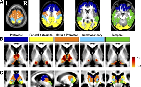
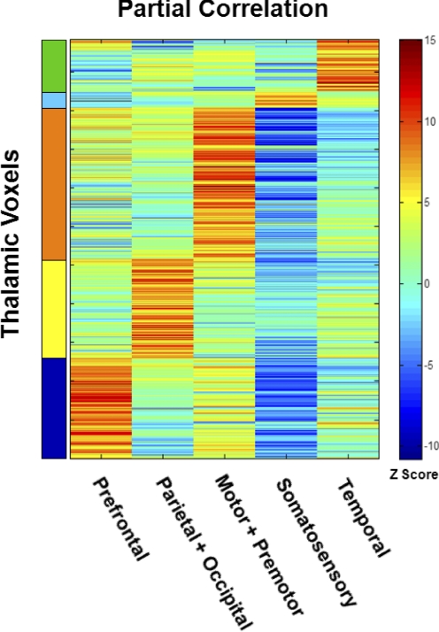
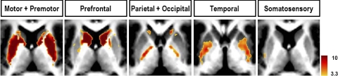
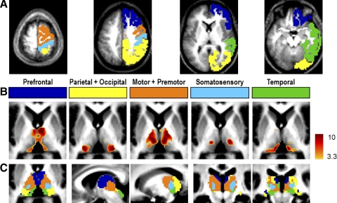
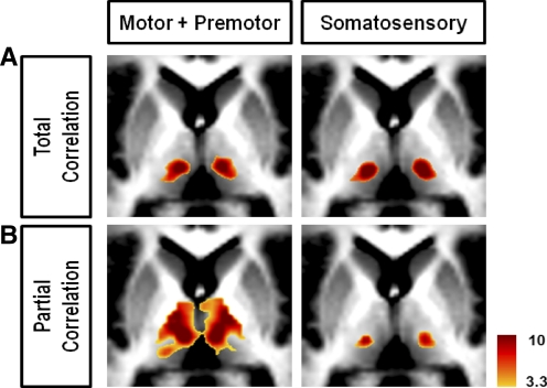
The brain is active even in the absence of explicit stimuli or overt responses. This activity is highly correlated within specific networks of the cerebral cortex when assessed with resting-state functional magnetic resonance imaging (fMRI) blood oxygen level-dependent (BOLD) imaging. The role of the thalamus in this intrinsic activity is unknown despite its critical role in the function of the cerebral cortex. Here we mapped correlations in resting-state activity between the human thalamus and the cerebral cortex in adult humans using fMRI BOLD imaging. Based on this functional measure of intrinsic brain activity we partitioned the thalamus into nuclear groups that correspond well with postmortem human histology and connectional anatomy inferred from nonhuman primates. This structure/function correspondence in resting-state activity was strongest between each cerebral hemisphere and its ipsilateral thalamus. However, each hemisphere was also strongly correlated with the contralateral thalamus, a pattern that is not attributable to known thalamocortical monosynaptic connections. These results extend our understanding of the intrinsic network organization of the human brain to the thalamus and highlight the potential of resting-state fMRI BOLD imaging to elucidate thalamocortical relationships.


Figures

FIG. 1.
FIG. 1.
Highly specific correlations are seen between the cerebral cortex and the thalamus in their intrinsic activity. A: bilateral cortical regions of interest (ROIs) illustrating partitioning according to major anatomical landmarks. Prefrontal: dark blue; Parietal + Occipital: yellow; Motor + Premotor: orange; Somatosensory: light blue; Temporal: green. This color code is consistently used in all figures. Selected transverse slices (z = 66, 39, 9, −18) are shown. B: partial correlation Z-score maps (fixed-effects analysis using all 17 subjects) generated for each of the cortical ROIs. Voxels meeting a criterion of P < 0.05 are colored. Transverse slice z = 6. Note: spatially distinct maps for each of the cortical ROIs. See supplemental data for localization of negative partial correlations and a discussion on the interpretation of negative partial correlations. C: winner-take-all (WTA) maps obtained by labeling each thalamic voxel according to the cortical ROI with the highest partial correlation value. Only voxels at which the most winning Z-score significance exceeded P < 0.05 are colored (color code as in A). From left to right: transverse slice z = 6; sagittal slices x = 5, 15; coronal slices y = −23, −27.
FIG. 2.
FIG. 2.
Each thalamic voxel is highly correlated with one cortical ROI. This matrix represents partial correlations computed for each thalamic voxel paired with each cortical ROI. First, correlation values were computed for each subject and transformed using Fisher's r-to-z transform. Then, descriptive Z-scores were generated at the population level using a fixed-effects model (see methods). Red and blue hues indicate, respectively, positive and negative correlations. Thalamic voxels within the same WTA bilateral thalamic partition (Fig. 1C) are grouped together on the vertical axis.
FIG. 3.
FIG. 3.
The basal ganglia system shows high specificity in correlational connectivity with the cortex. Motor and premotor cortex are most correlated with putamen and juxtaposing head of caudate nucleus (slice: z = 10). Prefrontal cortex is most correlated with caudate nucleus (slice: z = 10). Parietal and occipital cortex have weaker but seemingly specific correlations with parts of globus pallidus, mostly confined to the internal segment (slice: z = 2). Temporal cortex has weak correlations with parts of putamen (z = 0). Somatosensory (z = 10).
FIG. 4.
FIG. 4.
A: unilateral cortical ROIs in the right hemisphere demonstrate the same specificity as bilateral cortical ROIs in ipsilateral as well as contralateral thalamus. All else as in Fig. 1A. B: corresponding partial correlation Z-score maps. All else as in Fig. 1B. C: WTA maps. All else as in Fig. 1C.
FIG. 5.
FIG. 5.
Comparison of correlation mapping techniques illustrated using the Motor + Premotor and Somatosensory bilateral cortical ROIs shows the increased specificity using partial correlation. A: total correlation. B: partial correlation. All slices are transverse, z = 6.

References

    1. Anand A, Li Y, Wang Y, Wu J, Gao S, Bukhari L, Mathews VP, Kalnin A, Lowe MJ. Activity and connectivity of brain mood regulating circuit in depression: a functional magnetic resonance study. Biol Psychiatry 57: 1079–1088, 2005. - PubMed
    1. Beckmann CF, DeLuca M, Devlin JT, Smith SM. Investigations into resting-state connectivity using independent component analysis. Philos Trans R Soc Lond B Biol Sci 360: 1001–1013, 2005. - PMC - PubMed
    1. Behrens TE, Johansen-Berg H, Woolrich MW, Smith SM, Wheeler-Kingshott CA, Boulby PA, Barker GJ, Sillery EL, Sheehan K, Ciccarelli O, Thompson AJ, Brady JM, Matthews PM. Non-invasive mapping of connections between human thalamus and cortex using diffusion imaging. Nat Neurosci 6: 750–757, 2003. - PubMed
    1. Birn RM, Diamond JB, Smith MA, Bandettini PA. Separating respiratory-variation-related fluctuations from neuronal-activity-related fluctuations in fMRI. Neuroimage 31: 1536–1548, 2006. - PubMed
    1. Biswal B, Yetkin FZ, Haughton VM, Hyde JS. Functional connectivity in the motor cortex of resting human brain using echo-planar MRI. Magn Reson Med 34: 537–541, 1995. - PubMed

Publication types

LinkOut - more resources