

Immunogold labeling of amelogenin in developing porcine enamel revealed by field emission scanning electron microscopy
- PMID: 18701812
- PMCID: PMC2633245
- DOI: 10.1159/000151385
Immunogold labeling of amelogenin in developing porcine enamel revealed by field emission scanning electron microscopy
Abstract
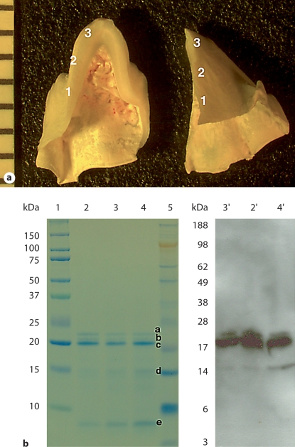
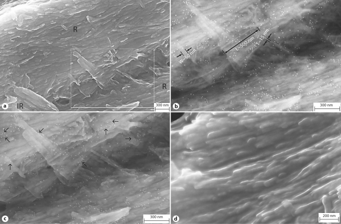
The present study describes a method using immunohistochemical labeling in combination with high-resolution imaging (field emission scanning electron microscopy) to investigate the spatial localization of amelogenins on apatite crystallites in developing porcine enamel. Cross-sections of developing enamel tissue from freeze-fractured pig third molar were treated with antiserum against recombinant mouse amelogenin and immunoreactivity confirmed by Western blot analysis. The samples were then treated with the goat anti-rabbit IgG conjugated with 10-nm gold particles. The control samples were treated with the secondary antibody only. The in-lens secondary electrons detector and quadrant back-scattering detector were employed to reveal the high-resolution morphology of enamel structures and gold particle distribution. The immunolabeling showed a preference of the gold particle localization along the side faces of the ribbon-like apatite crystals. The preferential localization of amelogenin in vivo on enamel crystals strongly supports its direct function in controlling crystal morphology.
Copyright 2008 S. Karger AG, Basel.
Figures
References
-
- Aoba T., Tanabe T., Moreno E.C. Proteins in the enamel fluid of immature porcine teeth. J Dent Res. 1987;66:1721–1726. - PubMed
-
- Du C., Falini G., Fermani S., Abbott C., Moradian-Oldak J. Supramolecular assembly of amelogenin nanospheres into birefringent microribbons. Science. 2005;307:1450–1454. erratum in Science 309: 2166. - PubMed
-
- Fincham A.G., Moradian-Oldak J., Sarte P.E. Mass-spectrographic analysis of a porcine amelogenin identifies a single phosphorylated locus. Calcif Tissue Int. 1994;55:398–400. - PubMed
-
- Fincham A.G., Moradian-Oldak J., Diekwisch T.G., Lyaruu D.M., Wright J.T., P. Bringas, Jr., H.C. Slavkin. Evidence for amelogenin ‘nanospheres’ as functional components of secretory-stage enamel matrix. J Struct Biol. 1995;115:50–59. - PubMed
Publication types
MeSH terms
Substances
Grants and funding
LinkOut - more resources
Full Text Sources
