. 2008 Sep 17;27(18):2422-31.
doi: 10.1038/emboj.2008.162. Epub 2008 Aug 14.

SUMO modification of PCNA is controlled by DNA

Affiliations

SUMO modification of PCNA is controlled by DNA

Joanne L Parker et al. EMBO J. .

Abstract

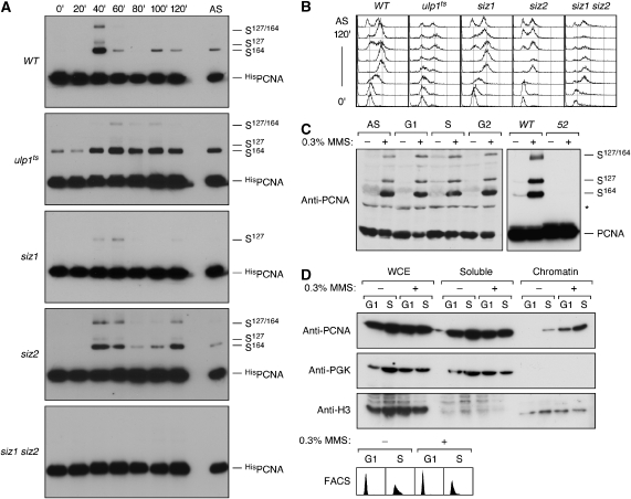
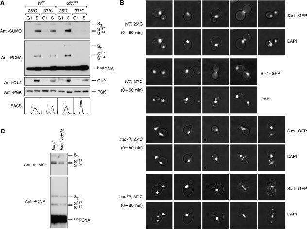
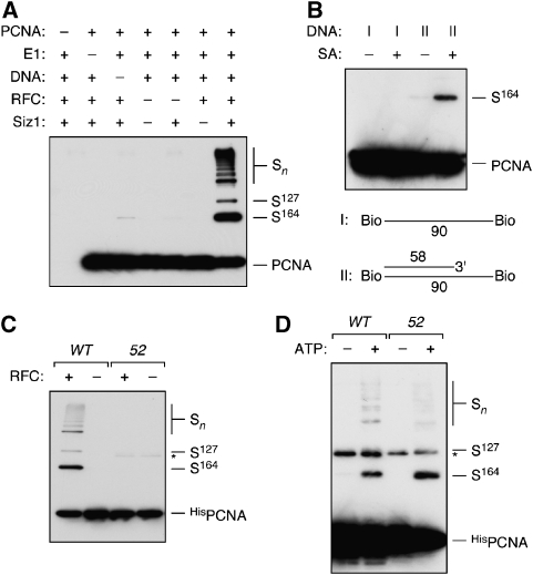
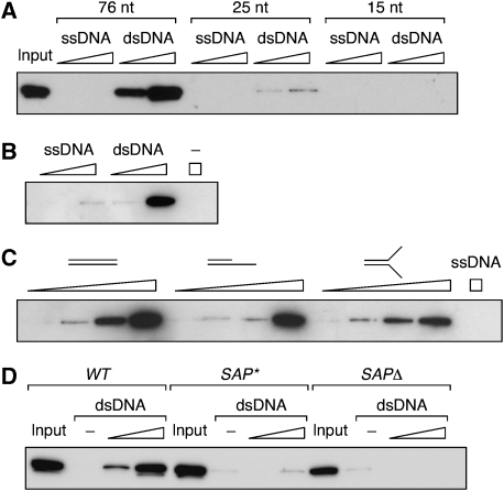
Post-translational modification by the ubiquitin-like protein SUMO is often regulated by cellular signals that restrict the modification to appropriate situations. Nevertheless, many SUMO-specific ligases do not exhibit much target specificity, and--compared with the diversity of sumoylation substrates--their number is limited. This raises the question of how SUMO conjugation is controlled in vivo. We report here an unexpected mechanism by which sumoylation of the replication clamp protein, PCNA, from budding yeast is effectively coupled to S phase. We find that loading of PCNA onto DNA is a prerequisite for sumoylation in vivo and greatly stimulates modification in vitro. To our surprise, however, DNA binding by the ligase Siz1, responsible for PCNA sumoylation, is not strictly required. Instead, the stimulatory effect of DNA on conjugation is mainly attributable to DNA binding of PCNA itself. These findings imply a change in the properties of PCNA upon loading that enhances its capacity to be sumoylated.


Figures

Figure 1
Figure 1
Cell cycle- and DNA damage-dependent sumoylation of PCNA. (A) HisPOL30 cells of the indicated genotypes were synchronised in G1 and released into the cell cycle. Samples were collected at the indicated times and analysed by Ni-NTA affinity chromatogaphy under denaturing conditions, followed by western blotting with PCNA-specific antibody. Asynchronous cultures (AS) were analysed in parallel. (B) Cell cycle profiles of the cultures shown in (A), determined by flow cytometry. (C) Lethal amounts of DNA damage cause PCNA hyper-sumoylation in WT, but not in pol30-52 cells. Cultures were arrested in G1, S or G2 phase or left asynchronous (AS) and treated where indicated with 0.3% methyl methanesulphonate (MMS) for 90 min. In the right-hand panel, both the WT and the pol30-52 strain (52) were treated during exponential growth. Total cell extracts prepared under denaturing conditions were analysed by western blotting using PCNA-specific antibody. The asterisk indicates a cross-reacting band visible with some batches of the antibody. (D) DNA damage leads to chromatin association of PCNA outside of S phase. G1 and S phase-arrested cells were treated with 0.3% MMS where indicated. Whole cell extracts (WCEs) were prepared by enzymatic lysis, separated into soluble and chromatin-associated fractions and analysed by western blotting for the presence of PCNA. Phosphoglycerate kinase (PGK) and histone H3 served as controls for soluble and chromatin-associated proteins. Arrests were confirmed by flow cytometry (FACS).
Figure 2
Figure 2
PCNA sumoylation during S phase requires active replication forks. (A) WT and cdc7ts cells bearing the HisPOL30 allele, grown at 25°C, were synchronised in G1 and either kept at 25°C or shifted to 37°C for 90 min before releasing them into the cell cycle at the indicated temperatures. Samples were taken before release (G1) and in mid-S phase (S) according to the budding pattern and Clb2 levels (30 min for WT at 25°C and cdc7ts at both temperatures, 15 min for WT at 37°C). PCNA sumoylation was detected as described in Figure 1, Clb2 and PGK were detected in total cell extracts, and the DNA content was monitored by flow cytometry (FACS; dashed line: G1 arrest; solid line: after release). (B) Subcellular distribution of Siz1 in WT and cdc7ts cells. Both strains expressing GFP-tagged Siz1 were synchronised in G1 and released into the cell cycle at 25 or 37°C as in (A). Samples were taken at 20-min intervals and analysed by fluorescence microscopy for Siz1–GFP and DNA (DAPI). Representative cells are shown as overlays of fluorescence with interference contrast images. (C) Cdc7 kinase is not required for PCNA sumoylation. Modification of HisPCNA was analysed in asynchronous cultures of bob1 and bob1 cdc7Δ mutants. The bob1 mutation affects the MCM5 gene and renders CDC7 non-essential.
Figure 3
Figure 3
PCNA sumoylation in vitro is stimulated by loading onto DNA. (A) In vitro sumoylation assays were performed with recombinant Ubc9 and SUMO in the presence or absence of PCNA, E1, RFC, Siz1 and circular, multiply primed ssDNA as indicated. Products were analysed by western blotting with PCNA-specific antibody. (B) In vitro sumoylation reactions were carried out with the complete set of proteins as in (A), but in the presence or absence of streptavidin (SA) and two different linear DNA structures (I and II) derivatised with biotin on both termini. (C) WT HisPCNA and the trimerisation-deficient protein encoded by the pol30-52 allele (52) were compared in sumoylation assays containing E1, Ubc9, Siz1, SUMO and circular primed ssDNA in the presence or absence of RFC. (D) Ubc9- and Siz1-dependent in vitro sumoylation of WT and mutant (52) HisPCNA in the absence of DNA. HisPCNA was used at 3 μM (compared with 50 nM in A–C).
Figure 4
Figure 4
The Siz1 SAP domain is required for DNA binding. (A) Siz1 binds to dsDNA, but not ssDNA. Equimolar amounts of biotinylated DNA fragments of the indicated lengths were immobilised on streptavidin Sepharose, and increasing amounts of GSTSiz1FLAGHis were added. Material retained after washing was analysed by western blotting with anti-FLAG antibody. (B) Binding to a 76-nt fragment of ssDNA or dsDNA was analysed as above in the presence of 1 mM EDTA. (C) Equimolar amounts of the indicated DNA structures were immobilised on streptavidin Sepharose, and Siz1 binding was analysed as above. (D) Mutation or deletion of the Siz1 SAP domain results in loss of DNA binding. Equal amounts of Siz1 WT, SAP* and SAPΔ were analysed on 76mer dsDNA as above.
Figure 5
Figure 5
The Siz1 SAP domain is dispensable for PCNA sumoylation in vivo. (A) Mutation or deletion of the SAP domain appears to result in partial or complete loss of PCNA sumoylation in vivo. Deletion mutants of siz1 were complemented with integrative plasmids bearing WT, SAP* or SAPΔ alleles of SIZ1 or empty vector (−), and modified PCNA was detected in denaturing extracts as described in Figure 1. (B) Mutation or deletion of the SAP domain appears to result in partial or complete loss of SIZ1 function. Sensitivities of the indicated strains to the DNA-damaging agents, methyl methanesulphonate (MMS) and 4-nitroquinoline oxide (NQO) were monitored by growth on plates containing the indicated concentrations of the drugs. Suppression of the damage sensitivity associated with the rad18 deletion indicates a loss of SIZ1 function. (C) Mutation or deletion of the SAP domain results in loss of the Siz1 protein in vivo, which can be rescued by overexpression. The indicated SIZ1 alleles were expressed from integrative plasmids under control of the SIZ1 or the galactose-inducible GAL1 promoter and tagged C-terminally by a 9myc-epitope. An empty plasmid (−) served as a control. Cells were grown in the presence of glucose or galactose, and total extracts were analysed for the presence of Siz19myc by western blotting. Detection of PGK served as a loading control. (D) Overexpression suppresses the sumoylation defects of the SIZ1 SAP domain mutants. The SIZ1 constructs shown in (C) were introduced into the HisPOL30 siz1 strain, and PCNA modifications were analysed as in Figure 1 after growth in galactose medium. (E) Overexpression of SIZ1 alleles suppresses the loss of function associated with mutation or deletion of the SAP domain. The SIZ1 constructs shown in (C) were introduced into rad18 siz1 strains, and SIZ1 function was analysed as described for (B) on glucose or galactose plates. (F) Expression of the siz1(SAPΔ) allele under control of the CUP1 promoter results in near physiological protein levels. The CUP1 promoter was induced by growth in 100 μM CuSO4, and 9myc-tagged versions of the indicated SIZ1 alleles were analysed as in (C). (G) Expression of siz1(SAPΔ) under control of the CUP1 promoter suppresses the siz1 phenotype. DNA damage sensitivity assays were carried out with the indicated SIZ1 alleles in rad18 siz1 as in (B, E) in the presence or absence of 100 μM CuSO4. (H) Expression of siz1(SAPΔ) under control of the CUP1 promoter restores PCNA sumoylation in vivo. The indicated SIZ1 alleles were analysed in HisPOL30 siz1 as in (A, D) in the presence or absence of 100 μM CuSO4.
Figure 6
Figure 6
PCNA loading stimulates sumoylation by Siz1 SAP mutants and by Ubc9 alone. (A) Mutation or deletion of the Siz1 SAP domain has no effect on PCNA sumoylation in the absence of DNA. In vitro sumoylation assays were performed at high substrate concentration (3 μM) with increasing amounts of Siz1 WT, SAP* or SAPΔ protein. GSTSiz1FLAGHis was detected by western blotting with anti-FLAG antibody. (B) PCNA loading stimulates sumoylation by Siz1 SAP mutants. In vitro sumoylation assays in the presence of RFC and circular primed ssDNA were performed at low substrate concentration (50 nM) with increasing amounts of Siz1 WT, SAP* or SAPΔ protein (∼1–40 nM). GSTSiz1FLAGHis was detected by western blotting with anti-FLAG antibody. (C) PCNA loading stimulates E3-independent sumoylation. In vitro sumoylation assays in the presence of RFC and circular primed ssDNA were performed with 10-fold elevated concentration of Ubc9 (5 μM). (D) PCNA mutants whose interactions with DNA are altered exhibit reduced sumoylation. PCNA modifications in vivo were analysed in WT, pol30(K20A) and pol30(K77A) as described in Figure 1. (E) Cell cycle distribution of the POL30 alleles shown in (D), determined by flow cytometry (FACS).

References

    1. Ayyagari R, Impellizzeri KJ, Yoder BL, Gary SL, Burgers PM (1995) A mutational analysis of the yeast proliferating cell nuclear antigen indicates distinct roles in DNA replication and DNA repair. Mol Cell Biol 15: 4420–4429 - PMC - PubMed
    1. Davies AA, Huttner D, Daigaku Y, Chen S, Ulrich HD (2008) Activation of ubiquitin-dependent DNA damage bypass is mediated by replication protein A. Mol Cell 29: 625–636 - PMC - PubMed
    1. Essers J, Theil AF, Baldeyron C, van Cappellen WA, Houtsmuller AB, Kanaar R, Vermeulen W (2005) Nuclear dynamics of PCNA in DNA replication and repair. Mol Cell Biol 25: 9350–9359 - PMC - PubMed
    1. Franco AA, Lam WM, Burgers PM, Kaufman PD (2005) Histone deposition protein Asf1 maintains DNA replisome integrity and interacts with replication factor C. Genes Dev 19: 1365–1375 - PMC - PubMed
    1. Fukuda K, Morioka H, Imajou S, Ikeda S, Ohtsuka E, Tsurimoto T (1995) Structure–function relationship of the eukaryotic DNA replication factor, proliferating cell nuclear antigen. J Biol Chem 270: 22527–22534 - PubMed

Publication types

MeSH terms