Review
. 2008 Oct;3(9-10):1157-71.
doi: 10.1002/biot.200800110.

Novel human antibody therapeutics: the age of the Umabs

Affiliations
Review

Novel human antibody therapeutics: the age of the Umabs

Sigrid R Ruuls et al. Biotechnol J. 2008 Oct.

Abstract

Monoclonal antibodies represent a major and increasingly important category of biotechnology products for the treatment of human diseases. The state-of-the-art of antibody technology has evolved to the point where therapeutic monoclonal antibodies, that are practically indistinguishable from antibodies induced in humans, are routinely generated. We depict how our science-based approach can be used to further improve the efficacy of antibody therapeutics, illustrated by the development of three monoclonal antibodies for various cancer indications: zanolimumab (directed against CD4), ofatumumab (directed against CD20) and zalutumumab (directed against epidermal growth factor receptor).


Figures

Figure 1
Figure 1
Classification of therapeutic antibodies in cancer. Progress in genetic engineering has facilitated the development of fully human therapeutic mAbs. Original mAb technologies yielded murine (and in some cases rat) molecules. Chimeric antibodies are genetically engineered mAb with murine variable regions (VL and VH) and constant regions derived from a human source. H umanized therapeutic mAb closely match the human germline sequence except for CDR, which are of murine (and occasionally rat) origin. The inset table explains the nomenclature of therapeutic antibodies in cancer indications, according to the system of International Nonproprietary Names (INN).
Figure 2
Figure 2
Mechanisms of action of zanolimumab, ofatumumab and zalutumumab. See text for further description.
Figure 3
Figure 3
Clinical improvement of mycosis fun-goides. Left panel shows baseline, right panel shows clinical response 4 weeks after start ofzanolimumab treatment. Adapted from [11].
Figure 4
Figure 4
CD20 expression in B cell ontogeny. B cell development is a multi-staged process that begins with a pluripotent hematopoietic stem cell and ends with the formation of an antibody-producing plasma cell. CD20 expression is restricted to the pre-B cell to memory B cell stage. B cell malignancies, as indicated, can occur at almost any stage of B cell development, producing a variety of distinct leukemias and lymphomas.
Figure 5
Figure 5
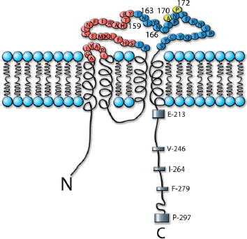
Epitope of ofatumumab. Peptide scanning and mutation studies revealed the binding epitope of ofatumumab on CD20. Amino acids contributing to ofatumumab binding are indicated in red [35]. Amino acids essential for rituximab, but not ofatumumab binding are indicated in yellow [36, 37].
Figure 6
Figure 6
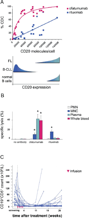
Potent B cell killing capacity of ofatumumab. (A) Anti-CD20-mediated CDC of CEM cells expressing varying amounts of CD20. The range of CD20 expression in follicular lymphoma and B cell chronic lymphatic leukemia is indicated, showing the potential for ofatumumab of killing cells that are resistant to rituximab. (B) Primary CLL cells are efficiently killed by ofatumumab with human effectors. Human blood (whole blood) was fractionated into polymorphonuclear (PMN) or mononuclear cells (MNC), or into complement containing plasma. Specific target cell lysis was assessed in 51Cr release assays (*, significant difference compared to “no antibody", p < 0.001). (C) Leukemic B cell counts in B-CLL patients receiving four weekly infusions (1×500 mg and 3×1000 mg) of ofatumumab. Adapted from [13, 33, 35].
Figure 7
Figure 7
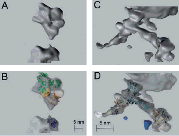
Conformation of zalutumumab-bound EGFR. Shown are tomograms of zalutumumab-bound EGFR. In the lower panels, the tethered crystal structure of sEGFR (PDB: 1 nql, shown as a ribbon representation) was superimposed into EGFR ectodomain tomograms. The crystal structure of human immunoglobulin 1 ﹛PDB: (A) 1 HZH [57], (B) 1 IGY [58]﹜ was superimposed into zalutumumab (green). Panels A and B show a tomogram of a zalutumumab molecule monovalently bound to EGFR. The complex was marked by anti-EGFR-3.5-nm colloidal gold-conjugated protein A intracellular labeling only. Dotted line in (B) marks the zalutumumab docking site on EGFR. The EGFR ectodomain structure is condensed and resembles the tethered EGFR conformation, when zalutumumab is bound (n=4). (C, D) Tomograms in which one zalutumumab antibody molecule binds two EGFR molecules. Zalutumumab binds one EGFR molecule with each of its Fab arms, spatially separating the two receptors (n=2). The extra volume present on EGFR domain I (white) likely represents carbohydrate chains. From [58].
Figure 8
Figure 8
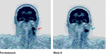
Clinical effect of zalutumumab. PET scan from a patient with SCCHN showing a partial metabolic response upon treatment with zalutumumab (8 mg/kg).

References

    1. Ortho Multicenter Transplant Study Group. A randomized clinical trial of OKT3 monoclonal antibody for acute rejection of cadaveric renal transplants. N. Engl. J. Med. 1985;313:337–342. - PubMed
    1. Morrison SL, Johnson MJ, Herzenberg LA, Oi VT. Chimeric human antibody molecules: Mouse antigen-binding domains with human constant region domains. Proc. Natl. Acad. Sci. USA. 1984;81:6851–6855. - PMC - PubMed
    1. Jones PT, Dear PH, Foote J, Neuberger MS, et al. Replacing the complementarity-determining regions in a human antibody with those from a mouse. Nature. 1986;321:522–525. - PubMed
    1. Lonberg N. Human antibodies from transgenic animals. Nat. Biotechnol. 2005;23:1117–1125. - PubMed
    1. Fishwild DM, O'Donnell SL, Bengoechea T, Hudson DV, et al. High-avidity human IgG kappa monoclonal antibodies from a novel strain of minilocus transgenic mice. Nat. Biotechnol. 1996;14:845–851. - PubMed

MeSH terms