

Mitochondrial NADH fluorescence is enhanced by complex I binding
- PMID: 18702505
- PMCID: PMC3515876
- DOI: 10.1021/bi800307y
Mitochondrial NADH fluorescence is enhanced by complex I binding
Abstract
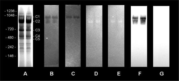
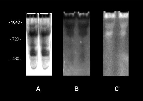
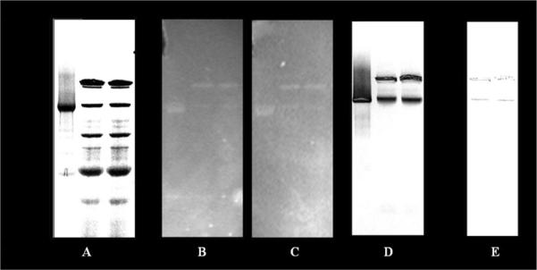
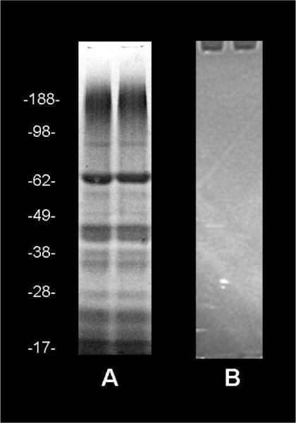
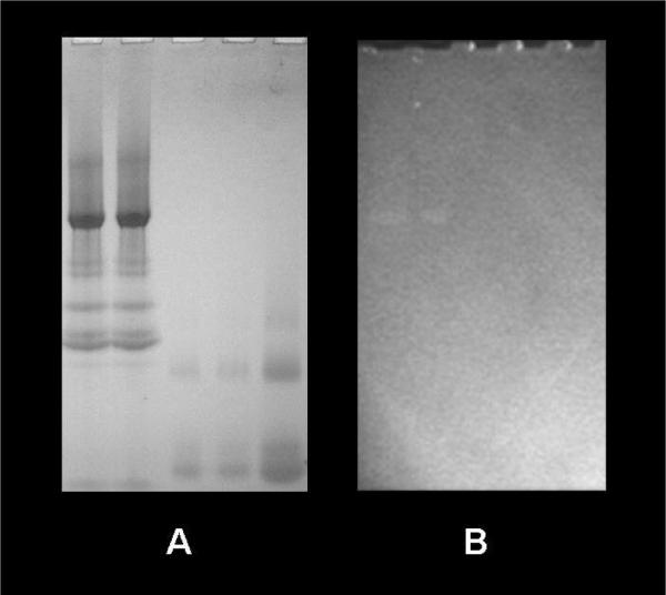
Mitochondrial NADH fluorescence has been a useful tool in evaluating mitochondrial energetics both in vitro and in vivo. Mitochondrial NADH fluorescence is enhanced several-fold in the matrix through extended fluorescence lifetimes (EFL). However, the actual binding sites responsible for NADH EFL are unknown. We tested the hypothesis that NADH binding to Complex I is a significant source of mitochondrial NADH fluorescence enhancement. To test this hypothesis, the effect of Complex I binding on NADH fluorescence efficiency was evaluated in purified protein, and in native gels of the entire porcine heart mitochondria proteome. To avoid the oxidation of NADH in these preparations, we conducted the binding experiments under anoxic conditions in a specially designed apparatus. Purified intact Complex I enhanced NADH fluorescence in native gels approximately 10-fold. However, no enhancement was detected in denatured individual Complex I subunit proteins. In the Clear and Ghost native gels of the entire mitochondrial proteome, NADH fluorescence enhancement was localized to regions where NADH oxidation occurred in the presence of oxygen. Inhibitor and mass spectroscopy studies revealed that the fluorescence enhancement was specific to Complex I proteins. No fluorescence enhancement was detected for MDH or other dehydrogenases in this assay system, at physiological mole fractions of the matrix proteins. These data suggest that NADH associated with Complex I significantly contributes to the overall mitochondrial NADH fluorescence signal and provides an explanation for the well established close correlation of mitochondrial NADH fluorescence and the metabolic state.
Figures
Similar articles
-
Distribution of mitochondrial NADH fluorescence lifetimes: steady-state kinetics of matrix NADH interactions.Biochemistry. 2005 Feb 22;44(7):2585-94. doi: 10.1021/bi0485124. Biochemistry. 2005. PMID: 15709771
-
Evaluation of functioning of mitochondrial electron transport chain with NADH and FAD autofluorescence.Ukr Biochem J. 2016 Jan-Feb;88(1):31-43. doi: 10.15407/ubj88.01.031. Ukr Biochem J. 2016. PMID: 29227076
-
Investigation of NADH binding, hydride transfer, and NAD(+) dissociation during NADH oxidation by mitochondrial complex I using modified nicotinamide nucleotides.Biochemistry. 2013 Jun 11;52(23):4048-55. doi: 10.1021/bi3016873. Epub 2013 May 30. Biochemistry. 2013. PMID: 23683271 Free PMC article.
-
Generation of superoxide by the mitochondrial Complex I.Biochim Biophys Acta. 2006 May-Jun;1757(5-6):553-61. doi: 10.1016/j.bbabio.2006.03.013. Epub 2006 Apr 17. Biochim Biophys Acta. 2006. PMID: 16678117 Review.
-
NADH/NAD+ interaction with NADH: ubiquinone oxidoreductase (complex I).Biochim Biophys Acta. 2008 Jul-Aug;1777(7-8):729-34. doi: 10.1016/j.bbabio.2008.04.014. Epub 2008 Apr 18. Biochim Biophys Acta. 2008. PMID: 18471432 Free PMC article. Review.
Cited by
-
Tissue Imaging and Quantification Relying on Endogenous Contrast.Adv Exp Med Biol. 2021;3233:257-288. doi: 10.1007/978-981-15-7627-0_12. Adv Exp Med Biol. 2021. PMID: 34053031 Review.
-
Reduced metabolic rate and oxygen radicals production in stored insect sperm.Proc Biol Sci. 2012 Jun 7;279(1736):2196-203. doi: 10.1098/rspb.2011.2422. Epub 2012 Jan 25. Proc Biol Sci. 2012. PMID: 22279170 Free PMC article.
-
Label-free, High-Resolution Optical Metabolic Imaging of Human Cervical Precancers Reveals Potential for Intraepithelial Neoplasia Diagnosis.Cell Rep Med. 2020 May 19;1(2):100017. doi: 10.1016/j.xcrm.2020.100017. Cell Rep Med. 2020. PMID: 32577625 Free PMC article.
-
Optical Redox Imaging of Lonidamine Treatment Response of Melanoma Cells and Xenografts.Mol Imaging Biol. 2019 Jun;21(3):426-435. doi: 10.1007/s11307-018-1258-z. Mol Imaging Biol. 2019. PMID: 30151646 Free PMC article.
-
Rapid, high-resolution, non-destructive assessments of metabolic and morphological homogeneity uniquely identify high-grade cervical precancerous lesions.bioRxiv [Preprint]. 2024 May 14:2024.05.10.593564. doi: 10.1101/2024.05.10.593564. bioRxiv. 2024. PMID: 38798665 Free PMC article. Preprint.
References
-
- Chance B, Williamson JR, Famieson D, Schoener B. Properties and kinetics of reduced pyridine nucleotide fluorescence of the isolated and in vivo rat heart. Biochemische Zeitschrift. 1965;341:357–377.
-
- Chance B, Cohen P, Jobsis F, Schoener B. Intracellular Oxidation-Reduction states in vivo. Science. 1962;137:499–508. - PubMed
-
- Chance B, THORELL B. Fluorescence measurements of mitochondrial pyridine nucleotide in aerobiosis and anaerobiosis. Nature. 1959;184:931–934. - PubMed
MeSH terms
Substances
Grants and funding
LinkOut - more resources
Full Text Sources
Other Literature Sources
