. 2008 Aug;28(3):510-20.
doi: 10.1111/j.1460-9568.2008.06345.x.

Brain-derived neurotrophic factor enhances fetal respiratory rhythm frequency in the mouse preBötzinger complex in vitro

Affiliations

Brain-derived neurotrophic factor enhances fetal respiratory rhythm frequency in the mouse preBötzinger complex in vitro

Julien Bouvier et al. Eur J Neurosci. 2008 Aug.

Abstract

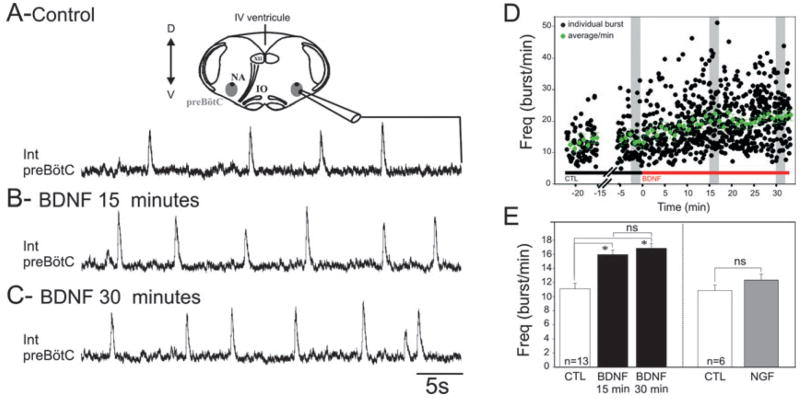
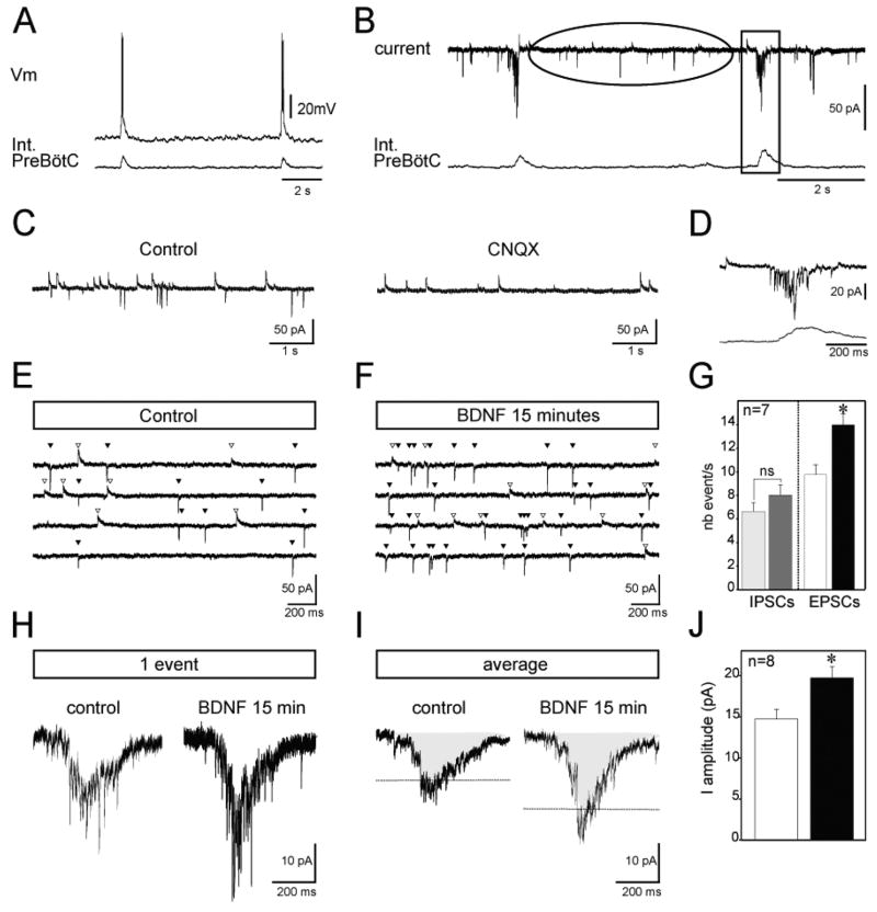
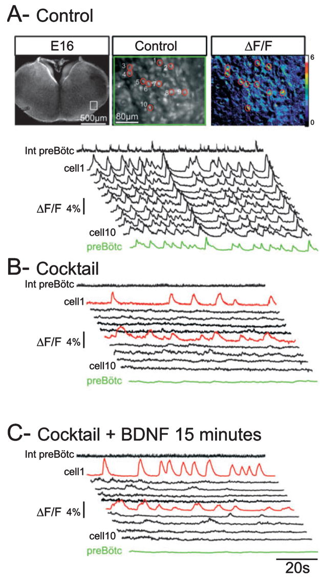
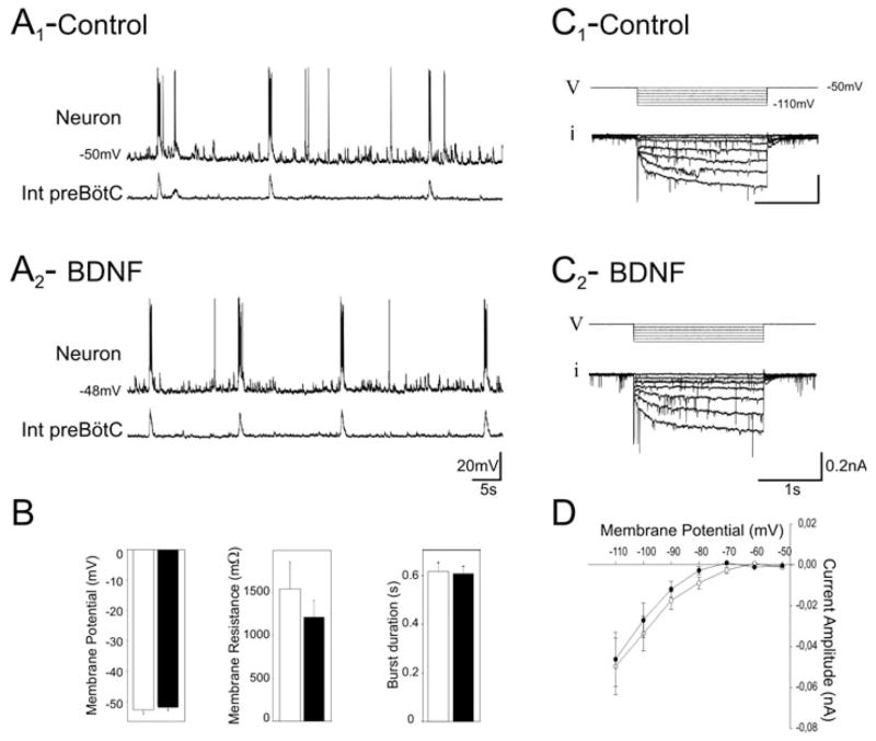
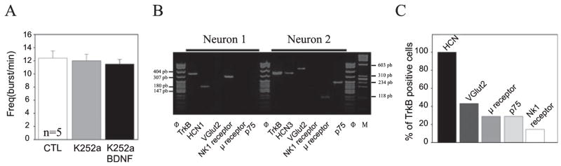
Brain-derived neurotrophic factor (BDNF) is required during the prenatal period for normal development of the respiratory central command; however, the underlying mechanisms remain unknown. To approach this issue, the present study examined BDNF regulation of fetal respiratory rhythm generation in the preBötzinger complex (preBötC) of the mouse, using transverse brainstem slices obtained from prenatal day 16.5 animals. BDNF application (100 ng/mL, 15 min) increased the frequency of rhythmic population activity in the preBötC by 43%. This effect was not observed when preparations were exposed to nerve growth factor (100 ng/mL, 30 min) or pretreated with the tyrosine kinase inhibitor K252a (1 h, 200 nm), suggesting that BDNF regulation of preBötC activity requires activation of its cognate tyrosine receptor kinase, TrkB. Consistent with this finding, single-cell reverse transcription-polymerase chain reaction experiments showed that one third of the rhythmically active preBötC neurons analysed expressed TrkB mRNA. Moreover, 20% expressed BDNF mRNA, suggesting that the preBötC is both a target and a source of BDNF. At the network level, BDNF augmented activity of preBötC glutamatergic neurons and potentiated glutamatergic synaptic drives in respiratory neurons by 34%. At the cellular level, BDNF increased the activity frequency of endogenously bursting neurons by 53.3% but had no effect on basal membrane properties of respiratory follower neurons, including the Ih current. Our data indicate that BDNF signalling through TrkB can acutely modulate fetal respiratory rhythm in association with increased glutamatergic drive and bursting activity in the preBötC.


Figures

Fig. 1
Fig. 1
BDNF increases respiratory frequency in the fetal mouse transverse brainstem slice preparation. Integrated population activity recorded from the preBötC respiratory network in control conditions (A) and after 15 min (B) and 30 min (C) exposure to 100 ng/mL BDNF. (A) Top: Schematic diagram of a transverse brainstem slice containing the preBötC from which population activity was recorded (see the integrated trace below). (D) Graph representing the kinetics of BDNF effects on individual burst frequency (black dots) and on frequency average/min (green dots) recorded from a single preparation. Grey rectangles indicate the time window during which measurements were performed for every experiment. (E) Bar histogram depicting the mean frequency of the preBötC rhythmic activity obtained from 13 preparations; control (CTL) (white bar), BDNF (15 and 30 min; two black bars) and nerve growth factor (NGF) (100 ng/mL; grey bar; n = 6). BDNF induced a significant increase in the frequency of the preBötC population activity, whereas NGF had no effect (*P < 0.05). IO, inferior olive; XII, hypoglossal nucleus; NA, nucleus ambiguous; D, dorsal; Int, integrated; IV, fourth (ventrical); V, ventral; ns, not significant.
Fig. 2
Fig. 2
BDNF enhances the activity of glutamatergic neurons in the fetal preBötC network. Intracellular recordings (top traces) from a rhythmic preBötC neuron in current-clamp (A) and voltage-clamp (B) mode recorded simultaneously with integrated population activity (bottom traces) under control conditions. The ellipse in B indicates the period between inspiratory bursts, represented at an extended time scale for a different neuron in C. Note that the application of CNQX blocks all excitatory glutamatergic events (right panel in C). The rectangle in B highlights one inspiratory burst, represented at an extended time scale in (D). Current traces recorded between inspiratory bursts at a holding potential of −50 mV are shown in control (E) and in the presence of BDNF (F). The four traces illustrated correspond to four samples obtained from the same neuron before and after BDNF application. White triangles indicate outward chloride-mediated synaptic currents and black triangles indicate inward glutamate-mediated synaptic currents. (G) Graph representing the mean number of inhibitory (IPSCs) and excitatory (EPSCs) events/s obtained from seven neurons in control conditions (light grey and white bars, respectively) and after BDNF treatment (dark grey and black bars, respectively). Synaptic glutamatergic drive (single event in H and average of 10 events in I) recorded from a fetal rhythmic neuron during a burst of population activity in control conditions (left panels in H and I) and after 15 min exposure to 100 ng/mL BDNF (right panels in H and I). Dashed lines indicating amplitude of the mean in the two conditions illustrate the potentiation of synaptic drive observed in the presence of BDNF. (J) Graph depicting the mean amplitude of the synaptic envelope obtained from eight neurons in control conditions (white bar) and after BDNF treatment (black bar). Int, integrated; nb, number; Vm, voltage. *P < 0.05.
Fig. 3
Fig. 3
BDNF modulates the frequency activity of isolated bursting neurons. (A) Top: Fluorescent images of an E16.5 transverse medullary slice taken at low (left) and high (middle) magnification following loading with the calcium indicator Calcium Green 1-AM. Note the recording electrode positioned at the surface of the slice in the preBötC region. The white rectangle delimits the area that contains rhythmic neurons and is shown at higher magnification in the middle panel with each of the cells numbered 1–10. An example of calcium transients visualized in these cells is shown in the right panel as relative changes in fluorescence (ΔF/F). The traces in A–C display the calcium transients recorded from neurons 1–10 in control conditions (A), blocker cocktail (20 μM CNQX, 50 μM AP5, 10 μM bicuculline, 5 μM strychnine and 50 μM carbenoxolone) (B) and blocker cocktail plus 100 ng/mL BDNF for 15 min (C). Calcium changes occurring in the preBötC area are shown in the green trace (preBötC) and electrical activity recorded in the contralateral preBötC is represented as the integrated trace (Int preBötC). In cocktail, population activity ceases (flat green trace and flat integrated recording) and synchronized calcium events disappear. The red traces highlight two neurons exhibiting endogenous bursting properties, neurons that remain rhythmically active after intra-network connectivity blockade. In the presence of BDNF the bursting frequency of these neurons increases (compare the red traces in B and C).
Fig. 4
Fig. 4
Membrane properties of inspiratory follower neurons are not affected by BDNF. (A) Intracellular recording from a rhythmic preBötC neuron (top traces) recorded simultaneously with integrated population activity (bottom traces) in control conditions (A1) and after 15 min exposure to 100 ng/mL BDNF (A2). (B) Bar histograms showing the mean values for membrane potential, membrane resistance and burst duration in control conditions (white bars) and in the presence of 100 ng/mL BDNF (black bars) obtained for eight neurons. Voltage stimulation protocol (V) used to evoke the Ih current (i) in control conditions (C1) and after 15 min exposure to BDNF (C2). (D) Graph of the Ih current amplitude vs. voltage. Mean curves were obtained from seven neurons and evoked currents were measured in control conditions (open circles) and after 15 min in the presence of 100 ng/mL BDNF (black circles). BDNF had no significant effect on any of the membrane properties tested. Int, integrated.
Fig. 5
Fig. 5
Evidence that TrkB signalling mediates BDNF regulation of fetal preBötC neuron activity. (A) Bar histograms showing the mean firing frequency of preBötC neurons (n = 5; extracellular activity from five preparations) in control (CTL) conditions (white bars), in the presence of 200 nM K252a for 1 h (light grey bar) and in the presence of 200 nM K252a plus 100 ng/mL BDNF for 15 min (black bar) following 1 h pretreatment with the tyrosine kinase inhibitor alone. In contrast to the increase in frequency observed in response to BDNF alone (Fig. 1), BDNF had no effect in the presence of K252a, consistent with a requirement for tyrosine kinase activation. (B) Agarose gel analysis of reverse transcription-PCR products amplified from two different rhythmic neurons. Φ and M correspond to molecular weight markers (indicated on the left and right of the gel). (C) Bar graph representing the proportion of TrkB-positive neurons expressing other markers, including HCNs, VGlut2, μ receptor, p75 receptor and NK1R.

References

    1. Arvanian VL, Mendell LM. Acute modulation of synaptic transmission to motoneurons by BDNF in the neonatal rat spinal cord. Eur J Neurosci. 2001;14:1800–1808. - PubMed
    1. Balkowiec A, Katz DM. Brain-derived neurotrophic factor is required for normal development of the central respiratory rhythm in mice. J Physiol. 1998;510(Pt 2):527–533. - PMC - PubMed
    1. Balkowiec A, Katz DM. Activity-dependent release of endogenous brain-derived neurotrophic factor from primary sensory neurons detected by ELISA in situ. J Neurosci. 2000;20:7417–7423. - PMC - PubMed
    1. Balkowiec A, Katz DM. Cellular mechanisms regulating activity-dependent release of native brain-derived neurotrophic factor from hippocampal neurons. J Neurosci. 2002;22:10399–10407. - PMC - PubMed
    1. Bienvenu T, Chelly J. Molecular genetics of Rett syndrome: when DNA methylation goes unrecognized. Nat Rev. 2006;7:415–426. - PubMed

Publication types

MeSH terms