. 2008 Nov;214(1):25-36.
doi: 10.1016/j.expneurol.2008.07.012. Epub 2008 Jul 22.

Maintaining epitheliopoietic potency when culturing olfactory progenitors

Affiliations

Maintaining epitheliopoietic potency when culturing olfactory progenitors

Woochan Jang et al. Exp Neurol. 2008 Nov.

Abstract

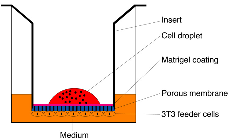
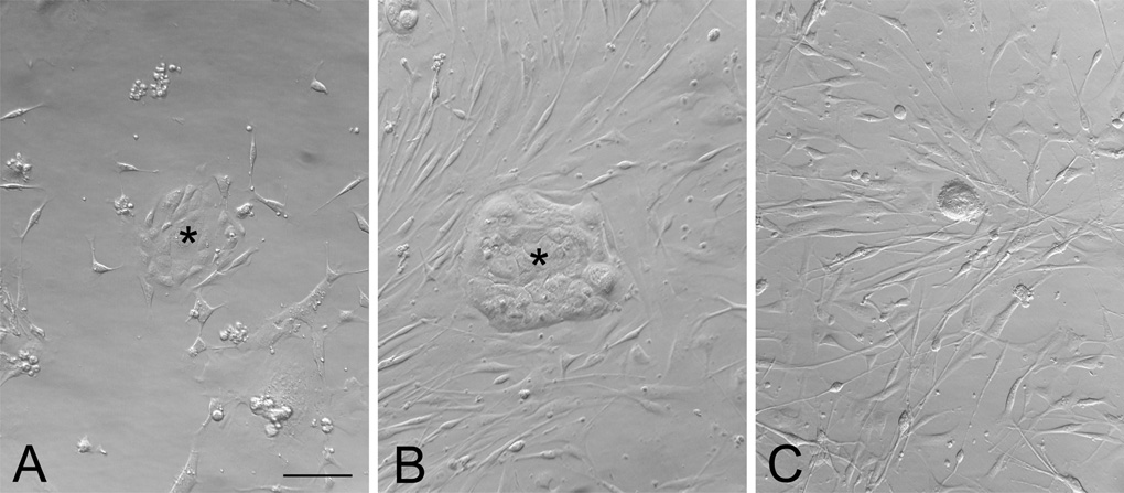
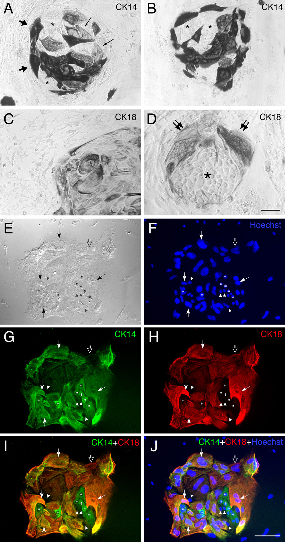
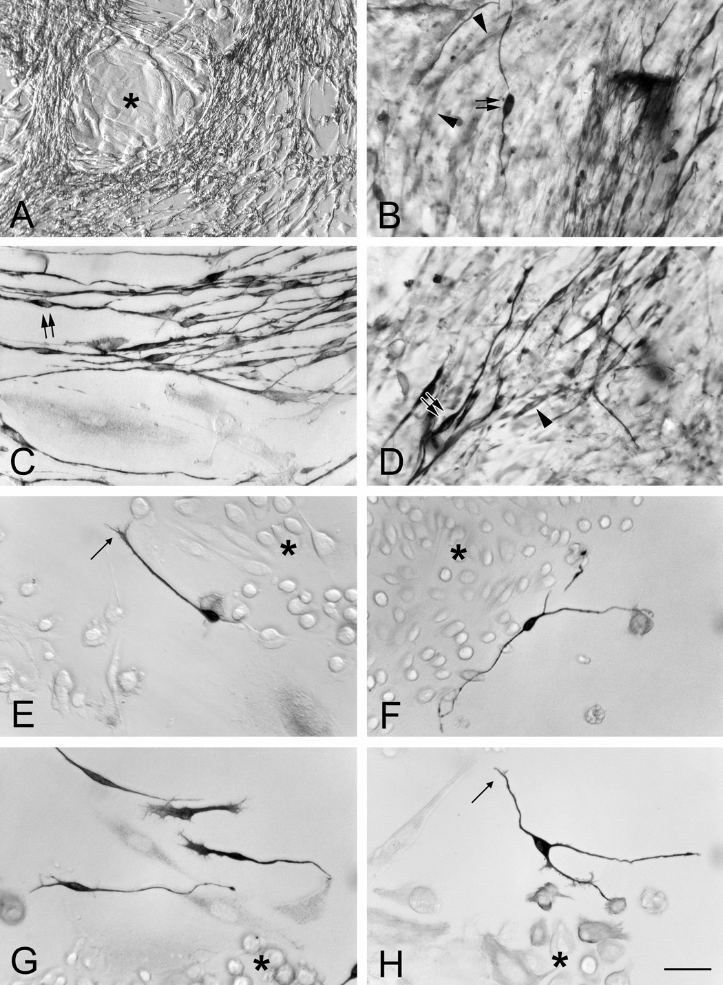
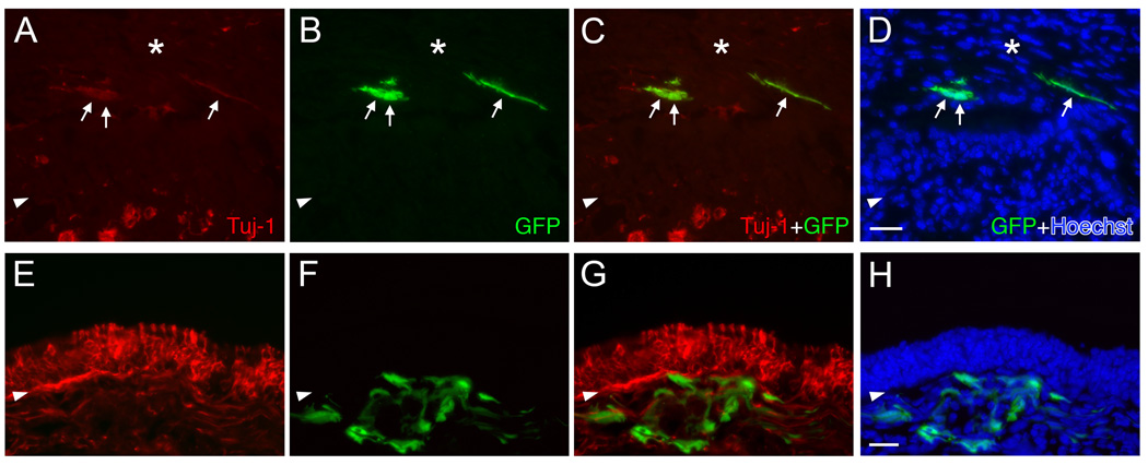
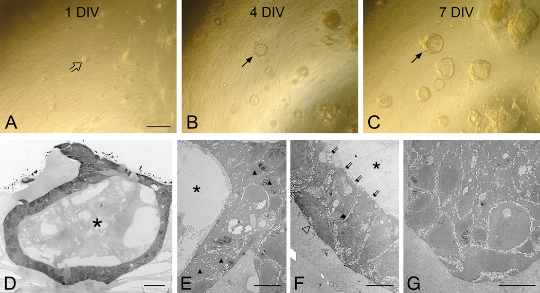
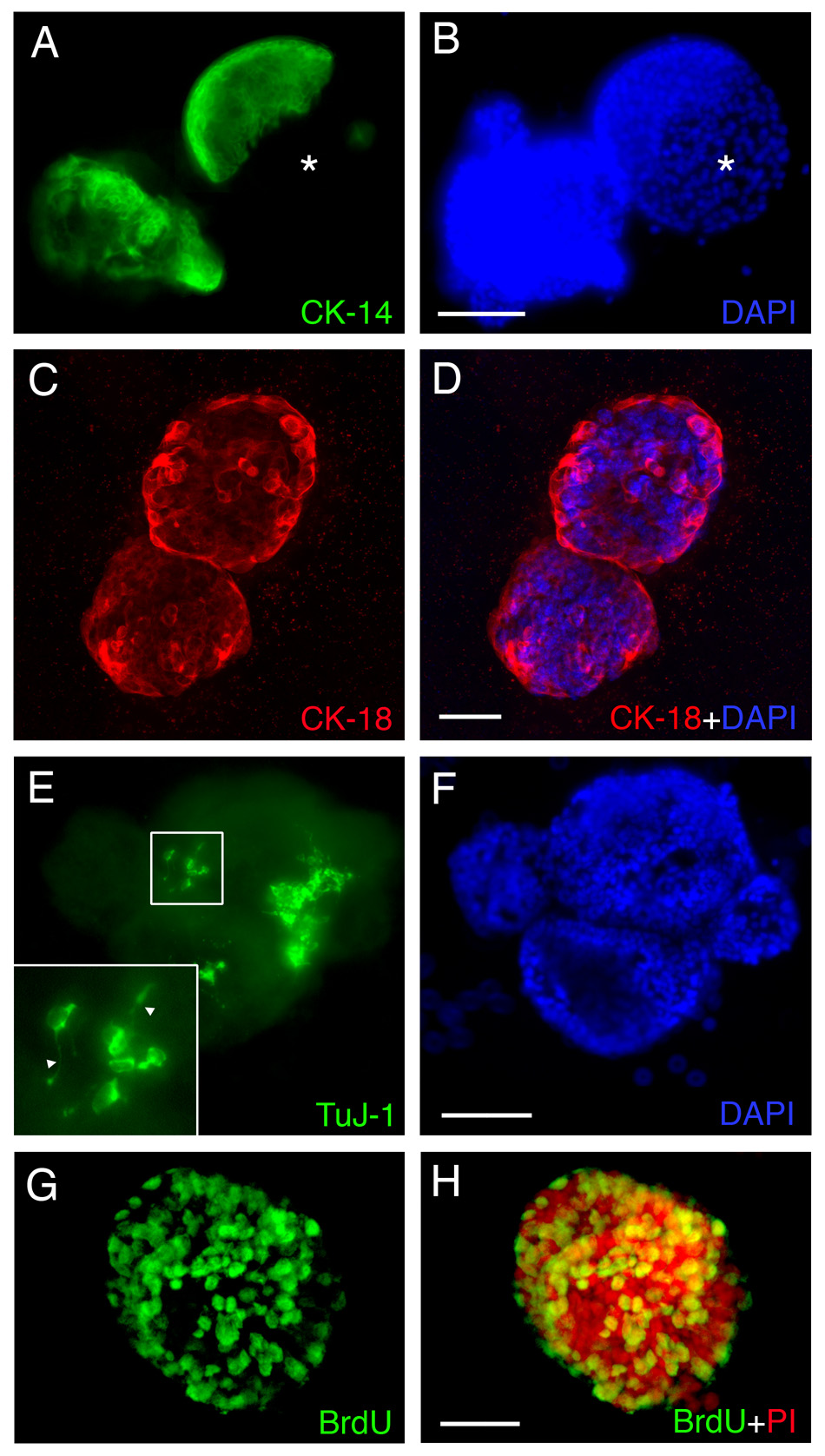
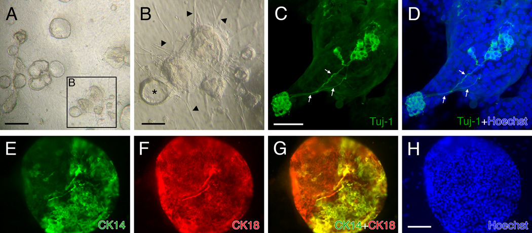
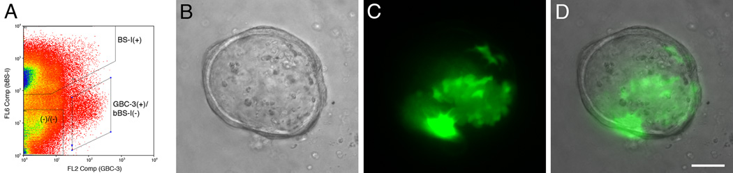
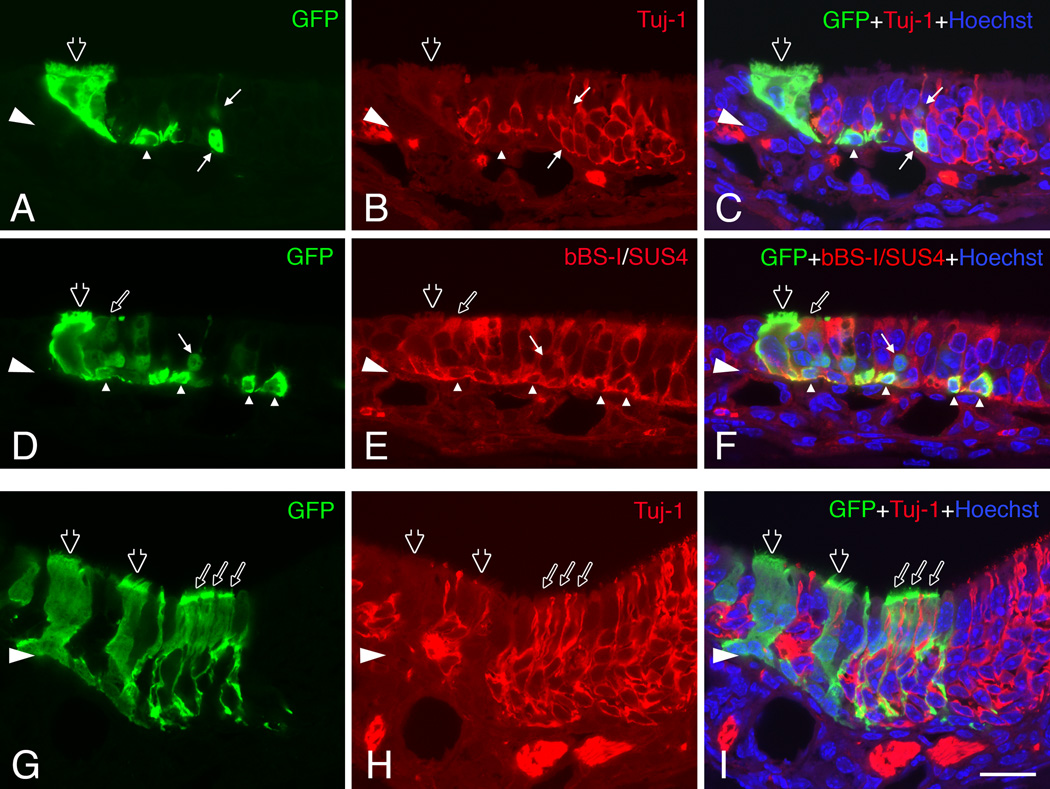
The olfactory epithelium is remarkable for the persistence of multipotent, neurocompetent progenitor and stem cells throughout life that can replace all of the various cell types of the epithelium following injury. The therapeutic exploitation of the neurocompetent stem cells of the adult olfactory epithelium would be facilitated by the development of a culture system that maintains the in vivo potency of the progenitors while they are expanded and/or manipulated. We have used an air-liquid interface culture protocol, in which a feeder cell layer of 3T3 cells is established on the underside of a culture insert and Facs-isolated or unsorted progenitor cells from the methyl bromide-lesioned adult rodent epithelium are seeded on upper side. Under these conditions, epithelial cells other than HBCs are capable of organizing themselves into complex three-dimensional, epithelium-lined spheres, which can be passaged. The spheres contain cells with the molecular phenotype of globose basal cells, horizontal basal cells, sustentacular cells and neurons. Spheres derived from mice that express the green fluorescent protein constitutively can be dissociated after 6 days in vitro and directly transplanted into the epithelium of wild-type, methyl bromide-lesioned mice via nasal infusion. The resulting clones contain the various cell types observed in aggregate when globose basal cells are transplanted acutely. In contrast, the same cells cultured as two-dimensional, submerged cultures undergo fibroblastic transition after transplantation and do not integrate into the epithelium. In conclusion, the culture system described here maintains the potency of progenitors, which can then participate in epitheliopoiesis in vivo.


Figures

Figure 1
Figure 1
Schematic diagram illustrating the three-dimensional air-liquid interface culture.
Figure 2
Figure 2
Epithelial islands develop from adult olfactory epithelium in submerged cultures. Bright field photomicrographs showing the two-dimensional submerged cultures of dissociated cells from the olfactory epithelium (OE) from normal (A), MeBr-exposed (B; at 2 days postlesion), and bulbectomized (C; at 5 days postlesion) mice grown on matrigel in serum-rich NIC medium. In normal and MeBr-exposed OE cultures, epithelioid islands are seen (asterisks in A and B) as well as scattered, elongated, spindle-shaped cells around the islands. In bulbectomized OE culture, the spindled cells are much more prevalent (C). Scale bar in A is 50 µm, and applies to B and C.
Figure 3
Figure 3
Epithelial islands contain cells expressing mixed phenotypic markers, i.e., CK14, a maker of HBCs in vivo, and CK18, a marker of sustentacular cells in vivo. (A, B) CK14(+) cells are prominent in cultures derived from MeBr-lesioned OE grown on laminin in serum-rich NIC medium. Some are densely positive (thick arrows), others are weakly positive (thin arrows) and still others are completely unstained (asterisks) within a single island. Elongated cells found outside of the islands are CK14(−). (C, D) CK-18(+) cells are also present in cultures of MeBr-lesioned OE maintained under identical conditions. Note the heterogeneity of size and shape as well as intensity of CK18 immunoreactivity. Furthermore, the proportion of CK18 cells also varies such that the island in C is dominated by CK18(+) cells, while the island in D is sharply divided with CK18(+) wedge-shaped cells at the surface (double arrows) and CK18(−) round cells in the interior (asterisks). Spindle-shaped cells outside of islands are CK18-negative. (E–J) Doubling labeling with CK14 (green) and CK18 (red) demonstrates that the island consists of CK14-positive presumptive HBCs (asterisks), CK18-positive presumptive sustentacular cells (open arrow), double positive cells (thin arrows) and unstained cells (triangles). The brightfield image and Hoechst staining of the same island are shown in E and F, respectively. Scale bar in D is 50 µm for A and B, 100 µm for C, and 25 µm for D. Scale bar in J is 50 µm and also applies to E–I.
Figure 4
Figure 4
TuJ-1(+) cells are present in the monolayer cultures of MeBr-lesioned OE grown on laminin in serum-rich NIC medium (A–D) and serum-free KGM (E–H). The TuJ-1(+) cells are elongated cells present surrounding epithelioid islands (asterisks in A and E–H). Note the difference in abundance of TuJ-1(+) cells in the two media conditions. In serum-rich condition, some TuJ-1(+) cells are heavily stained and clearly bipolar (double arrows in B, C and D), suggesting neuronal differentiation, while others are some weakly stained cells, and are usually spindle-shaped (arrowheads in B and D). In serum-free condition, such heavy labeled cells are scarce, but clearly neuronal-like in shape. Indeed, the leading process of some TuJ-1(+) cells closely resembles growth cones (thin arrow in E and H), particularly when found around the edges of the islands (asterisks). Note that not all scattered cells are TuJ-1(+). Scale bar in H is 50 µm for A and 25 µm for B–H.
Figure 5
Figure 5
Cells harvested from the MeBr-lesioned OE and maintained in submerged monolayer cultures do not integrate into the epithelium. The donor cells from the 1.5 days MeBr-lesioned OE of GFP-expressing mice were cultured as monolayers in serum-rich media for 6 days (A–D) or 15 days (E–H). In host wild type mice (see Materials and Methods), the donor-derived GFP-expressing cells (green) are found either in scar tissue (asterisks) (A–D), or below the basal lamina (arrowheads) (E–H) 11 days post-transplantation. Note that some engrafted cells are weakly TuJ-1(+) (arrows in A–D). (A, E) TuJ-1 only; (B, F) transplanted GFP cells; (C, G) merging of TuJ-1 and GFP; (D, H) merging of TuJ-1, GFP and Hoechst (blue). Scale bar in D is 25 µm and applies to A–C, and scale bar in H is 50 µm and applies to E–F.
Figure 6
Figure 6
Dissociated OE cells from MeBr-lesioned mice form complex spheres when cultured at the air-liquid interface with a 3T3 feeder cell layer on the underside of the insert. Cultures were photographed at (A) 1 day in vitro (1 DIV); (B) 4 DIV; and (C) 7 DIV. The dissociated cells aggregate within 24 hrs in culture (open arrow in A) and progressively form dome-like spheres (arrows in B and C). (D) Semithin sections of spheres examined with a light microscope. Electron microscopies (EM) of representative spheres (E–G). (D–F) The sphere shown here is hollow (asterisks in D–F) and consists of rounded (triangles in D), flattened (open triangle in F) and columnar (double arrows in F) cells. (G) Another sphere forms a solid ball-like structure composed of mostly rounded cells. Scale bar in A is 100 µm and applies to B and C. Scale bars in D–G are 20, 10, 5 ad 10 µm, respectively.
Figure 7
Figure 7
Cells in spheres derived from MeBr-lesioned rat OE express a variety of differentiated cell markers when maintained in air-liquid interface culture as in the regenerating OE, including CK14 (A, B), CK18 (C, D), TuJ-1 (E, F). (G–H) Some of the cells are labeled by their incorporation of BrdU added to the culture, and thus, are proliferating in vitro. (A, B) 10 DIV. CK14 positive cells are found in the sphere, and in some cases, the sphere is sharply divided into CK14 positive (green) and CK14 negative (asterisks) area. (C, D) 7 DIV. CK18 positive cells (red) are found in the spheres, and often times are found at the periphery of the spheres. (E, F) 10 DIV. TuJ-1 positive cells (green) are found clustered in the sphere, and often bear axon-like processes (triangles in higher magnification inset). (G, H) 6 DIV. A majority of cells in the sphere incorporate BrdU (anti-BrdU, green), indicating that spheres can grow via proliferation. Propidium iodide (PI, red) was used to stain the nuclei (H). Scale bars in B and F are 100 µm and apply to A and E. Scale bars in D and H are 50 µm and apply to C and G.
Figure 8
Figure 8
Cells harvested from rat OE on postnatal day 1 form spheres when maintained in air-liquid interface culture. (A) At 5 DIV, spheres appear and grow rapidly, indicating rapid proliferation. (B) Spheres appear either hollow with an empty cavity (asterisk) or clustered into a pile. Spheres are surrounded by a radiating array of elongated, spindle-shaped cells (arrowheads), which are rarely seen in the cultures derived from MeBr-lesioned OE. (C, D) Neurons are found with a greater frequency than in the cultures with adult OE cells. Confocal microscopy illustrates two areas with neuronal populations that are interconnected via axons (arrows). (E–H) Both CK14(+) (E) and CK18(+) (F) cells are found in a sphere and some cells are double positive with anti-CK14 and 18 (G). Scale bars in A and B are 200 µm and 100 µm, respectively. Scale bar in D is 50 µm and applies to C, and scale bar in H is 100 µm and applies to E, F and G.
Figure 9
Figure 9
FACS-sorted GBCs will participate in the formation of spheres in the air-liquid interface cultures. (A) FACS-profile of cells from 1.5 days post-MeBr lesioned rat OE sorted with GBC-3 (a marker selective for GBCs) and BS-I (an HBC marker). The GBC population corresponds to the GBC-3(+)/bBS-I(−) population. (B–D) Addition of FACS-sorted GBCs (green) to the non-GBC/HBC cells (double negative fraction, (−)/(−), shown in A) at a 1:99 ratio. The GFP-expressing cells are incorporated into the sphere (C, D), indicating that GBCs contribute to sphere formation. The sphere was photographed at 3 DIV and shown in brightfield (B), green channel epifluorescence (C) and merged (D). Scale bar in D is 50 µm and applies to A and B.
Figure 10
Figure 10
Cells harvested from spheres that formed in air-liquid interface cultures incorporate into the regenerating OE of the host after transplantation. (A–C) (D–F) Adjacent sections through a single graft-derived cluster 11 days post-transplantation. This cluster contains donor-derived (green) neurons (thin arrows) double labeled by Tuj-1 staining (red in B–C), sustentacular cells (thin open arrows) and HBCs (triangles) double labeled by bBS-I and SUS-4 staining (red in E–F) and ciliated respiratory epithelial cells (thick open arrows in A–F). (G–I) Another clone consists mainly of sustentacular cells (thin open arrows) positioned above the layer of neurons and marked by thin foot processes extending toward the basal lamina. Ciliated respiratory epithelial cells (thick open arrows in G–I) are also seen in this clone. The images were obtained with a confocal microscope. Scale bars in I is 20 µm and apply to A–H.

Similar articles

Cited by

References

    1. Asselineau D, Bernhard B, Bailly C, Darmon M. Epidermal morphogenesis and induction of the 67 kD keratin polypeptide by culture of human keratinocytes at the liquid-air interface. Exp Cell Res. 1985;159:536–539. - PubMed
    1. Caggiano M, Kauer JS, Hunter DD. Globose basal cells are neuronal progenitors in the olfactory epithelium: a lineage analysis using a replication-incompetent retrovirus. Neuron. 1994;13:339–352. - PubMed
    1. Calof AL, Chikaraishi DM. Analysis of neurogenesis in a mammalian neuroepithelium: proliferation and differentiation of an olfactory neuron precursor in vitro. Neuron. 1989;3:115–127. - PubMed
    1. Carter LA, MacDonald JL, Roskams AJ. Olfactory horizontal basal cells demonstrate a conserved multipotent progenitor phenotype. J Neurosci. 2004;24:5670–5683. - PMC - PubMed
    1. Cau E, Gradwohl G, Fode C, Guillemot F. Mash1 activates a cascade of bHLH regulators in olfactory neuron progenitors. Development. 1997;124:1611–1621. - PubMed

Publication types