Review
. 2008 Nov;135(5):1469-92.
doi: 10.1053/j.gastro.2008.05.047. Epub 2008 Aug 12.

Gastrointestinal neuroendocrine tumors: pancreatic endocrine tumors

Affiliations
Review

Gastrointestinal neuroendocrine tumors: pancreatic endocrine tumors

David C Metz et al. Gastroenterology. 2008 Nov.

Abstract

Pancreatic endocrine tumors (PETs) have long fascinated clinicians and investigators despite their relative rarity. Their clinical presentation varies depending on whether the tumor is functional or not, and also according to the specific hormonal syndrome produced. Tumors may be sporadic or inherited, but little is known about their molecular pathology, especially the sporadic forms. Chromogranin A appears to be the most useful serum marker for diagnosis, staging, and monitoring. Initially, therapy should be directed at the hormonal syndrome because this has the major initial impact on the patient's health. Most PETs are relatively indolent but ultimately malignant, except for insulinomas, which predominantly are benign. Surgery is the only modality that offers the possibility of cure, although it generally is noncurative in patients with Zollinger-Ellison syndrome or nonfunctional PETs with multiple endocrine neoplasia-type 1. Preoperative staging of disease extent is necessary to determine the likelihood of complete resection although debulking surgery often is believed to be useful in patients with unresectable tumors. Once metastatic, biotherapy is usually the first modality used because it generally is well tolerated. Systemic or regional therapies generally are reserved until symptoms occur or tumor growth is rapid. Recently, a number of newer agents, as well as receptor-directed radiotherapy, are being evaluated for patients with advanced disease. This review addresses a number of recent advances regarding the molecular pathology, diagnosis, localization, and management of PETs including discussion of peptide-receptor radionuclide therapy and other novel antitumor approaches. We conclude with a discussion of future directions and unsettled problems in the field.


Figures

Figure 1
Figure 1
Effect of widespread use of PPIs on diagnosis and referral of ZES patients in two centers (Italian-La Sapienza University [Rome, Italy] and NIH [Bethesda, Maryland]). The left panel shows the annual number of referrals of new cases before and after the widespread use of PPIs. The right panel shows the result for diagnosis of ZES at the NIH center. (Modified from66)
Figure 2
Figure 2
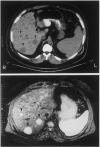
CT, MRI, EUS in patients with PETs. Panel A illustrates CT (top) and MRI(bottom) images of the abdomen in a patient with a metastatic gastrinoma. Liver metastases are indicated by arrowheads. Panel B illustrates an endoscopic ultrasound image of a pancreatic body insulinoma confirmed at subsequent surgery. The tumor is indicated by the black arrowheads.
Figure 2
Figure 2
CT, MRI, EUS in patients with PETs. Panel A illustrates CT (top) and MRI(bottom) images of the abdomen in a patient with a metastatic gastrinoma. Liver metastases are indicated by arrowheads. Panel B illustrates an endoscopic ultrasound image of a pancreatic body insulinoma confirmed at subsequent surgery. The tumor is indicated by the black arrowheads.
Figure 3
Figure 3
Comparison of conventional imaging (CT, MRI) and SRS to localize a primary gastrinoma (left) or metastatic disease (right) in two patients with ZES. In the left panel the patient had negative preoperative conventional imaging studies (CT, MRI) and angiography, but SRS showed a lesion in the pancreatic head area. At surgery a 2 cm tumor was resected and the patient has remained disease-free. In the right panel neither the MRI nor CT showed recurrent disease in this patient post resection of a gastrinoma, however the fasting gastrin was elevated and the SRS showed extensive metastases in lymph nodes and the liver. Both of these results show the greater sensitivity of SRS for localizing primary PETs as well as metastatic disease.
Figure 4
Figure 4
Comparison of the extent of liver metastases in a patient with a malignant PET on CT scanning (top panel) and positron emission tomographic scanning (bottom panel). This patient with a malignant PET had a few liver metastases seen on CT scanning (top) and SRS (not shown) but much more extensive disease on positron emission tomographic scanning with 11C-5-HTP demonstrating its greater sensitivity. (Images kindly provided by Prof. Anders Sundin, Department of Radiology, Uppsala University Hospital, Uppsala, Sweden).
Figure 5
Figure 5
Schematic diagram of a theoretical pancreatic endocrine tumor cell, smooth muscle cell (pericyte) or endothelial cell demonstrating the sites and mechanism of action of novel agents for the management of metastatic PETs. These cellular components of PETs all exhibit surface growth factor receptors (e.g., VEGFR, PDGFR, IGF-1R, c-KITR, etc) which when occupied by their respective growth factors (in an autocrine or paracrine manner) lead to autophosphorylation of the intracellular tyrosine kinase component of the receptor. Tyrosine kinase phosphorylation activates the PI3K-AKT-mTOR pathway (amongst others) ultimately promoting protein synthesis, cell cycle progression and cell survival which causes increased cellular proliferation, inhibition of apoptosis, cellular invasion, metastasis and tumor angiogenesis. This pathway can be inhibitied by monoclonal antibodies to growth factor receptors, tyrosine kinase inhibitors with specific activity against various growth factor receptors, or downstream mTOR inhibitors. Whilst mTOR inhibitors are active against both the tumor directly as well as its blood supply, tyrosine kinase inhibitors or antibodies directed against specific growth factors may predominantly effect the tumor itself or secondarily inhibit tumor cell growth by altering its blood supply-.

References

    1. Duerr EM, Chung DC. Molecular genetics of neuroendocrine tumors. Best Pract Res Clin Endocrinol Metab. 2007;21:1–14. - PubMed
    1. Corleto VD, Delle Fave G, Jensen RT. Molecular insights into gastrointestinal neuroendocrine tumors: importance and recent advances. Dig Liver Dis. 2002;34:668–680. - PubMed
    1. Arnold R, editor. Endocrine Tumors of the Gastrointestinal Tract: Part 11. 2005.
    1. Arnold R, editor. Endocrine tumors of the Gastrointestinal Tract: Part 1. 2005.
    1. Oberg K, Eriksson BE. Neuroendocrine tumors. Best Pract Res Clin Endocrinol Metab. 2007;21:1–172. - PubMed

Publication types