. 2008 Nov 1;423(2):116-24.
doi: 10.1016/j.gene.2008.07.018. Epub 2008 Jul 25.

Identification and characterization of PNRC splicing variants

Affiliations

Identification and characterization of PNRC splicing variants

Yuanzhong Wang et al. Gene. .

Abstract

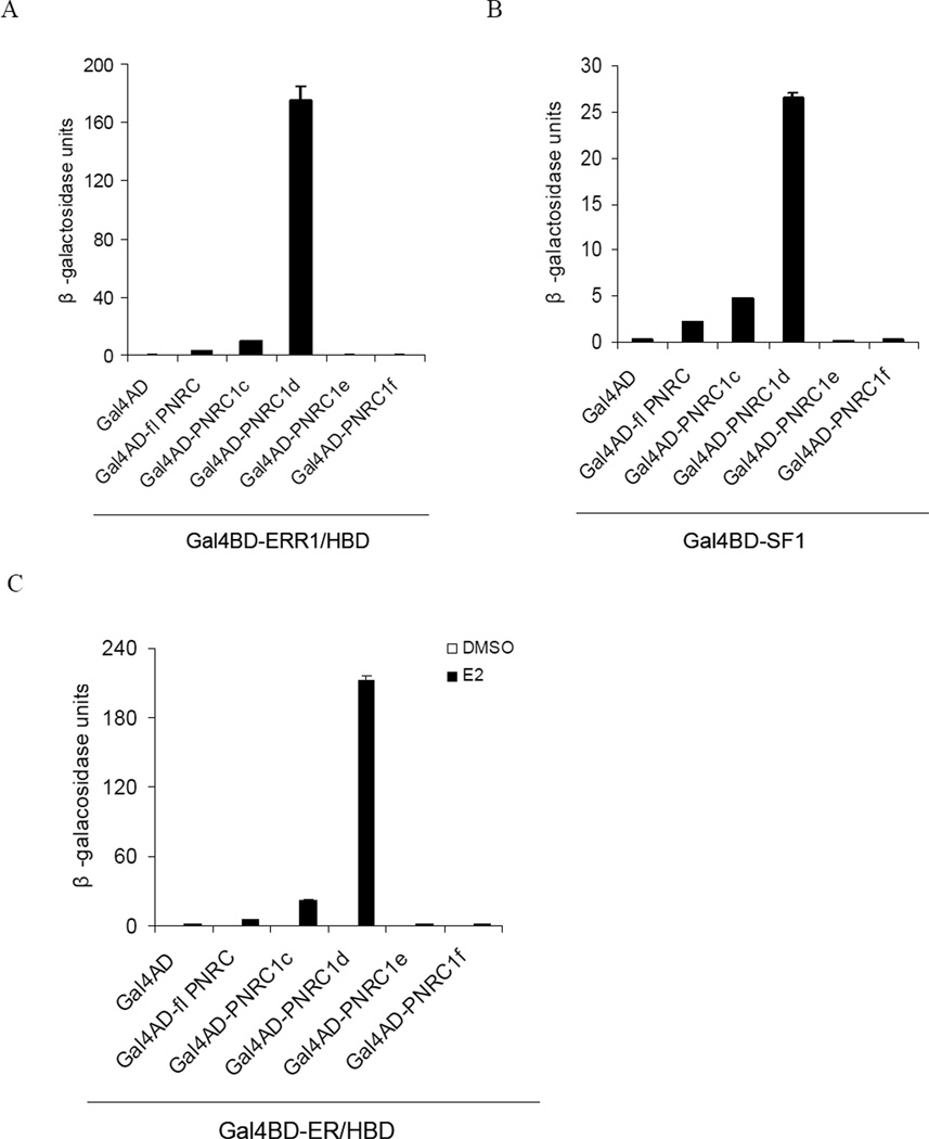
Nuclear receptor (NR) dependent transcriptional action requires recruitment of diverse factors characterized as coregulators. PNRC (proline-rich nuclear receptor coregulatory protein) is a member of coregulators that are capable of potentiating the transcriptional activity of NRs. Here we identified three human PNRC splicing variants designated PNRC1c, PNRC1d and PNRC1f. PNRC1c and PNRC1f are generated through alternative recognition of the 3'-splice site in exon 1, leading to in-frame deletion of 79 amino acids (aa) and an altered reading frame, respectively. PNRC1d is generated through the alternate promoter usage and forms a truncated protein containing C-terminus 142 aa of full-length PNRC. These isoforms differ in their abilities to bind NRs and potentiate NR mediated transcriptions. Moreover, PNRC1d can modulate the activity of full-length PNRC in enhancing ER mediated transcription. Our results suggest that PNRC exists as functionally distinct isoforms and alternative splicing serves as a regulatory mechanism of PNRC coactivator activity.


Figures

Fig 1
Fig 1. Schematic representation of PNRC alternative splicing products
(A). Schematic representation of human PNRC genomic structure, PNRC alternatively spliced transcript variants, and the position of the primers used in RT-PCR for identification of splicing variants. PNRC gene locates on chromosome 6 at 6q15 and contains two exons. Alternative splicing at exon 1 (termed 1a) or 2 of PNRC gene generates splicing variants PNRC1c, PNRC1f and PNRC1e. Alternative promoter usage generates splicing variant PNRC1d. The open boxes represent the putative amino acid-coding region of each transcript, the broken lines represent the part alternatively spliced out in the transcript, and the arrows P1–P5 indicate the positions of the primers used in RT-PCR for identification of PNRC splicing variants. (B). Schematic representation of deduced protein structure of PNRC isoforms. Full-length PNRC (fl PNRC) is composed of 327 amino acids (aa), containing two proline-rich Src homology domain-3 (SH3) binding motifs (aa 38–44, aa 285–291), one LXXLL motif (NR box, aa 319–323) and one putative nuclear localization signal (NLS, aa 94–101). PNRC1c lacks the middle 79 aa of fl PNRC (aa 103–181), PNRC1d is the carboxyl terminus 142 aa of fl PNRC (aa 186–327) due to noncoding of exon1b, whereas PNRC1e lacks 99 aa of fl PNRC (aa 229–327) but has 5 unique aa at the carboxyl terminus, and PNRC1f lacks 300 aa of fl PNRC (aa 28–327) but has 25 unique aa at the carboxyl terminus due to an altered reading frame and a stop codon resulting from alterative splicing.
Fig 2
Fig 2. RT-PCR analysis of PNRC splicing variants in various human cell lines
Total RNAs from the indicated cell lines were analyzed by RT-PCR using primer pairs of P1 and P2 (upper panel) or P3 and P2 (lower panel). P1 and P2 were used to amplify simultaneously the coding sequence (CDS) of fl PNRC (984bp), CDS of PNRC1c (747bp), and CDS plus part of 3’-UTR of PNRC1f (523bp). P3 and P2 were used to amplify part of 5’-UTR plus CDS of PNRC1d (474bp). Arrows indicate fl PNRC and its splicing variants PNRC1c, PNRC1f and PNRC1d, respectively.
Fig 3
Fig 3. Differential expression of PNRC splicing variants in human cell lines
Total RNAs were isolated from the indicated cell lines and subjected to reverse transcription quantitative PCR. Real-time PCR was carried out using an isoform specific forward primer and a common reverse primer for each of the isoforms. Each sample was assayed in triplicate and normalized to β-actin. (A). Real-time quantitative PCR analysis of human PNRC splicing variants in MCF-10A and MCF-7. Expression of each PNRC splicing variant was plotted relative to β-actin levels. (B). Relative expression of each PNRC splicing variant in various cell lines as indicated.
Fig 4
Fig 4. Subcellular localization of GFP-tagged PNRC isoforms
HeLa cells were transfected with plasmids expressing GFP-tagged PNRC isoform, respectively. Twenty-four hours after transfection, cells were fixed and stained with DAPI. Subcellular localization of GFP-tagged PNRC isoforms was examined under confocal fluorescence microscopy. GFP-tagged PNRC isoforms are shown in green, and nuclei stained with DAPI are shown in blue.
Fig 5
Fig 5. PNRC isoforms differ in their activity to interact with nuclear receptors in yeast
Yeast strain Y187 was cotransformed with Gal4DBD fusion expression vector for nuclear receptor SF1, ERRα/HBD or ER/HBD (pGBT9-ERR1/HBD, pGBT9-SF1 or pGBT9-ER/HBD) and Gal4AD fusion expression vector for PNRC isoform (pACT2-PNRC isoform). The positive yeast clones were selected by growth on SD/-Trp/-Leu agar plate. The interactions between PNRC isoforms and ERR1/HBD (A), SF1 (B) or ERα/HBD (C) were analyzed by measuring β-galacosidase activity of liquid cultures of the positive yeast clones bearing both vectors. Relative β-galactosidase activities in liquid cultures were expressed in Miller units as mean ± SD of three independent assays.
Fig 6
Fig 6. PNRC isoforms differ in their activity to stimulate nuclear receptor mediated transcription by ERα or SF1
Cos-7 cells were maintained in phenol-red-free DMEM with 10 % charcoal stripped FBS (A and C), or in the regular DMEM with 10 % FBS (B). Cells were transiently co-transfected with 0.125µg reporter pGL3(ERE)3-SV40 or pGL3(SF1)3-SV40, 25ng pCI-hERα or 0.2µg pSG5-SF1, and 1µg pCI-PNRC isoform or pCI. The total amount of transfected DNA was kept constant by adding the corresponding empty vectors. The transfected cells were added with DMSO or the ligand. Twenty-four hours after transfection, the cells were harvested and assayed for the protein concentrations and their luciferase activities using the Luciferase Reporter Assay System. The activity of PNRC isoforms to stimulate ERα (A) or SF1 (B) meditated transcription was analyzed by measuring relative luciferase activity of 10 µg cell extracts. The data are expressed as Mean ± SD of triplicate transfections. (C), Comparison of the ability of PNRC isoforms to potentiate ERα mediated transcription in the presence of the ligand at different concentration. Cos-7 cells were transiently cotransfected with 0.2 µg reporter plasmid pGL3(ERE)3-SV40, 10ng pCI-hERα, and different amount of PNRC isoform expression vectors (0.05µg, 0.2µg and 0.5µg). The transfection and the luciferase assays were performed as described for (A) and (B).
Fig 7
Fig 7. PNRC1d modulates the activity of fl PNRC in enhancing ERα mediated transcription
Cos-7 cells grown in phenol-red-free DMEM with 10 % charcoal stripped FBS were transiently cotransfected with reporter pGL3(ERE)3-SV40 and ERα expression plasmid and additional plasmids for fl PNRC and PNRC1d as indicated. Five hours after transfection, cells were changed to culture in the presence of 10 nM 17β-estrodail. Twenty-four hours after transfection, the cells were harvested and assayed for the protein concentrations and their luciferase activities using the Luciferase Reporter Assay System. The data are expressed as mean of triplicate transfections ± standard error. (A). Cells were cotransfected with different equal amounts of fl PNRC and PNRC1d expression vectors (ratio 1:1) as indicated in addition to reporter gene (0.2µg) and ERα expression plasmid (10 ng). (B). Cells were cotransfected with increasing amounts of PNRC1d expression vectors (0.05µg, 0.1µg, 0.2 µg and 0.5 µg) and a constant amount of fl PNRC expression vector (0.5 µg) in addition to reporter gene (0. 2 µg) and ERα expression plasmid (25 ng).
Fig 8
Fig 8. Analysis of the interaction between PNRC isoforms by yeast two-hybrid
Yeast strain Y187 was cotransformed with Gal4DBD fusion expression vector for fl PNRC or PNRC1d (pGBKT7-fl PNRC or pGBKT7-PNRC1d) and Gal4AD fusion expression vector for PNRC isoform (pACT2-PNRC isoform). The positive yeast clones were selected by growth on SD/-Trp/-Leu agar plate. The interactions between PNRC isoforms were analyzed by measuring β-galacosidase activity of liquid cultures of the yeast clones bearing both vectors. Relative β-galactosidase activities in liquid cultures were expressed in Miller units as mean ± SD of three independent assays.

Similar articles

Cited by

References

    1. Albers M, Kranz H, Kober I, Kaiser C, Klink M, Suckow J, Kern R, Koegl M. Automated yeast two-hybrid screening for nuclear receptor-interacting proteins. Mol Cell Proteomics. 2005;4:205–213. - PubMed
    1. Auboeuf D, Dowhan DH, Li X, Larkin K, Ko L, Berget SM, O'Malley BW. CoAA, a nuclear receptor coactivator protein at the interface of transcriptional coactivation and RNA splicing. Mol Cell Biol. 2004;24:442–453. - PMC - PubMed
    1. Baek SH, Rosenfeld MG. Nuclear receptor coregulators: their modification codes and regulatory mechanism by translocation. Biochem Biophys Res Commun. 2004;319:707–714. - PubMed
    1. Brinkman BM. Splice variants as cancer biomarkers. Clin Biochem. 2004;37:584–594. - PubMed
    1. Chen J, Liu L, Pohajdak B. Cloning a cDNA from human NK/T cells which codes for a protein with high proline content. Biochim Biophys Acta. 1995;1264:19–22. - PubMed

Publication types

MeSH terms

LinkOut - more resources