. 2008 Oct 10;380(1):99-108.
doi: 10.1016/j.virol.2008.07.012. Epub 2008 Aug 13.

Dissection and identification of regions required to form pseudoparticles by the interaction between the nucleocapsid (N) and membrane (M) proteins of SARS coronavirus

Affiliations

Dissection and identification of regions required to form pseudoparticles by the interaction between the nucleocapsid (N) and membrane (M) proteins of SARS coronavirus

Seisuke Hatakeyama et al. Virology. .

Abstract

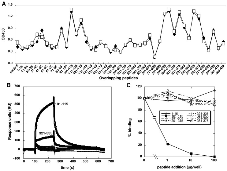
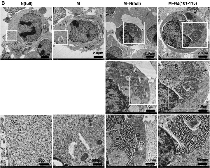
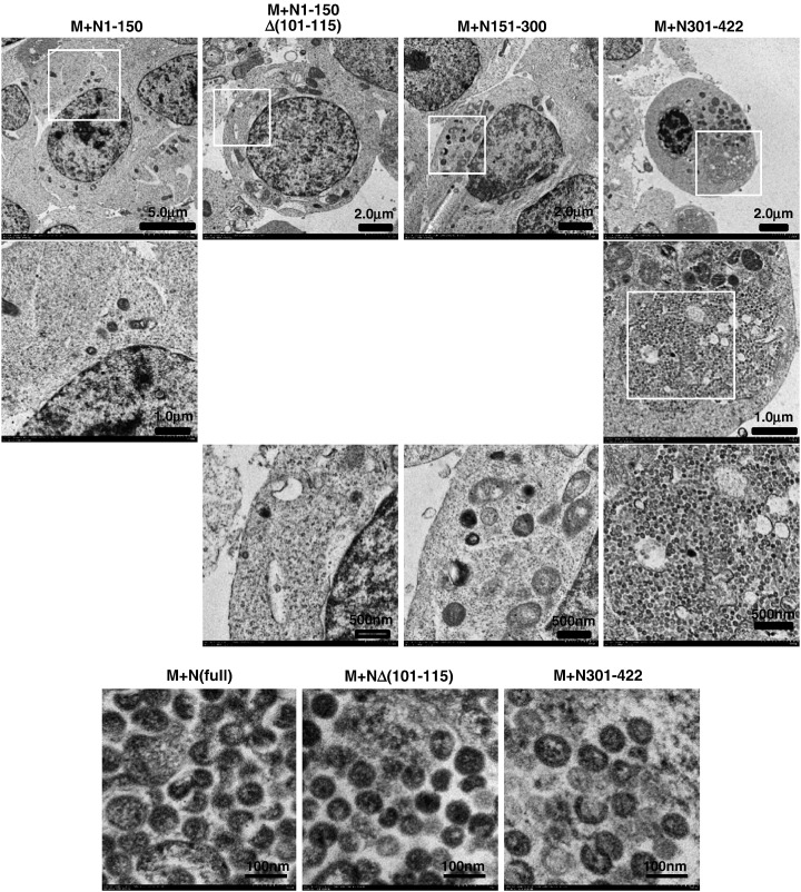
When expressed in mammalian cells, the nucleocapsid (N) and membrane (M) proteins of the severe acute respiratory syndrome coronavirus (SARS-CoV) are sufficient to form pseudoparticles. To identify region(s) of the N molecule required for pseudoparticle formation, we performed biochemical analysis of the interaction of N mutants and M in HEK293 cells. Using a peptide library derived from N, we found that amino acids 101-115 constituted a novel binding site for M. We examined the ability of N mutants to interact with M and form pseudoparticles, and our observations indicated that M bound to NDelta(101-115), N1-150, N151-300, and N301-422, but not to N1-150Delta(101-115). However, pseudoparticles were formed when NDelta(101-115) or N301-422, but not N1-150 or N151-300, were expressed with M in HEK293 cells. These results indicated that the minimum portion of N required for the interaction with M and pseudoparticle formation consists of amino acids 301-422.


Figures

Fig. 1
Fig. 1
Interaction of N-derived peptides with M protein. (A) Each peptide (10 μg/mL) was immobilized on ELISA plates. After blocking, the plates were incubated with M63 (final 1 μg/mL), and binding of M63 to N protein was detected with rabbit anti-M63 plus HRP-goat anti-rabbit IgG. The two data points for each peptide are the results of two independent experiments. (B) Each peptide (10 μg/mL) was injected into a BIAcore flow cell containing immobilized M116. The injection was started at 100 s, stopped at 240 s, and dissociated for 300 s. (C) Competitive inhibition of binding between N and M63 proteins by N-derived peptides. M63 was preincubated with each N peptide, and each mixture was added to a plate coated with N protein. Binding of M63 to N protein was detected as in (A). Binding in the absence of peptide was set at 100%. Data are presented as means ± SD.
Fig. 2
Fig. 2
Schematic diagram of the recombinant N and M proteins used in this study. N mutants are numbered according to the amino acid positions in the full-length N. M116 and M63 consist of the C-terminal 116 and 63 amino acids of M, respectively.
Fig. 3
Fig. 3
Analyses of interactions between N mutants and M and the inhibition by peptide 101–115. (A) ELISA of the interaction between N mutants and M116. Each N peptide (0.01–10 μg/mL) was immobilized onto a plate. After blocking, the plate was incubated with M116 (1 μg/mL), and bound M116 was detected with rabbit anti-M63 and HRP-goat anti-rabbit IgG as described in Materials and methods. (B) Inhibition of the interaction between N mutants and M116 was observed with peptide 101–115. The experimental procedure was identical to that described in the legend to Fig. 1C. Data are presented as means ± SD.
Fig. 4
Fig. 4
Immunoprecipitation analyses of the interaction between N mutants and M116 in mammalian cells. (A) Western blotting of the lysates of HEK293 cells expressing N mutants 3 days post-transfection was performed as described in Materials and methods. (B,C) Immunoprecipitation analyses using control antiserum (C), anti-M63 (M63), or anti-N (N) antibodies of lysates of HEK293 cells coexpressing N mutants and M116. As both of the antibodies used for immunoprecipitation and detection of the target proteins were raised in rabbits, the immunoglobulin light chain (ca. 25 kDa) was observed due to cross-reaction with the 2nd antibody.
Fig. 5
Fig. 5
Assembly of pseudoparticles in transfected HEK293 cells. (A) Gradient sedimentation analyses of pseudoparticles from cell lysates transfected with M and N mutants or transfected N mutants alone were performed 3 days after transfection, as described in Materials and methods. Fractions are numbered from the top of each tube (lower density). Arrows indicate the positions of M and N mutants. In the top panel, N and M were detected simultaneously by anti-M63 antiserum and anti-N serum, while only anti-N serum was used in the bottom panel. (B) Analysis of viral assembly of mutants of N and M proteins by TEM. TEM analysis was performed on HEK293 cells transfected with N mutants and M as described in Materials and methods. The areas in the white squares indicate the areas magnified and shown in the respective lower panels.
Fig. 5
Fig. 5
Assembly of pseudoparticles in transfected HEK293 cells. (A) Gradient sedimentation analyses of pseudoparticles from cell lysates transfected with M and N mutants or transfected N mutants alone were performed 3 days after transfection, as described in Materials and methods. Fractions are numbered from the top of each tube (lower density). Arrows indicate the positions of M and N mutants. In the top panel, N and M were detected simultaneously by anti-M63 antiserum and anti-N serum, while only anti-N serum was used in the bottom panel. (B) Analysis of viral assembly of mutants of N and M proteins by TEM. TEM analysis was performed on HEK293 cells transfected with N mutants and M as described in Materials and methods. The areas in the white squares indicate the areas magnified and shown in the respective lower panels.
Fig. 6
Fig. 6
Determination of the critical residues in N peptide 101–115 that inhibit the interaction between M and N. (A) Peptide sequences used in the analysis. Pep1 is the 101—115 peptide itself. Pep6 to Pep15 are 10-mer peptides spanning amino acids 101–115 of N. Pep16 to Pep18 are sequence-scrambled versions of Pep10. Amino acid sequences of Pep8 to Pep10, which showed strong inhibitory effects on the interaction between N and M in (B) are indicated in a box. (B) Ability of the 10-mer peptides derived from N peptide 101–115 to inhibit the interaction between N and M. The indicated peptides were analyzed for their ability to inhibit the interaction between N and M, as described in Materials and methods. (C) Abilities of the 10-mer scrambled versions of the peptides to inhibit the interaction between N and M. Binding in the absence of peptide was set at 100% (not shown). Data are presented as means ± SD.
Fig. 7
Fig. 7
Schematic model for the assembly of pseudoparticles by N and M of SARS-CoV. Three portions of N have been shown to interact with M, corresponding to residues 101–115 (hatched), 168–208 (striped), and 351–422 (stippled) (He et al., 2004a, He et al., 2004b, Luo et al., 2006b), but only interaction via the C-terminal of N led to formation of pseudoparticles. The 351–422 region can also form N tetramers, and thus may provide a scaffold for the stable interaction between N and M, allowing pseudoparticle formation.

Similar articles

Cited by

References

    1. Almazan F., Galan C., Enjuanes L. The nucleoprotein is required for efficient coronavirus genome replication. J. Virol. 2004;78:12683–12688. - PMC - PubMed
    1. Bos E.C., Luytjes W., Van Der Meulen H.V., Koerten H.K., Spaan W.J. The production of recombinant infectious DI-particles of a murine coronavirus in the absence of helper virus. Virology. 1996;218:52–60. - PMC - PubMed
    1. Chang C.K., Sue S.C., Yu T.H., Hsieh C.M., Tsai C.K., Chiang Y.C., Lee S.J., Hsiao H.H., Wu W.J., Chang W.L., Lin C.H., Huang T.H. Modular organization of SARS coronavirus nucleocapsid protein. J. Biomed. Sci. 2006;13:59–72. - PMC - PubMed
    1. Chen C.Y., Chang C.K., Chang Y.W., Sue S.C., Bai H.I., Riang L., Hsiao C.D., Huang T.H. Structure of the SARS coronavirus nucleocapsid protein RNA-binding dimerization domain suggests a mechanism for helical packaging of viral RNA. J. Mol. Biol. 2007;368:1075–1086. - PMC - PubMed
    1. Dediego M.L., Alvarez E., Almazan F., Rejas M.T., Lamirande E., Roberts A., Shieh W.J., Zaki S.R., Subbarao K., Enjuanes L. A severe acute respiratory syndrome coronavirus that lacks the E gene is attenuated in vitro and in vivo. J. Virol. 2007;81:1701–1713. - PMC - PubMed

Publication types

MeSH terms