Review
. 2008 Sep;18(9):414-20.
doi: 10.1016/j.tcb.2008.07.003. Epub 2008 Aug 14.

Cytonemes and tunneling nanotubules in cell-cell communication and viral pathogenesis

Affiliations
Review

Cytonemes and tunneling nanotubules in cell-cell communication and viral pathogenesis

Nathan M Sherer et al. Trends Cell Biol. 2008 Sep.

Abstract

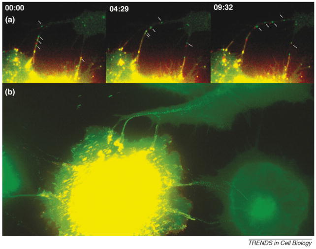
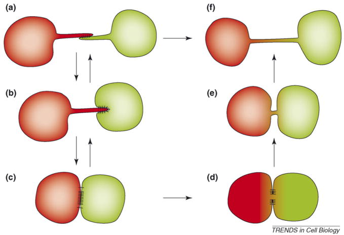
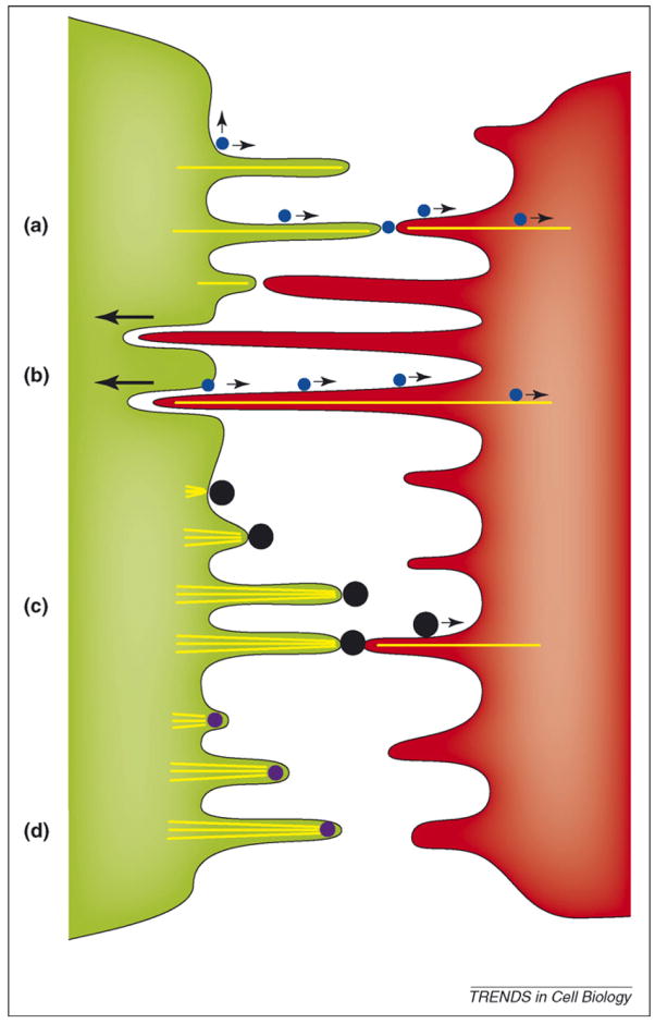
Cells use a variety of intercellular structures, including gap junctions and synapses, for cell-cell communication. Here, we present recent advances in the understanding of thin membrane bridges that function in cell-cell signaling and intercellular transport. Cytonemes or filopodial bridges connect neighboring cells via mechanisms of adhesion, which enable ligand-receptor-mediated transfer of surface-associated cargoes from cell to cell. By contrast, tunneling nanotubes establish tubular conduits between cells that provide for the exchange of both cell-surface molecules and cytoplasmic content. We propose models for the biogenesis of both types of membrane bridges and describe how viruses use these structures for the purpose of cell-to-cell spread.


Figures

Figure 1
Figure 1
Cytonemes and nanotubes in comparison to classical forms of cell–cell communication. (a) Neurological and immunological synapses transmit cell–cell signals through the extracellular space, relying on neurotransmitter release and receptor signaling through a tight cell–cell junction. (b) Cytonemes as thin filopodial bridges topologically identical to stretched out synaptic contacts. (c) By contrast, cells communicating using gap junctions establish direct connectivity between two cytoplasms. (d) Tunneling nanotubes represent thin cell–cell contacts with communicating cytoplasms.
Figure 2
Figure 2
Retroviruses establish filopodial bridges for the purpose of cell-to-cell transmission. (a) Filopodial bridges between virally infected cells (yellow) and target cells (green) support the transport of fluorescently labeled retroviruses from cell to cell. Time intervals are given in min:sec. (b) All frames from the time-lapse video depicted in (a) are superimposed into a single image to illustrate the individual tracks of viruses as they ‘walked’ along a filopodial bridge to infect neighboring cells.
Figure 3
Figure 3
Biogenesis of cytonemes and nanotubes in relationship to tight cell–cell contacts. Transient filopodial contacts (a) can stabilize to form elongated stable cytonemes (b). In the presence of additional adhesion proteins that favor strong cell–cell interfaces, cytonemes could mature into broad synaptic cell–cell interfaces (c). Downregulation of these cell–cell contacts could lead to cytoneme regeneration (c⃗a). At some synapses, cytoplasmic connectivity is established [61,62] (d). Downregulation of these cell–cell interfaces generates tunneling nanotubes with connected cytoplasms (e,f). Under specific circumstances, filopodial contacts might directly mature into tunneling nanotubes (a or b⃗f transition).
Figure 4
Figure 4
Viral use and manipulation of filopodia for the purpose of viral spread. (a) Viruses (blue circles) accumulating at the surface of infected cells (green) can be passed on by transient contact to filopodia of target cells (red) where they use the underlying retrograde flow of filamentous actin to infect cells at the cell body [25]. (b) Continued filopodial contact leads to the internalization of target cell filopodia into the infected cell [4]. Viruses then bud at the infected–target cell interface and move along the filopodial bridge powered by the underlying actin flow (actin is depicted by yellow lines). (c) Vaccinia virus (black circles) induces the formation of actin comet tails beneath the released virus particle, from ‘outside in’ to propel itself towards the target cell [39]. Once in contact with the target cell, particles can still engage the underlying actin flow to further advance towards the cell body [45]. (d) By contrast, African swine fewer virus induces the formation of propulsive filopodia while still residing inside the cytoplasm of the infected cell [40]. The mechanism by which the virus is transferred to target cells is not known.

Similar articles

Cited by

References

    1. Ramirez-Weber FA, Kornberg TB. Signaling reaches to new dimensions in Drosophila imaginal discs. Cell. 2000;103:189–192. - PubMed
    1. Davis DM, Sowinski S. Membrane nanotubes: dynamic long-distance connections between animal cells. Nat Rev Mol Cell Biol. 2008;9:431–436. - PubMed
    1. Gerdes HH, Carvalho R. Intercellular transfer mediated by tunneling nanotubes. Curr Opin Cell Biol. 2008;20:470–475. - PubMed
    1. Sherer NM, et al. Retroviruses can establish filopodial bridges for efficient cell-to-cell transmission. Nat Cell Biol. 2007;9:310–315. - PMC - PubMed
    1. Rustom A, et al. Nanotubular highways for intercellular organelle transport. Science. 2004;303:1007–1010. - PubMed

Publication types

MeSH terms