. 2008 Dec;79(6):1046-53.
doi: 10.1095/biolreprod.108.070664. Epub 2008 Aug 13.

Association of the protein D and protein E forms of rat CRISP1 with epididymal sperm

Affiliations

Association of the protein D and protein E forms of rat CRISP1 with epididymal sperm

Kenneth P Roberts et al. Biol Reprod. 2008 Dec.

Abstract

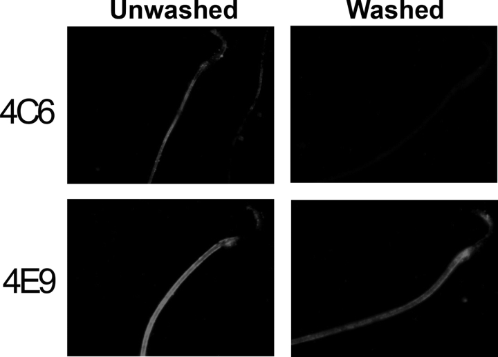
Cysteine-rich secretory protein 1 (CRISP1) is a secretory glycoprotein produced by the rat epididymal epithelium in two forms, referred to as proteins D and E. CRISP1 has been implicated in sperm-egg fusion and has been shown to suppress capacitation in rat sperm. Several studies have suggested that CRISP1 associates transiently with the sperm surface, whereas others have shown that at least a portion of CRISP1 persists on the surface. In the present study, we demonstrate that protein D associates transiently with the sperm surface in a concentration-dependent manner, exhibiting saturable binding to both caput and cauda sperm in a concentration range that is consistent with its capacitation-inhibiting activity. In contrast, protein E persists on the sperm surface after all exogenous protein D has been dissociated. Comparison of caput and cauda sperm reveal that protein E becomes bound to the sperm in the cauda epididymidis. We show that protein E associates with caput sperm, which do not normally have it on their surfaces, in vitro in a time- and temperature-dependent manner. These studies demonstrate that most CRISP1 interacts with sperm transiently, possibly with a specific receptor on the sperm surface, consistent with its action in suppressing capacitation during epididymal storage of sperm. These studies also confirm a tightly bound population of protein E that could act in the female tract.


Figures

FIG. 1.
FIG. 1.
The binding of CRISP1 forms to sperm as determined by Western blot analysis with antibodies to the far carboxyl terminus (4C6) and specifically to the protein E form of CRISP1 (4E9). After swimming out of the caput or cauda epididymidis, sperm were incubated with the dilute epididymal fluid in which they swam out. The sperm were washed at the times indicated and further incubated for the times indicated. At the end of the incubation, total sperm proteins (sperm) or an aliquot of the wash (supernatant) were analyzed by Western blotting. Each blot was stripped and reprobed with all antibodies. The transient nature of binding by the large molecular weight form of CRISP1 (protein D) is apparent. The smallest molecular weight form of protein E detected by 4E9 cannot be removed from sperm by washing. There is no CRISP1 in caput fluid or bound to caput sperm. E7 is an antibody against rat tubulin and was included here as a control for the presence of sperm and as a loading control in the sperm lanes.
FIG. 2.
FIG. 2.
To further characterize the binding of CRISP1 forms to rat sperm, Western blot analysis was carried out on whole-cell samples and detergent extracts (OBG) of epididymal sperm from the caput (Cp) or cauda (Cd), or of cells from the testis (T). Again, the large molecular weight form of CRISP1 is easily removed by washing, and the small molecular weight form of protein E is not. However, the E form of CRISP1 can be removed with detergent. Purified CRISP1 is included as a control (C1).
FIG. 3.
FIG. 3.
Immunocytochemical analysis of unwashed and washed sperm from the caput or cauda epididymis. CRISP1 staining is observed with 4C6 only in unwashed epididymal sperm. The 4E9 staining appears the same for washed and unwashed cauda sperm, confirming the tight binding of protein E. No protein E is detected on caput sperm or testicular sperm. Original magnification ×600.
FIG. 4.
FIG. 4.
A) Saturation binding of CRISP1 to caput (squares) and cauda (diamonds) sperm. Sperm were incubated in increasing concentrations of purified CRISP1. Following washing to remove unbound CRISP1, total sperm proteins were subjected to Western blot analysis for detection of CRISP1. Relative amounts of bound CRISP1 were determined semiquantitatively by densitometric scanning of the Western blot films. Saturation of CRISP1 binding to the sperm occurs at a concentration of 400 μg/ml. B) Dose-dependent inhibition of capacitation of rat sperm as determined by progesterone-inducible acrosome reaction. Cauda sperm were capacitated in the presence of increasing concentrations of purified CRISP1, followed by induction of the acrosome reaction with progesterone. Acrosome-reacted sperm (AR) were determined by staining with Coomassie Blue. The data are presented as percentage of control (sperm incubated under noncapacitating conditions) and clearly show a complete inhibition of capacitation at 200 μg/ml CRISP1, consistent with the dose range of CRISP1 saturation binding to the sperm. Errors bars indicate SEM.
FIG. 5.
FIG. 5.
Characteristics of binding of the protein E form of CRISP1 to rat sperm. A) Aliquots of proximal caput epididymal sperm were incubated with epididymal fluid at 33°C for increasing amounts of time. At the designated times (5 min, 30 min, 2 h, or 18 h), aliquots of sperm were washed, and the sperm-bound protein E was determined by ELISA with monoclonal antibody 4E9. Protein E binds to sperm in vitro to nearly the same saturation as that found in vivo, as illustrated by sperm isolated from the cauda epididymidis (CS) that served as a control. B) The strength of the interaction between protein E and sperm was determined by washing (W) proximal caput sperm that had been preloaded in epididymal fluid for 5 min or 2 h. Following a wash incubation of 18 h in PBS, the bound protein E was determined by ELISA with antibody 4E9. The protein E that binds in vitro is as tightly bound as that loaded in vivo. Error bars indicate SEM. C) The loading of protein E determined by ELISA was confirmed visually by immunocytochemistry using antibody 4E9. Original magnification ×600.
FIG. 6.
FIG. 6.
Temperature dependence of protein E binding to sperm. Proximal caput sperm were incubated in epididymal fluid at 33°C or 4°C, or at 33°C in epididymal fluid that had been preheated to 100°C. Bound protein E was determined by ELISA with monoclonal antibody 4E9. Protein E binds to sperm at both 33°C and 4°C, but loading only occurs at the physiological temperature of 33°C. Heating the epididymal fluid destroys binding activity. Error bars indicate SEM.
FIG. 7.
FIG. 7.
Forms of protein E, as determined by Western blot, produced by region-specific cultured epididymal epithelial cells (EC; middle panel) compared with forms produced regionally by the epididymis in vivo (EF; upper panel). The regions of the epididymis used are proximal caput (PCp), distal caput (DCp), corpus (Co), proximal cauda (PCd), and distal cauda (DCd). The three forms of protein E are produced primarily by the cauda epididymal epithelium, and this differentiation is maintained in short-term culture. When proximal caput sperm are cocultured with cauda epithelial cells (CC), protein E loads on these sperm in a manner indistinguishable from the loading observed using epididymal fluid (Fig. 5) and mimics the binding found to distal cauda sperm (lower panel). Original magnification ×600.

References

    1. Gibbs GM, Scanlon MJ, Swarbrick J, Curtis S, Gallant E, Dulhunty AF, O'Bryan MK.The cysteine-rich secretory protein domain of Tpx-1 is related to ion channel toxins and regulates ryanodine receptor Ca2+ signaling. J Biol Chem 2006; 281: 4156–4163. - PubMed
    1. Guo M, Teng M, Niu L, Liu Q, Huang Q, Hao Q.Crystal structure of the cysteine-rich secretory protein stecrisp reveals that the cysteine-rich domain has a K+ channel inhibitor-like fold. J Biol Chem 2005; 280: 12405–12412. - PubMed
    1. Shikamoto Y, Suto K, Yamazaki Y, Morita T, Mizuno H.Crystal structure of a CRISP family Ca2+ -channel blocker derived from snake venom. J Mol Biol 2005; 350: 735–743. - PubMed
    1. Wang F, Li H, Liu MN, Song H, Han HM, Wang QL, Yin CC, Zhou YC, Qi Z, Shu YY, Lin ZJ, Jiang T.Structural and functional analysis of natrin, a venom protein that targets various ion channels. Biochem Biophys Res Commun 2006; 351: 443–448. - PubMed
    1. Wang J, Shen B, Guo M, Lou X, Duan Y, Cheng XP, Teng M, Niu L, Liu Q, Huang Q, Hao Q.Blocking effect and crystal structure of natrin toxin, a cysteine-rich secretory protein from Naja atra venom that targets the BKCa channel. Biochemistry 2005; 44: 10145–10152. - PubMed

Publication types

MeSH terms