. 2008 Nov;79(5):906-13.
doi: 10.1095/biolreprod.108.070474. Epub 2008 Aug 13.

UBE2I (UBC9), a SUMO-conjugating enzyme, localizes to nuclear speckles and stimulates transcription in mouse oocytes

Affiliations

UBE2I (UBC9), a SUMO-conjugating enzyme, localizes to nuclear speckles and stimulates transcription in mouse oocytes

Motomasa Ihara et al. Biol Reprod. 2008 Nov.

Abstract

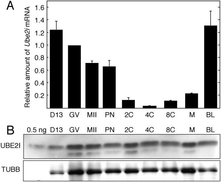
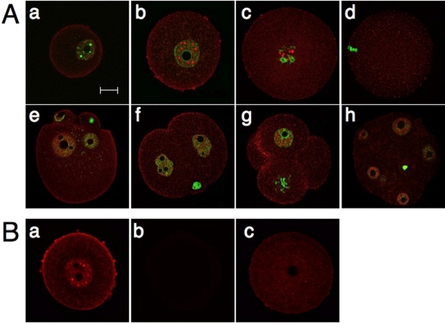
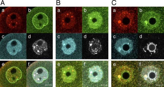
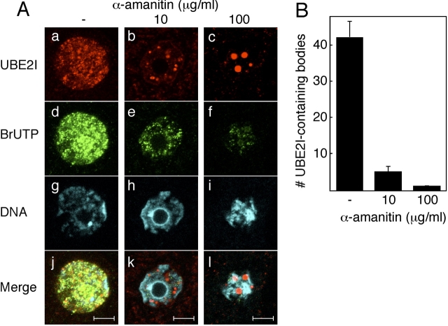
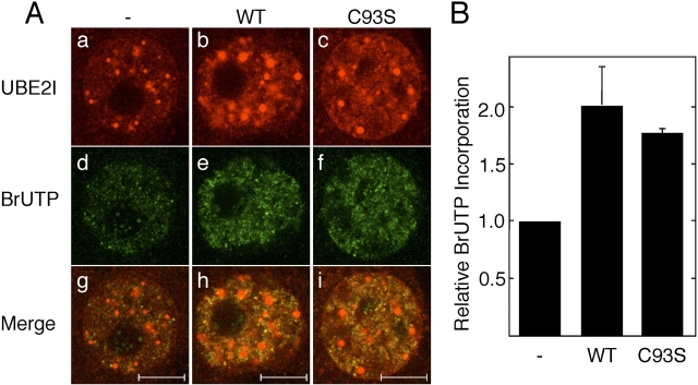
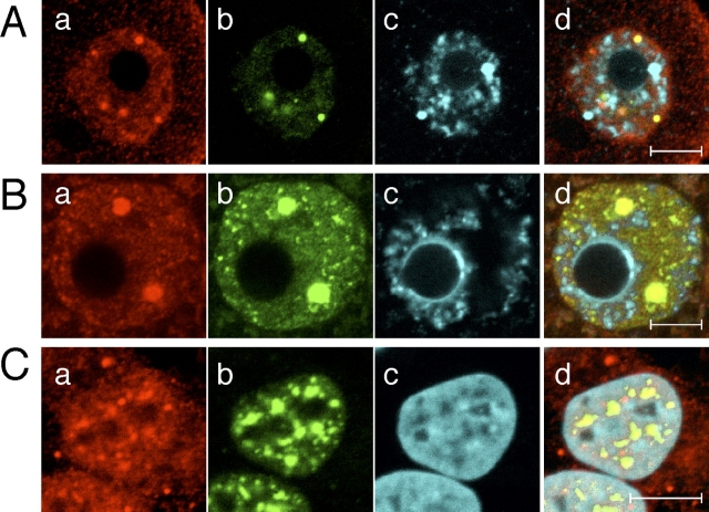
Sumoylation is a posttranslational modification in which SUMO (small ubiquitin-related modifier) proteins are covalently attached to their substrates. In vertebrates, developmental roles for sumoylation have been studied, but the function of sumoylation during mammalian oocyte growth and maturation is not known. As a prelude to conducting studies on the role of sumoylation during oocyte development, we analyzed the temporal and spatial pattern of expression of UBE2I, a SUMO-conjugating E2 enzyme. Immunocytochemical analysis of UBE2I revealed a punctate nuclear staining pattern, with transcriptionally quiescent, fully grown, GV-intact oocytes having larger UBE2I-containing bodies than transcriptionally active, meiotically incompetent growing oocytes. Inhibiting transcription in incompetent oocytes resulted in an increase in the size of the UBE2I-containing bodies. Overexpression of either wild-type UBE2I or catalytically inactive UBE2I resulted in an increase in the size of the UBE2I-containing bodies but also an increase in BrUTP incorporation, suggesting that transcriptional activation by UBE2I is independent of its catalytic activity. Although UBE2I-containing bodies did not completely colocalize with SUMO1 or SUMO2 and SUMO3, which were localized mainly on the nuclear membrane and in the nucleoplasm, UBE2I strikingly colocalized with SFRS2, which is a component of nuclear speckles and critical for mRNA processing. These results suggest a novel function for UBE2I and therefore sumoylation in gene expression.


Figures

FIG. 1.
FIG. 1.
Temporal pattern of Ube2i mRNA and UBE2I protein expression during oocyte growth and preimplantation development. A) Ube2i transcript abundance was determined by quantitative RT-PCR and expressed relative to fully grown GV-intact oocytes. The results shown are the mean ± SEM from three independent experiments. B) Immunoblot analysis of UBE2I in oocyte and preimplantation embryos. Immunoblots were performed using 0.5 ng of UBE2I recombinant protein or extracts derived from 100 oocytes or embryos. The blots were probed with either anti-UBE2I antibody (upper panel) or anti-TUBB antibody (lower panel). The results shown are representative of three independent experiments. 0.5 ng, 0.5 ng of recombinant UBE2I protein; D13, Day 13 incompetent oocyte; GV, fully grown oocyte; MII, metaphase II; PN, pronuclear embryo; 2C, 4C, 8C, 2-cell, 4-cell, 8-cell stage embryo, respectively; M, morula; BL, blastocyst.
FIG. 2.
FIG. 2.
Subcellular localization of UBE2I in oocytes and preimplantation embryos. A) UBE2I was stained with the anti-UBE2I antibody (red). The nuclei were stained with SYTOX green (green). ah) Day 13 incompetent oocyte (a), GV oocyte (b), GVBD oocyte (c), MII (d), pronuclear embryo (e), 2-cell embryo (f), 4-cell embryo (g), and morula (h). The results shown are representative of three independent experiments in which at least 30 oocytes or embryos were examined. B) Antibody specificity. Oocytes were stained with (a) or without (b) the anti-UBE2I antibody or prior to immunostaining (c); the anti-UBE2I antibody was initially incubated with recombinant UBE2I. Bar = μm.
FIG. 3.
FIG. 3.
Subcellular localization of UBE2I, SUMO1, and SUMO2 and SUMO3 in the nucleus of oocytes at different stages of development. A) Day 13 meiotically incompetent oocyte. B) Fully grown NSN oocyte. C) Fully grown SN oocyte. In each panel, oocytes were stained with anti-UBE2I antibody (a), anti-SUMO1 antibody (b), and anti-SUMO2 and -SUMO3 antibody (c), sequentially. The nuclei were stained with DAPI (d). e) Merge of UBE2I and SUMO1 image. f) Merge of UBE2I, SUMO1, SUMO2 and SUMO3, and DAPI staining. The experiment was conducted at least four times, and shown are representative images; at least eight images were taken for each type of oocyte. Bars = 10 μm.
FIG. 4.
FIG. 4.
Reorganization of UBE2I-containing bodies in response to α-amanitin. A) Localization of UBE2I-containing bodies and sites of transcription. Day 13 incompetent oocytes were incubated for 12 h without (a, d, g, and j) or with 10 μg/ml (b, e, h, and k) or 100 μg/ml (c, f, i, and l) of α-amanitin. ac) UBE2I was stained with anti-UBE2I antibody. df) BrUTP was stained with anti-BrUTP antibody. gi) Chromatin was stained with DAPI. jl) Merge of UBE2I, BrUTP, and DAPI staining images. The experiment was conducted three times, and shown are representative images; at least five images were taken for each type of oocyte. Bar = 10 μm. B) Quantification of the effect of α-amanitin on the number of UBE2I-containing bodies. The number of UBE2I-containing dots in the nucleus of each ooocyte was counted after reconstruction of 3D images. Three oocytes were examined in each of three independent experiments. The data are presented as mean ± SEM.
FIG. 5.
FIG. 5.
UBE2I stimulates BrUTP incorporation independent of catalytic activity. A) Effect of UBE2I on BrUTP incorporation. Day 13 meiotically incompetent oocytes were injected with water (a, d, and g), wild-type Ube2i mRNA (b, e, and h), or Ube2i C93S mRNA (c, f, and i). The oocytes were then cultured for 18 h and then immunostained for UBE2I (ac) and an anti-BrUTP antibody (df). gi) Merged images of UBE2I and BrUTP staining. The experiment was performed three times, and shown are representative images. At least 50 oocytes were examined. The bars represent 10 μm. B) Quantification of BrUTP incorporation. For each experiment, the control was set to a value of 1, and the data (mean ± SEM) are expressed relative to the control, water-injected oocytes.
FIG. 6.
FIG. 6.
Colocalization of UBE2I with SFRS2. Subcellular localization of UBE2I and SFRS2 in the nucleus of a Day 13 incompetent oocyte (A), a fully grown oocyte (B), and Hela S3 cells (C). ad) UBE2I (a), SFRS2 (b), DNA (DAPI) (c), and merged image (d) of UBE2I, SFRS2, and DAPI staining. The experiment was performed three times, and shown are representative images. At least 30 oocytes were examined. Bars = 10 μm.

Similar articles

Cited by

References

    1. Johnson E.Protein modification by SUMO. Annu Rev Biochem 2004; 73: 355–382.. - PubMed
    1. Zhao J.Sumoylation regulates diverse biological processes. Cell Mol Life Sci 2007; 64: 3017–3033.. - PMC - PubMed
    1. Geiss-Friedlander R, Melchior F.Concepts in sumoylation: a decade on. Nat Rev Mol Cell Biol 2007; 8: 947–956.. - PubMed
    1. Hayashi T, Seki M, Maeda D, Wang W, Kawabe Y, Seki T, Saitoh H, Fukagawa T, Yagi H, Enomoto T.Ubc9 is essential for viability of higher eukaryotic cells. Exp Cell Res 2002; 280: 212–221.. - PubMed
    1. Nowak M, Hammerschmidt M.Ubc9 regulates mitosis and cell survival during zebrafish development. Mol Biol Cell 2006; 17: 5324–5336.. - PMC - PubMed

Publication types

MeSH terms

Substances