

Impaired follicle development and infertility in female mice lacking steroidogenic factor 1 in ovarian granulosa cells
- PMID: 18703422
- PMCID: PMC2780474
- DOI: 10.1095/biolreprod.108.069435
Impaired follicle development and infertility in female mice lacking steroidogenic factor 1 in ovarian granulosa cells
Abstract
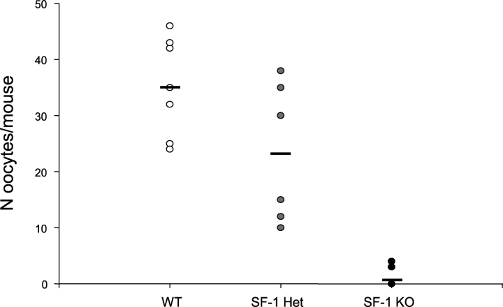
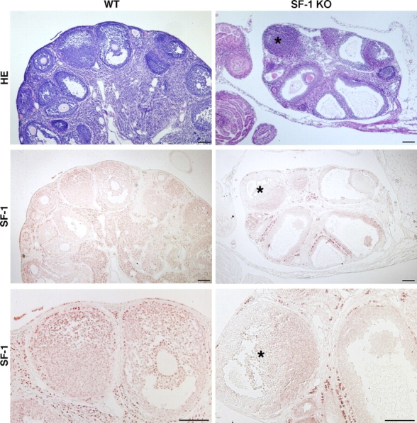
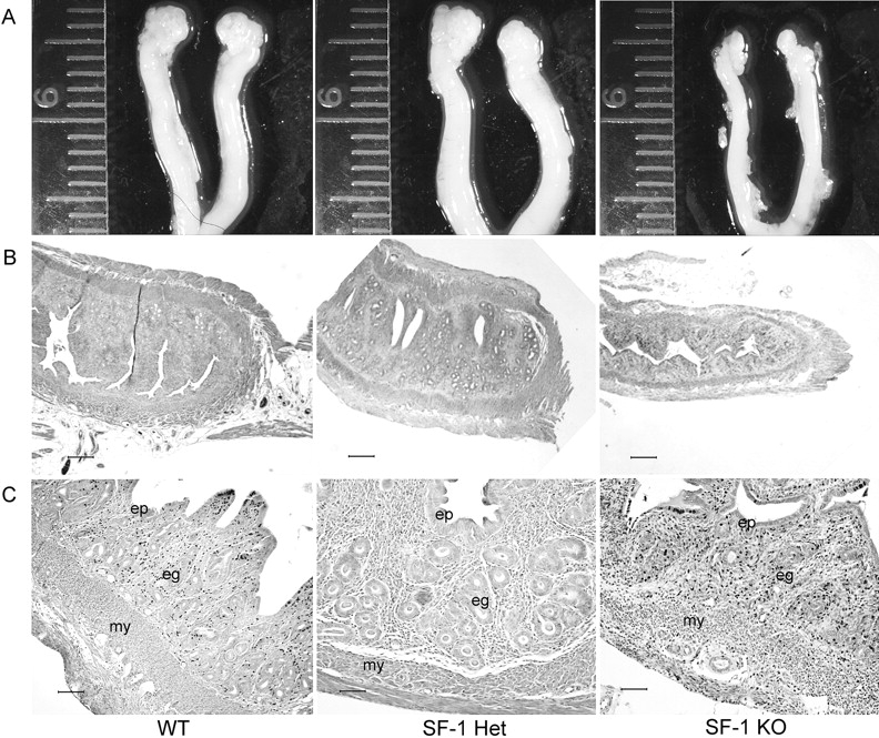
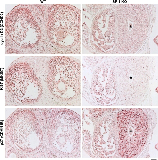
The nuclear receptor steroidogenic factor 1 (SF-1 [officially designated NR5A1]) is essential for fetal gonadal development, but its roles in postnatal ovarian function are less well defined. Herein, we have extended our analyses of knockout (KO) mice with markedly decreased SF-1 expression in granulosa cells. As described, these SF-1 KO mice had hypoplastic ovaries that contained a decreased number of follicles and lacked corpora lutea. In the present study, we showed that SF-1 KO mice exhibited abnormal estrous cycles, were infertile, and released significantly fewer oocytes in response to a standard superovulation regimen. Moreover, they had blunted induction of plasma estradiol in response to gonadotropins. The granulosa cell-specific SF-1 KO also significantly affected ovarian expression of putative SF-1 target genes. Consistent with their decreased follicle number, these mice had reduced ovarian expression of anti-müllerian hormone (Amh), which correlates with the reserve pool of ovarian follicles, as well as decreased gonadotropin-induced ovarian expression of aromatase (Cyp19a1) and cyclin D2 (Ccnd2). In contrast, perhaps because of their abnormal cyclicity, SF-1 KO ovaries had higher basal expression of inhibin-alpha. They also had decreased immunoreactivity for genes related to proliferation (Ccnd2 and Mki67 [also known as Ki67]) and increased expression of Cdkn1b, also known as p27, which inhibits cyclin-dependent kinases, arguing for a role of SF-1 in granulosa cell proliferation. These findings demonstrate that SF-1 has a key role in female reproduction via essential actions in granulosa cells.
Figures


Similar articles
-
The expression of Steroidogenic Factor-1 and its role in bovine steroidogenic ovarian cells during the estrus cycle and first trimester of pregnancy.Anim Reprod Sci. 2013 Apr;138(1-2):74-81. doi: 10.1016/j.anireprosci.2013.01.008. Epub 2013 Feb 18. Anim Reprod Sci. 2013. PMID: 23481593
-
Cell-specific knockout of steroidogenic factor 1 reveals its essential roles in gonadal function.Mol Endocrinol. 2004 Jul;18(7):1610-9. doi: 10.1210/me.2003-0404. Epub 2004 Apr 29. Mol Endocrinol. 2004. PMID: 15118069
-
Steroidogenic Factor 1 Regulation of the Hypothalamic-Pituitary-Ovarian Axis of Adult Female Mice.Endocrinology. 2022 Apr 1;163(4):bqac028. doi: 10.1210/endocr/bqac028. Endocrinology. 2022. PMID: 35247045 Free PMC article.
-
Interactions between androgens, FSH, anti-Müllerian hormone and estradiol during folliculogenesis in the human normal and polycystic ovary.Hum Reprod Update. 2016 Nov;22(6):709-724. doi: 10.1093/humupd/dmw027. Epub 2016 Aug 27. Hum Reprod Update. 2016. PMID: 27566840 Review.
-
Physiology and endocrinology symposium: Anti-Müllerian hormone: a biomarker for the ovarian reserve, ovarian function, and fertility in dairy cows.J Anim Sci. 2019 Apr 3;97(4):1446-1455. doi: 10.1093/jas/skz022. J Anim Sci. 2019. PMID: 30668706 Free PMC article. Review.
Cited by
-
The bHLH-PAS transcriptional complex Sim:Tgo plays active roles in late oogenesis to promote follicle maturation and ovulation.Development. 2023 Jun 15;150(12):dev201566. doi: 10.1242/dev.201566. Epub 2023 Jun 14. Development. 2023. PMID: 37218521 Free PMC article.
-
Oocyte-somatic cell communication and microRNA function in the ovary.Ann Endocrinol (Paris). 2010 May;71(3):144-8. doi: 10.1016/j.ando.2010.02.020. Epub 2010 Apr 2. Ann Endocrinol (Paris). 2010. PMID: 20362967 Free PMC article.
-
FOXL2 Is an Essential Activator of SF-1-Induced Transcriptional Regulation of Anti-Müllerian Hormone in Human Granulosa Cells.PLoS One. 2016 Jul 14;11(7):e0159112. doi: 10.1371/journal.pone.0159112. eCollection 2016. PLoS One. 2016. PMID: 27414805 Free PMC article.
-
Lrh1 can help reprogram sexual cell fate and is required for Sertoli cell development and spermatogenesis in the mouse testis.PLoS Genet. 2022 Feb 22;18(2):e1010088. doi: 10.1371/journal.pgen.1010088. eCollection 2022 Feb. PLoS Genet. 2022. PMID: 35192609 Free PMC article.
-
NR5A1 and cell population heterogeneity: Insights into developmental and functional disparities and regulatory mechanisms.Reprod Med Biol. 2025 Feb 17;24(1):e12621. doi: 10.1002/rmb2.12621. eCollection 2025 Jan-Dec. Reprod Med Biol. 2025. PMID: 39968346 Free PMC article. Review.
References
-
- Parker KL, Schimmer BP.Steroidogenic factor 1: a key determinant of endocrine development and function. Endocr Rev 1997; 18: 361–377.. - PubMed
-
- Hinshelwood MM, Repa JJ, Shelton JM, Richardson JA, Mangelsdorf DJ, Mendelson CR.Expression of LRH-1 and SF-1 in the mouse ovary: localization in different cell types correlates with differing function. Mol Cell Endocrinol 2003; 207: 39–45.. - PubMed
-
- Ikeda Y, Lala DS, Luo X, Kim E, Moisan MP, Parker KL.Characterization of the mouse FTZ-F1 gene, which encodes a key regulator of steroid hydroxylase gene expression. Mol Endocrinol 1993; 7: 852–860.. - PubMed
-
- Tajima K, Dantes A, Yao Z, Sorokina K, Kotsuji F, Seger R, Amsterdam A.Down-regulation of steroidogenic response to gonadotropins in human and rat preovulatory granulosa cells involves mitogen-activated protein kinase activation and modulation of DAX-1 and steroidogenic factor-1. J Clin Endocrinol Metab 2003; 88: 2288–2299.. - PubMed
Publication types
MeSH terms
Substances
Grants and funding
LinkOut - more resources
Full Text Sources
Medical
Molecular Biology Databases
Research Materials
Miscellaneous
