. 2008 Nov 15;17(22):3509-20.
doi: 10.1093/hmg/ddn243. Epub 2008 Aug 14.

The cell adhesion molecule nectin-1 is critical for normal enamel formation in mice

Affiliations

The cell adhesion molecule nectin-1 is critical for normal enamel formation in mice

Martin J Barron et al. Hum Mol Genet. .

Abstract

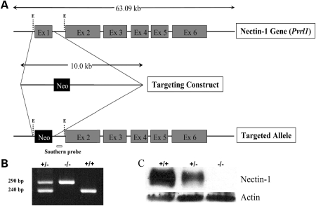
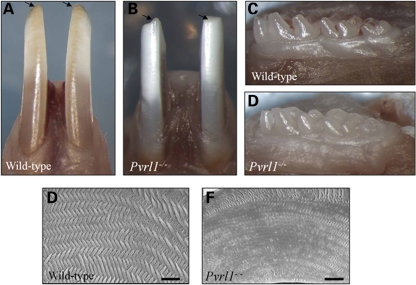
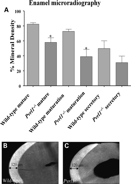
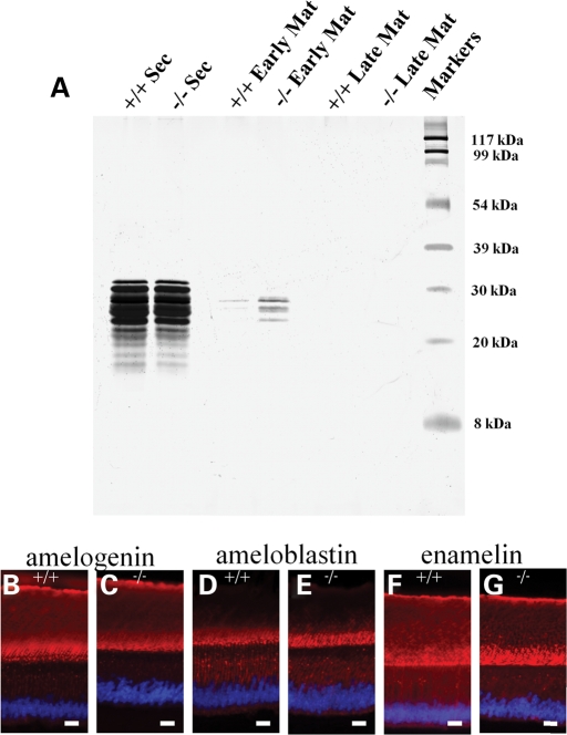
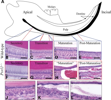
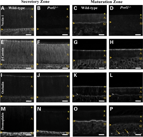
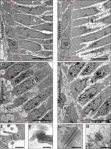
Nectin-1 is a member of a sub-family of immunoglobulin-like adhesion molecules and a component of adherens junctions. In the current study, we have shown that mice lacking nectin-1 exhibit defective enamel formation in their incisor teeth. Although the incisors of nectin-1-null mice were hypomineralized, the protein composition of the enamel matrix was unaltered. While strong immunostaining for nectin-1 was observed at the interface between the maturation-stage ameloblasts and the underlying cells of the stratum intermedium (SI), its absence in nectin-1-null mice correlated with separation of the cell layers at this interface. Numerous, large desmosomes were present at this interface in wild-type mice; however, where adhesion persisted in the mutant mice, the desmosomes were smaller and less numerous. Nectins have been shown to regulate tight junction formation; however, this is the first report showing that they may also participate in the regulation of desmosome assembly. Importantly, our results show that integrity of the SI-ameloblast interface is essential for normal enamel mineralization.


Figures

Figure 1.
Figure 1.
Generation of Pvrl1−/− mice. (A) Structure of Pvrl1, gene targeting vector and targeted allele following homologous recombination. Exons are depicted as grey boxes, whereas the neomycin-resistance cassette (neo) is shown as a black box. The EcoR1 sites used for cloning, E, and the position of the probe used for Southern blot analysis are also shown. (B) Confirmation of the genotypes of wild-type (+/+), heterozygous (±) and homozygous mutant (−/−) mice by duplex PCR analysis in which the wild-type and targeted alleles are represented by 240 and 290 bp products, respectively. (C) Western blot analysis of protein extracts from wild-type (+/+), heterozygous (±) and homozygous mutant (−/−) mouse embryos. No nectin-1 protein was detected in samples obtained from Pvrl1−/− embryos. Actin-1 loading controls are also shown.
Figure 2.
Figure 2.
Morphology of the dentition of Pvrl1−/− mice. (A) The lower incisor teeth of wild-type mice are stained yellow and show a sharp incisal edge (arrows), whereas (B) the lower incisor teeth of Pvrl1−/− mice are chalky white in colour and often show signs of mechanical attrition and/or fracture (arrows). In contrast, the molar teeth of the wild-type (C) and Pvrl1−/− mice (D) appear grossly normal. (E and F) Scanning electron microscopy reveals that the normal ‘herring-bone’ pattern of hydroxyapatite prisms of the wild-type incisor (E) is absent from the Pvrl1−/− incisor (F). Scale bars, 10 µm.
Figure 3.
Figure 3.
Micro-radiography and thickness of Pvrl1−/− enamel. (A) Micro-densitometric analysis of wild-type and Pvrl1−/− enamel revealed statistically significant differences (P < 0.05; asterisks) in mineral density between mature and maturation stage enamel. No statistically significant difference in mineral density was observed at the secretory stage of enamel formation. (B and C) Comparison of the thickness of mature enamel showed no differences between (B) wild-type and (C) Pvrl1−/− lower incisor teeth.
Figure 4.
Figure 4.
Biochemical and immunohistochemical analysis of the enamel extracellular matrix. (A) SDS–PAGE analysis of whole enamel extracts from the lower incisor teeth showed identical protein profiles between the secretory stage enamel of wild-type (+/+) and Pvrl1−/− (−/−) mice. In the early maturation stage enamel, the total protein content is much reduced in both wild-type and Pvrl1−/− lower incisor teeth. The differences in intensity between the wild-type and Pvrl1−/− samples at this stage of amelogenesis reflect the difficulty in sampling this region of the lower incisor. No enamel proteins were detected in the late maturation stage enamel of wild-type and Pvrl1−/− samples. (BG) Immunohistochemical analysis of the major extracellular matrix proteins amelogenin (B, C), ameloblastin (D, E) and enamelin (F, G) showed no discernable differences between wild-type and Pvrl1−/− lower incisor teeth. Scale bars, 25 µm.
Figure 5.
Figure 5.
Histological analysis of the lower incisor teeth. (A) Diagrammatic representation of the mouse lower incisor tooth within the mandible. Amelogenesis commences at the apical region of the tooth and proceeds towards the incisal edge. The position of the regions concerned with enamel formation is shown in black. (BI) The secretory (B), transition (C), maturation (D) and post-maturation (E) stages of amelogenesis showed characteristically normal morphology in the wild-type lower incisor. In the Pvrl1−/− lower incisor the secretory (F) and transition (G) stages also show normal morphology; however, the maturation (H) and post-maturation (I) stages show a separation between the ameloblasts and stratum intermedium forming a blister-like structure. (JL) Separation of the ameloblasts from the stratum intermedium (arrow) begins soon after the start of the maturation stage (J), and worsens progressively in the incisal direction (K). Once a blister-like structure has formed (L), the ameloblasts lose their columnar morphology and adopt a cuboidal shape reminiscent of post-maturation ameloblasts. A, ameloblasts; SI, stratum intermedium; P, papillary layer. Scale bars: (A–I), 50 µm; (J–L), 10 µm.
Figure 6.
Figure 6.
Immunohistochemical analysis of the lower incisor tooth. (AD) Nectin-1 showed strong immunoreactivity around the cells of the stratum intermedium of the secretory zone and at the proximal junctional complex of the ameloblasts in wild-type mice (A). The immunoreactivity for nectin-1 shifted from around the stratum intermedium cells to the SI–ameloblast interface in the wild-type maturation zone (C). No nectin-1 expression was detected in the Pvrl1−/− secretory or maturation zones (B and D). (EF) β-Catenin showed strong immunostaining of the stratum intermedium and ameloblast proximal junctional complexes of the secretory zone ameloblasts; moderate immunostaining for β-catenin was also seen between these ameloblasts in both wild-type and Pvrl1−/− mice (E and F). In the maturation zone, β-catenin expression appeared to decrease but remained strong at the proximal junctional complex of the ameloblast in both wild-type and Pvrl1−/− mice (G and H). (IL) Occludin immunostaining in the secretory zone was similar in wild-type and Pvrl1−/− mice being strong at the ameloblast proximal junctional complex (I and J). In the maturation zone, occludin immunoreactivity was again similar between wild-type and Pvrl1−/− animals with strong labelling of the proximal and distal (arrows) junctional complexes of the ameloblasts (K and L). (MP) Desmoplakin expression was strong at the SI–ameloblast interface in the secretory and maturation zones of wild-type mice (M and O). In Pvrl1−/− mice, however, strong expression was seen at the SI–ameloblast interface of the secretory zone but this was largely lost in the maturation zone (N and P). The yellow arrows indicate autofluorescence from erythrocytes, the yellow arrowheads mark the position of the SI–ameloblast interface and the yellow asterisks mark the position of the ameloblast proximal junctional complex. A, ameloblast, SI, stratum intermedium. Scale bar, 50 µm.
Figure 7.
Figure 7.
Transmission electron microscopy of the lower incisor enamel organ. (A and B) Low power electron micrographs of the secretory zone SI–ameloblast interface of (A) wild-type and (B) Pvrl1−/− mice. In both wild-type and Pvrl1−/− mice, there is close contact between the ameloblasts and stratum intermedium cells. (C and D) Low power electron micrographs of maturation zone SI–ameloblast interface of (C) wild-type and (D) Pvrl1−/− mice. In wild-type mice, numerous contacts are made between the stratum intermedium cells and the ameloblasts, whereas in the Pvrl1−/− mice little contact is made between these cells. The red arrows indicate the position of the SI–ameloblast interface. A, ameloblast, SI, stratum intermedium. Scale bars in A–D = 8 µm. (EH) Examples of desmosomes seen at the SI–ameloblast interface in the secretory and maturation zones of wild-type and Pvrl1−/− mice. In the secretory zone of wild-type and Pvrl1−/− mice the desmosomes were of typical size (E and F, respectively). In contrast, while the desmosomes observed in the maturation zone of wild-type mice (G) were usually larger than those observed in the secretory zone, those observed in the maturation zone of Pvrl1−/− mice were of a similar size to those found in the secretory zone (H). Scale bar in: (E), 150 nm; (F–H), 160 nm.
Figure 8.
Figure 8.
Apoptosis, proliferation and iron transport in Pvrl1−/− mice (A and B) Occasional TUNEL-positive nuclei (A), and activated caspase-3 positive cells (B), were observed in the papillary layer of Pvrl1−/− mice beneath the regions where ameloblast separation had occurred (arrows). Orange areas are artefactual and correspond to autofluorescence from erythrocytes. (C) Occasional BrdU-positive nuclei were observed in the papillary layer of wild-type mice (arrow). (D) BrdU-positive nuclei were more numerous in the papillary layer of Pvrl1−/− mice. (E) Quantitative analysis of the BrdU-positive nuclei in the papillary layer of wild-type and Pvrl1−/− mice indicated a statistically significant increase (P < 0.05) in the latter of ∼3%. (F) Perl’s Prussian blue reaction of wild-type maturation zone showed strong reactivity (blue) in a supra-nuclear position within the ameloblasts. (G) Similar preparation as (F), showing the maturation zone of a Pvrl1−/− mouse. Reactivity was not concentrated in the ameloblasts as it was in wild-type mice but remained diffuse throughout the whole layer. Above the vascular bed that underlies the enamel organ, cells showing strong reactivity were often seen (arrows). Scale bar in (A–D), (F) and (G) is 50 µm.

Similar articles

Cited by

References

    1. Miyoshi J., Takai Y. Nectin and nectin-like molecules: biology and pathology. Am. J. Nephrol. 2007;27:590–604. - PubMed
    1. Ogita H., Takai Y. Nectins and nectin-like molecules: roles in cell adhesion, polarization, movement, and proliferation. I.U.B.M.B. Life. 2006;58:334–343. - PubMed
    1. Satoh-Horikawa K., Nakanishi H., Takahashi K., Miyahara M., Nishimura M., Tachibana K., Mizoguchi A., Takai Y. Nectin-3, a new member of immunoglobulin-like cell adhesion molecules that shows homophilic and heterophilic cell–cell adhesion activities. J. Biol. Chem. 2000;275:10291–10299. - PubMed
    1. Mandai K., Nakanishi H., Satoh A., Obaishi H., Wada M., Nishioka H., Itoh M., Mizoguchi A., Aoki T., Fujimoto T., et al. Afadin: A novel actin filament-binding protein with one PDZ domain localized at cadherin-based cell-to-cell adherens junction. J. Cell Biol. 1997;139:517–528. - PMC - PubMed
    1. Takai Y., Nakanishi H. Nectin and afadin: novel organizers of intercellular junctions. J. Cell Sci. 2003;116:17–27. - PubMed

Publication types

MeSH terms