. 2008 Oct 24;283(43):28852-9.
doi: 10.1074/jbc.M805376200. Epub 2008 Aug 14.

TDP-43 overexpression enhances exon 7 inclusion during the survival of motor neuron pre-mRNA splicing

Affiliations

TDP-43 overexpression enhances exon 7 inclusion during the survival of motor neuron pre-mRNA splicing

Jayarama Krishnan Bose et al. J Biol Chem. .

Abstract

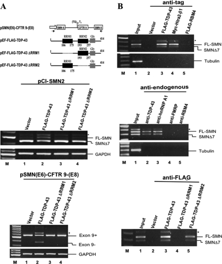
TDP-43 is a highly conserved, 43-kDa RNA-binding protein implicated to play a role in transcription repression, nuclear organization, and alternative splicing. More recently, this factor has been identified as the major disease protein of several neurodegenerative diseases, including frontotemporal lobar degeneration with ubiquitin-positive inclusions and amyotrophic lateral sclerosis. For the splicing activity, the factor has been shown to be mainly an exon-skipping promoter. In this study using the survival of motor neuron (SMN) minigenes as the reporters in transfection assay, we show for the first time that TDP-43 could also act as an exon-inclusion factor. Furthermore, both RNA-recognition motif domains are required for its ability to enhance the SMN2 exon 7 inclusion. Combined protein-immunoprecipitation and RNA-immunoprecipitation experiments also suggested that this exon inclusion activity might be mediated by multimeric complex(es) consisting of this protein interacting with other splicing factors, including Htra2-beta1. Our data further evidence TDP-43 as a multifunctional RNA-binding protein for a diverse set of cellular activities.


Figures

FIGURE 1.
FIGURE 1.
Enhancement of SMN2 exon 7 inclusion by overexpressed TDP-43. A. Left panel, schematic representations of the two pEF-based plasmids expressing FLAG-TDP-43 and Myc-Htra2-β1, and pCI-based SMN minigene constructs, respectively. The small arrows indicate the locations of the primer pair used for RT-PCR analysis of SMN minigene expression. The location of C → T change at position 6 of the exon 7 of SMN2 is indicated by an asterisk. Right panel, RT-PCR analysis of the total RNAs isolated from pCI-SMN2-transfected 293 cells without (lane 1) or with cotransfection of the plasmid pEF-FLAG-TDP-43 (lanes 2–5) or pEF-Myc-Htra2-β1(lanes 6–9). The amounts of the cotransfected plasmids (μg) are indicated. B. Left panel, RT-PCR analysis of the expression of the SMN1 (lanes 1 and 2) or SMN2 minigene (lanes 3–5) as affected by coexpression of FLAG-TDP-43 (lanes 2 and 4), Myc-Htra2-β1(lane 5), or a combination of both (lane 6). 1 μg of each of the reporter plasmid and 1 μg of each of the expression plasmids were used in transfection. Asterisk indicates the mismatched heterodimer formed between the RT-PCR products from the SMN1 and SMN2 templates. The band on the top of the gel, which is most abundant in lane 5, is the incomplete splicing product containing intron 7. Both of the above were identified by cloning and sequencing of the bands excised from the gel. Right panel, histobar graph comparing the enhancing effects of SMN2 exon 7 inclusion by overexpression of TDP-43 and/or Htra2-β1. The effects are expressed as the ratios of the RT-PCR band signals of FL-SMN/SMNΔ7 averaged from three independent cotransfection experiments as exemplified in the gel in the left panel.
FIGURE 2.
FIGURE 2.
Requirement of the RRM domains for TDP-43 to enhance SMN2 exon 7 inclusion and to associate with the SMN mRNAs. A. Top panel, schematic representations of the inserts of the pCMV-based CFTR minigene and pEF-based plasmids expressing the two RRM domain-deletion mutants of TDP-43. Middle panel, RT-PCR analysis of the total RNA isolated from 293 cells transfected with the SMN2 minigene plus the pEF vector (lane 1) or one of the three expression plasmids as indicated in lanes 2–4. FL, full-length. In the bottom panel, 293 cells were transfected with pSMN(E6)-CFTR9-(E8) together with the pEF-vector or with one of the three expression plasmids as indicated. Note that only the full-length TDP-43 could enhance the SMN2 exon 7 inclusion or the CFTR exon 9-skipping. B. RNA association assays. Top panel, 293 cells were transfected with plasmids expressing FLAG-TDP-43 (lane 3), Myc-Htra2-β1(lane 4), and FLAG-RBM4 (lane 5), respectively, and then immunoprecipitated with anti-FLAG or anti-Myc. The RNAs in the immunoprecipitates were then analyzed by RT-PCR using primers specific for the SMN1/2 transcripts or the tubulin mRNA. Middle panel, RNA-IP analysis of 293 cell lysate with the use of antibodies against the endogenous TDP-43 (lane 2), hnRNP A1 (lane 3), FMRP (lane 4), and RBM4 (lane 5), respectively. Note that only the use of anti-TDP-43 (lane 2) and hnRNP A1 (lane 3) antibodies showed enrichment of SMN1 and SMN2 RNAs. Bottom panel, RNA-IP of cells transfected with pEF-FLAG-TDP-43 (lane 3), pEF-FLAG-TDP-43 ΔRRM1 (lane 4), or pEF-FLAG-TDP-43 ΔRRM2 (lane 5).
FIGURE 3.
FIGURE 3.
Requirement of AG-rich SE2 element for TDP-43-induced SMN2 exon 7 inclusion. Upper panel, schematic representation of the SMN exon 7. The nucleotide difference between SMN1 and SMN2 at position 6 (*) in exon 7 and the stop codon (underlined) are indicated. Nucleotides mutated into U in the SE1, SE2, and SE3 elements, respectively, of the three mutant minigene plasmids are indicated with bold letters. Lower panel, data of RT-PCR analysis of RNAs from 293 cells transfected with the three mutant minigenes pCI-SMN1 (SE1) (lanes 1 and 2), pCI-SMN1 (SE2) (lanes 3 and 4), and pCI-SMN1 (SE3) (lanes 5 and 6), respectively, without (lanes 1, 3, and 5) or with (lanes 2, 4, and 6) coexpression of FLAG-TDP-43. The splicing patterns of the wild-type SMN1 and SMN2 minigene transcripts are shown (lanes 7 and 8) for comparison.
FIGURE 4.
FIGURE 4.
Protein pulldown by biotinylated RNA. Biotin-labeled RNA oligonucleotides corresponding to sequences of the ex 7 SMN1, ex 7 SMN2, ex 6 SMN, ex 7 SMN1 (SE2 mut), 3× SE2, (UG)12 repeats, and a region of the tubulin mRNA (for the oligonucleotide sequences, see “Experimental Procedures”) were incubated with the nuclear extracts prepared from 293 cells transiently transfected with pEF-FLAG-TDP-43, pEF-Myc-Htra2-β1, pEF-FLAG-hnRNP G, and pEF-FLAG-Fyn, respectively. The RNA oligonucleotide–protein complexes were then pulled down with streptavidin beads, and the proteins were resolved by SDS-PAGE and probed with the anti-FLAG or anti-Myc antibody.
FIGURE 5.
FIGURE 5.
Interaction of TDP-43 with Htra2-β1. A, coimmunoprecipitation (IP) was performed with 293 cell lysates using preimmune serum (lane 1) or with anti-TDP-43 antibody without (lane 2) or with prior RNase A treatment (lane 3). The precipitated samples were resolved by SDS-PAGE and analyzed by Western blotting with anti-TDP-43, anti-Htra2-β1, or antitubulin. B, domain requirement of TDP-43 interaction with Htra2-β1. Cell lysates prepared from 293 cells exogenously expressing Myc-Htra2-β1 plus FLAG-TDP-43, FLAG-TDP-43 ΔRRM1, or FLAG-TDP-43 ΔRRM2 were immunoprecipitated with anti-FLAG, and the precipitates were then subjected to Western blotting (IB) with use of anti-Myc and anti-FLAG, respectively.
FIGURE 6.
FIGURE 6.
Effects of RNAi of TDP-43 and Htra2-β1 on alternative splicing. A, 293 cells were transfected with siRNA oligonucleotides specific for TDP-43 (lanes 1 and 2), Htra2-β1(lanes 3 and 4), and hnRNP A1 (lanes 5 and 6), respectively. The in vivo splicing patterns of the SMN1/2 minigene transcripts were then analyzed by RT-PCR. The positions of the full-length (FL) and exon 7-skipped PCR products are indicated on the right. The lower Western blotting panel validates the depletion of the individual RNAi target proteins. B, requirement of TDP-43 for the CFTR exon 9 skipping. The assay was carried out as in A above, except that the pSMN(E6)-CFTR9-(E8)-T3 minigene and only the RNAi oligonucleotides specific for TDP-43 were used.
FIGURE 7.
FIGURE 7.
Models of involvement of TDP-43 and other factors in the alternative splicing of SMN1 and SMN2 pre-mRNAs. The regulation of the SMN pre-mRNAs by TDP-43, compared with other splicing factors, under different conditions is modeled in diagram presentation. In model 1, TDP-43, Htra2-β1, and hnRNP G participate in the SMN splicing under the normal condition. In model 2, they do not. The possible molecular basis for the consequences upon overexpression and RNAi knockdown of TDP-43, respectively, as shown in Figs. 1, 2, 3 and 6 are also depicted by the diagrams. Note that in either model, the SE2 element in exon 7 is required for the splicing reactions. For details, see “Discussion.”

Similar articles

Cited by

References

    1. Ou, S. H., Wu, F., Harrich, D., Garcia-Martinez, L. F., and Gaynor, R. B. (1995) J. Virol. 69 3584–3596 - PMC - PubMed
    1. Buratti, E., Dork, T., Zuccato, E., Pagani, F., Romano, M., and Baralle, F. E. (2001) EMBO J. 20 1774–1784 - PMC - PubMed
    1. Wang, I. F., Reddy, N. M., and Shen, C. K. (2002) Proc. Natl. Acad. Sci. U. S. A. 99 13583–13588 - PMC - PubMed
    1. Wang, I. F., Wu, L. S., Chang, H. Y., and Shen, C. K. (2008) J. Neurochem. 105 797–806 - PubMed
    1. Dreyfuss, G., Matunis, M. J., Pinol-Roma, S., and Burd, C. G. (1993) Annu. Rev. Biochem. 62 289–321 - PubMed

Publication types

Substances

LinkOut - more resources