

Differential gene expression in human conducting airway surface epithelia and submucosal glands
- PMID: 18703793
- PMCID: PMC2633141
- DOI: 10.1165/rcmb.2008-0240OC
Differential gene expression in human conducting airway surface epithelia and submucosal glands
Abstract
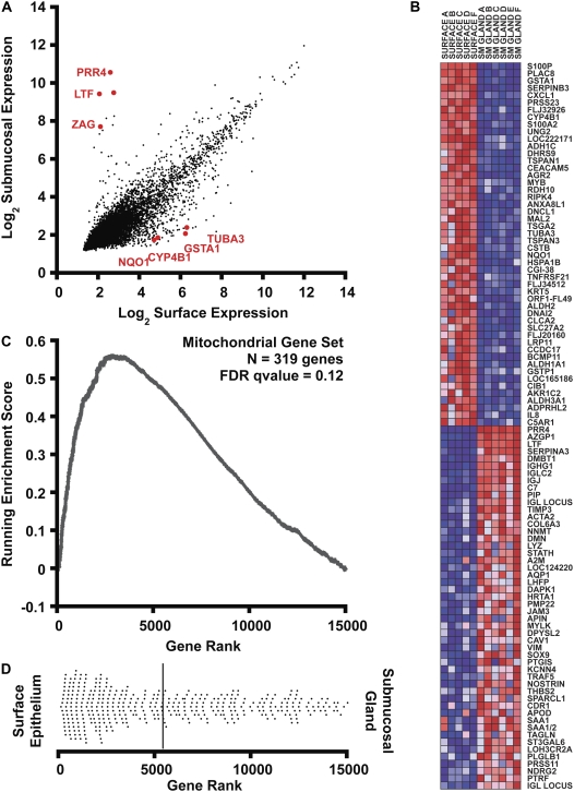
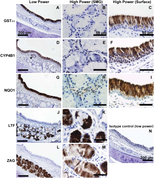
Human conducting airways contain two anatomically distinct epithelial cell compartments: surface epithelium and submucosal glands (SMG). Surface epithelial cells interface directly with the environment and function in pathogen detection, fluid and electrolyte transport, and mucus elevation. SMG secrete antimicrobial molecules and most of the airway surface fluid. Despite the unique functional roles of surface epithelia and SMG, little is known about the differences in gene expression and cellular metabolism that orchestrate the specialized functions of these epithelial compartments. To approach this problem, we performed large-scale transcript profiling using epithelial cell samples obtained by laser capture microdissection (LCM) of human bronchus specimens. We found that SMG expressed high levels of many transcripts encoding known or putative innate immune factors, including lactoferrin, zinc alpha-2 glycoprotein, and proline-rich protein 4. By contrast, surface epithelial cells expressed high levels of genes involved in basic nutrient catabolism, xenobiotic clearance, and ciliated structure assembly. Selected confirmation of differentially expressed genes in surface and SMG epithelia demonstrated the predictive power of this approach in identifying genes with localized tissue expression. To characterize metabolic differences between surface epithelial cells and SMG, immunostaining for a mitochondrial marker (isocitrate dehydrogenase) was performed. Because greater staining was observed in the surface compartment, we predict that these cells use significantly more energy than SMG cells. This study illustrates the power of LCM in defining the roles of specific anatomic features in airway biology and may be useful in examining how disease states alter transcriptional programs in the conducting airways.
Figures
Similar articles
-
Wnt Signaling Regulates Airway Epithelial Stem Cells in Adult Murine Submucosal Glands.Stem Cells. 2016 Nov;34(11):2758-2771. doi: 10.1002/stem.2443. Epub 2016 Jul 11. Stem Cells. 2016. PMID: 27341073 Free PMC article.
-
Airway glandular development and stem cells.Curr Top Dev Biol. 2004;64:33-56. doi: 10.1016/S0070-2153(04)64003-8. Curr Top Dev Biol. 2004. PMID: 15563943 Review.
-
Human LPLUNC1 is a secreted product of goblet cells and minor glands of the respiratory and upper aerodigestive tracts.Histochem Cell Biol. 2010 May;133(5):505-15. doi: 10.1007/s00418-010-0683-0. Epub 2010 Mar 18. Histochem Cell Biol. 2010. PMID: 20237794 Free PMC article.
-
Myoepithelial Cells of Submucosal Glands Can Function as Reserve Stem Cells to Regenerate Airways after Injury.Cell Stem Cell. 2018 May 3;22(5):668-683.e6. doi: 10.1016/j.stem.2018.03.018. Epub 2018 Apr 12. Cell Stem Cell. 2018. PMID: 29656943 Free PMC article.
-
Ca²⁺ signaling and fluid secretion by secretory cells of the airway epithelium.Cell Calcium. 2014 Jun;55(6):325-36. doi: 10.1016/j.ceca.2014.02.001. Epub 2014 Feb 11. Cell Calcium. 2014. PMID: 24703093 Review.
Cited by
-
Stem Cell-derived Respiratory Epithelial Cell Cultures as Human Disease Models.Am J Respir Cell Mol Biol. 2021 Jun;64(6):657-668. doi: 10.1165/rcmb.2020-0440TR. Am J Respir Cell Mol Biol. 2021. PMID: 33428856 Free PMC article. Review.
-
Identification of key genes and pathways in chronic rhinosinusitis with nasal polyps and asthma comorbidity using bioinformatics approaches.Front Immunol. 2022 Aug 17;13:941547. doi: 10.3389/fimmu.2022.941547. eCollection 2022. Front Immunol. 2022. PMID: 36059464 Free PMC article.
-
Mucus concentration-dependent biophysical abnormalities unify submucosal gland and superficial airway dysfunction in cystic fibrosis.Sci Adv. 2022 Apr;8(13):eabm9718. doi: 10.1126/sciadv.abm9718. Epub 2022 Apr 1. Sci Adv. 2022. PMID: 35363522 Free PMC article.
-
Physiology and pathophysiology of human airway mucus.Physiol Rev. 2022 Oct 1;102(4):1757-1836. doi: 10.1152/physrev.00004.2021. Epub 2022 Jan 10. Physiol Rev. 2022. PMID: 35001665 Free PMC article. Review.
-
Dissecting the cellular specificity of smoking effects and reconstructing lineages in the human airway epithelium.Nat Commun. 2020 May 19;11(1):2485. doi: 10.1038/s41467-020-16239-z. Nat Commun. 2020. PMID: 32427931 Free PMC article.
References
-
- Wine JJ, Joo NS. Submucosal glands and airway defense. Proc Am Thorac Soc 2004;1:47–53. - PubMed
-
- Scheetz TE, Zabner J, Welsh MJ, Coco J, Eyestone Mde F, Bonaldo M, Kucaba T, Casavant TL, Soares MB, McCray PB Jr. Large-scale gene discovery in human airway epithelia reveals novel transcripts. Physiol Genomics 2004;17:69–77. - PubMed
Publication types
MeSH terms
Substances
Grants and funding
LinkOut - more resources
Full Text Sources
Miscellaneous
