. 2009 Jan;40(1):108-18.
doi: 10.1165/rcmb.2007-0454OC. Epub 2008 Aug 14.

Role of small GTPases and alphavbeta5 integrin in Pseudomonas aeruginosa-induced increase in lung endothelial permeability

Affiliations

Role of small GTPases and alphavbeta5 integrin in Pseudomonas aeruginosa-induced increase in lung endothelial permeability

Michael T Ganter et al. Am J Respir Cell Mol Biol. 2009 Jan.

Abstract

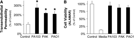
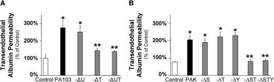
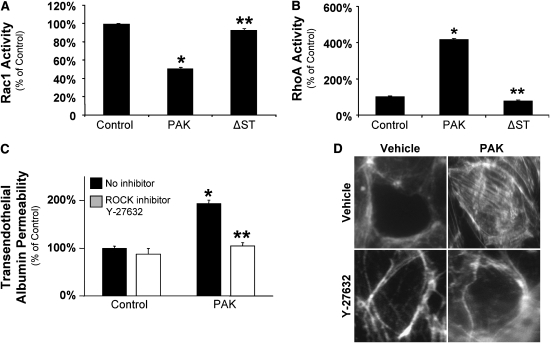
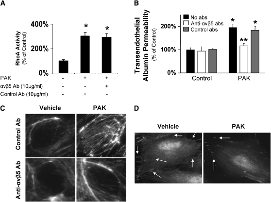
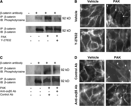
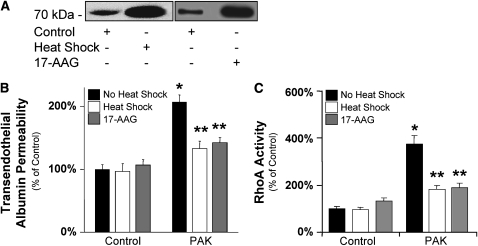
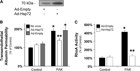
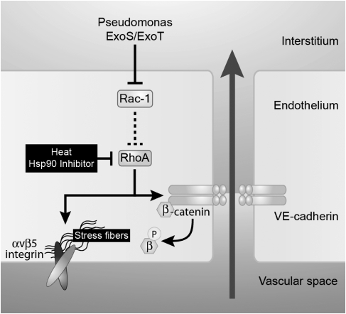
Pseudomonas aeruginosa is an opportunistic pathogen that can cause severe pneumonia associated with airspace flooding with protein-rich edema in critically ill patients. The type III secretion system is a major virulence factor and contributes to dissemination of P. aeruginosa. However, it is still unknown which particular bacterial toxin and which cellular pathways are responsible for the increase in lung endothelial permeability induced by P. aeruginosa. Thus, the first objective of this study was to determine the mechanisms by which this species causes an increase in lung endothelial permeability. The results showed that ExoS and ExoT, two of the four known P. aeruginosa type III cytotoxins, were primarily responsible for bacterium-induced increases in protein permeability across the lung endothelium via an inhibition of Rac1 and an activation of the RhoA signaling pathway. In addition, inhibition of the alphavbeta5 integrin, a central regulator of lung vascular permeability, prevented these P. aeruginosa-mediated increases in albumin flux due to endothelial permeability. Finally, prior activation of the stress protein response or adenoviral gene transfer of the inducible heat shock protein Hsp72 also inhibited the damaging effects of P. aeruginosa on the barrier function of lung endothelium. Taken together, these results demonstrate the critical role of the RhoA/alphavbeta5 integrin pathway in mediating P. aeruginosa-induced lung vascular permeability. In addition, activation of the stress protein response with pharmacologic inhibitors of Hsp90 may protect lungs against P. aeruginosa-induced permeability changes.


Figures

<b>Figure 1.</b>
Figure 1.
Pseudomonas aeruginosa increases protein permeability across bovine lung endothelial cell monolayers without affecting cell viability. (A) Bovine pulmonary arterial endothelial cells (BPAEC) monolayers were treated with the following P. aeruginosa strains: PA103 (bacterial to bovine cell ratio: 1:25), PAK (bacterial to bovine cell ratio: 1:5), or PAO1 (bacterial to bovine cell ratio: 1:5) or their vehicle for 3 hours. Paracellular protein permeability was measured with 125I-albumin. All experiments were performed at least in triplicate and repeated three times. Data are shown as percentage of controls; results are shown as means ± SEM; *P ≤ 0.05 from controls. (B) BPAEC monolayers were treated with the following P. aeruginosa strains: PA103 (bacterial to bovine cell ratio: 1:25), PAK (bacterial to bovine cell ratio: 1:5), or PAO1 (bacterial to bovine cell ratio: 1:5) or their vehicle for 3 hours. Cell viability was measured with the Alamar Blue assay. All experiments were performed at least in triplicate and repeated three times. Data are shown as percentage of controls; results are shown as means ± SEM.
<b>Figure 2.</b>
Figure 2.
P. aeruginosa–mediated increase in protein permeability across bovine lung endothelial cell monolayers is ExoS and ExoT dependent. (A) BPAEC monolayers were treated with the wild-type P. aeruginosa strain PA103, its isogenic mutants with single or combined deletion for ExoU, ExoT, or ExoU and ExoT (bacterial to bovine cell ratio: 1:25) or their vehicle for 3 hours. Paracellular protein permeability was measured with 125I-albumin. All experiments were performed at least in triplicate and repeated three times. Data are shown as percentage of controls; results are shown as means ± SEM; *P ≤ 0.05 from controls; **P ≤ 0.05 from wild-type PA103. (B) BPAEC monolayers were treated with the wild-type P. aeruginosa strain PAK, its isogenic mutants with single or combined deletion for ExoS; ExoT; ExoY or ExoS and ExoT; or ExoS, ExoT, and ExoY (bacterial to bovine cell ratio: 1:5) or their vehicle for 3 hours. Paracellular protein permeability was measured with 125I-albumin. All experiments were performed at least in triplicate and repeated three times. Data are shown as percentage of controls; results are shown as means ± SEM; *P ≤ 0.05 from controls; **P ≤ 0.05 from wild-type PAK.
<b>Figure 3.</b>
Figure 3.
The distal lung epithelium is more susceptible than the lung endothelium to ExoU-mediated cytotoxicity. (A) A549 human alveolar epithelial and BPAEC cell monolayers were exposed to either ExoU-producing P. aeruginosa strain PA103 or the ExoU-deficient PA103ΔU for 4 hours. Cytotoxicity was assessed by the release of lactate dehydrogenase (LDH) from lysed cells. Cytotoxicity was expressed as a ratio of observed LDH release from lysed cells in experimental wells to the LDH release by maximally lysed cells incubated with Trizol. ExoU-specific cytotoxicity was expressed as the difference between the cytotoxicity caused by wild-type PA103 and ExoU-deficient PA103ΔU. (B) A549 human alveolar epithelial and BPAEC cell monolayers were exposed to 25 pmol of recombinant ExoU or its vehicle for 1 hour. Lysophospholipase activity was measured as described in Materials and Methods. ExoU-mediated lysophospholipase activity was expressed as the ratio between the baseline lysophospholipase activity and that measured in the presence of ExoU. (C) Basal SOD activity was measured in confluent A549 human alveolar epithelial and BPAEC cell monolayers. SOD activity was assessed by measuring the dismutation of superoxide radicals generated by xanthine oxidase and hypoxanthine and quantified by an SOD standard curve. One SOD unit is defined as the amount of enzyme needed to exhibit 50% dismutation of the superoxide radical. For all panels, results are shown as means ± SEM; *P ≤ 0.05 from values measured in A549 cells.
<b>Figure 4.</b>
Figure 4.
P. aeruginosa–mediated increase in protein permeability across bovine lung endothelial cell monolayers is RhoA dependent. (A) BPAEC cell monolayers were treated with the wild-type P. aeruginosa strain PAK (bacterial to bovine cell ratio: 1:5), its isogenic mutant with a combined deletion for ExoS and ExoT, or its vehicle for 10 minutes. Rac1 activity was measured as described in Materials and Methods. All experiments were performed at least in triplicate and repeated three times. Data are shown as percentage of controls; results are shown as means ± SEM; *P ≤ 0.05 from controls; **P ≤ 0.05 from wild-type PAK. (B) BPAEC cell monolayers were treated with the wild-type P. aeruginosa strain PAK (bacterial to bovine cell ratio: 1:5), its isogenic mutant with a combined deletion for ExoS and ExoT, or its vehicle for 10 minutes. RhoA activity was measured as described in Materials and Methods. All experiments were performed at least in triplicate and repeated three times. Data are shown as percentage of controls; results are shown as means ± SEM; *P ≤ 0.05 from controls; **P ≤ 0.05 from wild-type PAK. (C) BPAEC cell monolayers were treated with the wild-type P. aeruginosa strain PAK (bacterial to bovine cell ratio: 1:5) or its vehicle for 3 hours. Some cell monolayers were pretreated with a RhoA kinase inhibitor (Y-27632) (10 μM) or its vehicle before exposure to PAK or its vehicle. Paracellular protein permeability was measured with 125I-albumin. All experiments were performed at least in triplicate and repeated three times. Data are shown as percentage of controls; results are shown as means ± SEM; *P ≤ 0.05 from controls; **P ≤ 0.05 from cell monolayers treated with PAK alone. (D) BPAEC cell monolayers were treated with the wild-type P. aeruginosa strain PAK (bacterial to bovine cell ratio: 1:5) or its vehicle for 10 minutes. Some cell monolayers were pretreated with a RhoA kinase inhibitor (Y-27632) (10 μM) or its vehicle before exposure to PAK or its vehicle. Cells were then fixed, permeabilized, and stained with rhodamin-phalloidin. One representative blot of four experiments is shown.
<b>Figure 5.</b>
Figure 5.
P. aeruginosa–mediated increase in protein permeability across bovine lung endothelial cell monolayers is αvβ5 integrin dependent. (A) BPAEC cell monolayers were treated with the wild-type P. aeruginosa strain PAK (bacterial to bovine cell ratio: 1:5) or its vehicle for 10 minutes. Some cell monolayers were pretreated with blocking Ab to the αvβ5 integrin or isotype control Ab before exposure to PAK or its vehicle. RhoA activity was measured as described in Materials and Methods. Data are shown as percentage of controls; results are shown as means ± SEM; *P ≤ 0.05 from controls. (B) BPAEC cell monolayers were treated with the wild-type P. aeruginosa strain PAK (bacterial to bovine cell ratio: 1:5) or its vehicle for 3 hours. Some cell monolayers were pretreated with a blocking antibody to αvβ5 integrin or its isotype control antibody before exposure to PAK or its vehicle. Paracellular protein permeability was measured with 125I-albumin. All experiments were performed at least in triplicate and repeated three times. Data are shown as percentage of controls; results are shown as means ± SEM; *P ≤ 0.05 from controls; **P ≤ 0.05 from cell monolayers treated with PAK alone. (C) BPAEC cell monolayers were treated with the wild-type P. aeruginosa strain PAK (bacterial to bovine cell ratio: 1:5) or its vehicle for 10 minutes. Some cell monolayers were pretreated with a blocking antibody to αvβ5 integrin or its isotype control antibody before exposure to PAK or its vehicle. Cells were then fixed, permeabilized, and stained with rhodamin-phalloidin. One representative blot of four experiments is shown.
<b>Figure 6.</b>
Figure 6.
P. aeruginosa causes adherens junction disassembly and formation of paracellular gaps in bovine pulmonary arterial endothelial cell monolayers. (A) BPAEC cell monolayers were treated with the wild-type P. aeruginosa strain PAK (bacterial to bovine cell ratio: 1:5) or its vehicle for 10 minutes. Some cell monolayers were pretreated with a RhoA kinase inhibitor (Y-27632) (10 μM) or its vehicle before exposure to PAK or its vehicle. Cells were then harvested and cell extracts were subjected to immunoprecipitation with an antibody against β-catenin and immunoblotted with an antibody to phosphotyrosine. The same blots were then reprobed with an antibody to β-catenin. One representative blot of four experiments is shown. (B) BPAEC cell monolayers were treated with the wild-type P. aeruginosa strain PAK (bacterial to bovine cell ratio: 1:5) or its vehicle for 3 hours. Some cell monolayers were pretreated with a RhoA kinase inhibitor (Y-27632) (10 μM) or its vehicle before exposure to PAK or its vehicle. Cells were then fixed, permeabilized, and incubated with a primary Ab against β-catenin and a fluorescein isothiocyanate (FITC)-conjugated secondary antibody. (C) BPAEC cell monolayers were treated with the wild-type P. aeruginosa strain PAK (bacterial to bovine cell ratio: 1:5) or its vehicle for 10 minutes. Some cell monolayers were pretreated with a blocking antibody to αvβ5 integrin or its isotype control antibody before exposure to PAK or its vehicle. Cells were then harvested and cell extracts were subjected to immunoprecipitation with an antibody against β-catenin and immunoblotted with an antibody to phosphotyrosine. The same blots were then reprobed with an antibody to β-catenin. (D) BPAEC cell monolayers were treated with the wild-type P. aeruginosa strain PAK (bacterial to bovine cell ratio: 1:5) or its vehicle for 3 hours. Some cell monolayers were pretreated with blocking Ab to the αvβ5 integrin or isotype control Ab before exposure to PAK or its vehicle. Cells were then fixed, permeabilized, and incubated with a primary Ab against β-catenin and an FITC-conjugated secondary antibody. For all experiments, one representative blot is shown. Three additional experiments gave comparable results.
<b>Figure 7.</b>
Figure 7.
P. aeruginosa–mediated increase in protein permeability across bovine lung endothelial cell monolayers is prevented by the activation of the heat shock response. (A) BPAEC cell monolayers were either treated with 17-AAG (10 ng/ml) or its vehicle for 8 hours or were pretreated with heat (43°C for 60 min), then recovered overnight at 37°C before being harvested. Control cell monolayers were maintained at 37°C. The expression of Hsp72 protein was determined by Western blotting. One representative blot is shown. Three additional experiments gave comparable results. (B) BPAEC cell monolayers were either treated with 17-AAG (10 ng/ml) or its vehicle for 8 hours or were pretreated with heat (43°C for 60 min), then recovered overnight at 37°C before exposure to wild-type P. aeruginosa strain PAK (bacterial to bovine cell ratio: 1:5) or its vehicle for 3 hours. Control cell monolayers were maintained at 37°C. Paracellular protein permeability was measured with 125I-albumin. All experiments were performed at least in triplicates and repeated three times. Data are shown as percentage of controls; results are shown as means ± SEM; *P ≤ 0.05 from controls; **P ≤ 0.05 from cell monolayers treated with PAK alone. (C) BPAEC cell monolayers were either treated with 17-AAG (10 ng/ml) or its vehicle for 8 hours or were pretreated with heat (43°C for 60 min), then recovered overnight at 37°C before exposure to wild-type P. aeruginosa strain PAK (bacterial to bovine cell ratio: 1:5) or its vehicle for 10 minutes. Control cell monolayers were maintained at 37°C. RhoA activity was measured as described in Materials and Methods. All experiments were performed at least in triplicate and repeated three times. Data are shown as percentage of controls; results are shown as means ± SEM; *P ≤ 0.05 from controls; **P ≤ 0.05 from cell monolayers treated with PAK alone.
<b>Figure 8.</b>
Figure 8.
P. aeruginosa–mediated increase in protein permeability across bovine lung endothelial cell monolayers is prevented by the adenoviral gene transfer of Hsp72. (A) BPAEC cell monolayers were infected with a recombinant adenovirus encoding Hsp72 (multiplicity of infection [MOI] = 50) or an empty adenovirus and harvested 48 hours after infection. The expression of Hsp72 protein was determined by Western blotting. One representative blot is shown. Three additional experiments gave comparable results. (B) BPAEC cell monolayers were infected with a recombinant adenovirus encoding Hsp72 (MOI = 100) or an empty adenovirus for 48 hours and were then exposed to wild-type P. aeruginosa strain PAK (bacterial to bovine cell ratio: 1:5) or its vehicle for 3 hours. Paracellular protein permeability was measured with 125I-albumin. All experiments were performed at least in triplicate and repeated three times. Data are shown as percentage of controls; results are shown as means ± SEM; *P ≤ 0.05 from controls; **P ≤ 0.05 from cell monolayers treated with PAK alone. (C) BPAEC cell monolayers were infected with a recombinant adenovirus encoding Hsp72 (MOI = 100) or an empty adenovirus for 48 hours and were then exposed to wild-type P. aeruginosa strain PAK (bacterial to bovine cell ratio: 1:5) or its vehicle for 10 minutes. RhoA activity was measured as described in Materials and Methods. All experiments were performed at least in triplicate and repeated three times. Data are shown as percentage of controls; results are shown as means ± SEM; *P ≤ 0.05 from controls; **P ≤ 0.05 from cell monolayers treated with PAK alone.
<b>Figure 9.</b>
Figure 9.
Schematics of the effect of Pseudomonas aeruginosa on the lung endothelial barrier. Our model diagrams the P. aeruginosa–induced signaling pathway that leads to an increase in lung endothelial permeability. ExoS and ExoT, two cytotoxins from the type III secretion system of P. aeruginosa, increase lung endothelial permeability via the inhibition of Rac1 and the activation of the RhoA/αvβ5 signaling.

Similar articles

Cited by

References

    1. Rello J, Rue M, Jubert P, Muses G, Sonora R, Valles J, Niederman MS. Survival in patients with nosocomial pneumonia: impact of the severity of illness and the etiologic agent. Crit Care Med 1997;25:1862–1867. - PubMed
    1. Gupta SK, Berk RS, Masinick S, Hazlett LD. Pili and lipopolysaccharide of Pseudomonas aeruginosa bind to the glycolipid asialo GM1. Infect Immun 1994;62:4572–4579. - PMC - PubMed
    1. Shaver CM, Hauser AR. Relative contributions of Pseudomonas aeruginosa ExoU, ExoS, and ExoT to virulence in the lung. Infect Immun 2004;72:6969–6977. - PMC - PubMed
    1. Finck-Barbancon V, Goranson J, Zhu L, Sawa T, Wiener-Kronish JP, Fleiszig SM, Wu C, Mende-Mueller L, Frank DW. ExoU expression by Pseudomonas aeruginosa correlates with acute cytotoxicity and epithelial injury. Mol Microbiol 1997;25:547–557. - PubMed
    1. Kipnis E, Sawa T, Wiener-Kronish J. Targeting mechanisms of Pseudomonas aeruginosa pathogenesis. Med Mal Infect 2006;36:78–91. - PubMed

Publication types

MeSH terms