Comparative Study
. 2008 Oct;9(10):983-9.
doi: 10.1038/embor.2008.151. Epub 2008 Aug 15.

Chondroitin sulphate-modified neuropilin 1 is expressed in human tumour cells and modulates 3D invasion in the U87MG human glioblastoma cell line through a p130Cas-mediated pathway

Affiliations
Comparative Study

Chondroitin sulphate-modified neuropilin 1 is expressed in human tumour cells and modulates 3D invasion in the U87MG human glioblastoma cell line through a p130Cas-mediated pathway

Paul Frankel et al. EMBO Rep. 2008 Oct.

Abstract

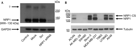
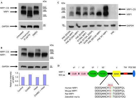
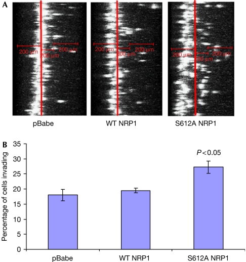
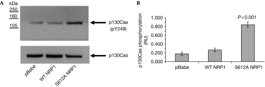
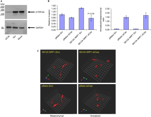
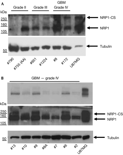
Neuropilin 1 (NRP1), a non-tyrosine kinase receptor for vascular endothelial growth factor and class 3 Semaphorins, is highly expressed in many human tumour cell lines, but its function is poorly understood. Here, we describe the expression of a new chondroitin sulphate-modified NRP1 (NRP1-CS) in human tumour cell lines. Expression of a non-modifiable NRP1 mutant (S612A) in U87MG human glioma cells results in enhanced invasion in three dimensions (3D), whereas wild-type NRP1 has no effect. Furthermore, the S612A NRP1 cells show a significant increase in p130Cas tyrosine phosphorylation compared with control and wild-type NRP1 cells. Silencing of p130Cas in S612A NRP1 cells resulted in a loss of increased invasive phenotype. Interestingly, p130Cas silencing does not inhibit basal 3D invasion, but leads to a mesenchymal to amoeboid transition. Biopsies from both low- and high-grade human gliomas show strong expression of NRP1, and little expression of NRP1-CS. Our data establish distinct roles for NRP1 and NRP1-CS in modulating a new NRP1-p130Cas signalling pathway contributing to glioblastoma cell invasion in 3D.


Conflict of interest statement

This study was financially assisted in part by Ark Therapeutics Plc, which is developing therapies targeted at inhibition of neuropilin. I.C.Z. is a consultant to Ark Therapeutics Plc. P.F., L.C. and P.L. are employees of Ark Therapeutics Plc. M.L.T. was a past employee of Ark Therapeutics Plc.

Figures

Figure 1
Figure 1
Identification of a high molecular weight NRP1 species in human tumour cell lines. (A) Identification of a high molecular weight band (>250 kDa) in Western blot analysis of NRP1, which is eliminated by NRP1 siRNA treatment. (B) The high molecular weight NRP1 species is expressed in several human tumour cell lines. Blots shown here and in all subsequent figures are representative of at least three separate experiments. NRP1, neuropilin 1; NRP1-CS, chondroitin sulphate-modified NRP1; siRNA, short interfering RNA.
Figure 2
Figure 2
NRP1 is post-translationally modified by N-linked glycosylation and by the addition of O-linked chondroitin sulphate to Ser 612 (S612). (A) A549 cells were untreated or pretreated with 5 μg/ml tunicamycin or control vehicle (0.1% DMSO) for 16 h, lysed and blotted with NRP1 antibody. (B) A549 cells were untreated or treated with 1 U/ml ChABC, 1 U/ml heparitinase (Hase), or both for 2 h before lysis and blotting for NRP1. A graphical representation of the relative intensity of the NRP1 band measured by densitometry is shown below. (C) Transient transfection of PAE cells with WT and NRP1 mutants, which show that only S612A NRP1 transfection results in the expression of non-CS-modified NRP1. (D) S612 in the region between the b2 and MAM domains of NRP1 is conserved between several vertebrate species. ChABC, chondroitinase ABC; CS, chondroitin sulphate; NRP1, neuropilin 1; PAE, porcine aortic endothelium; WT, wild type.
Figure 3
Figure 3
Expression of S612A NRP1 increases three-dimensional invasion. (A) xz reconstructions of U87MG pBabe, WT NRP1 and S612A NRP1 cells labelled with sytox green in collagen I invasion assays, performed as described in the Methods. (B) Quantification of invasion of U87MG pBabe, WT NRP1 and S612A NRP1 cells. Analysis was carried out as described in the supplementary information online. Data are expressed as the means±s.e.m. from three independent experiments. NRP1, neuropilin 1; WT, wild type.
Figure 4
Figure 4
Effect of expression of wild-type and S612A NRP1 on p130Cas phosphorylation in U87MG cells. (A) U87MG pBabe, WT NRP1 and S612A NRP1 cells grown on collagen I-coated plates and incubated in 1% FCS for 18 h were lysed and blotted for total and phosphor-Tyr 249 (pY239) p130Cas. (B) Quantification of p130Cas phosphorylation was performed by densitometry using Image J. Data from three independent experiments are presented as p130Cas phosphorylation relative units (RUs) normalized to total p130Cas and expressed as means±s.e.m. NRP1, neuropilin 1; WT, wild type.
Figure 5
Figure 5
p130Cas is required for S612A NRP1-mediated elongated invasion in three-dimensions. (A) S612A NRP1 cell lysates were immunoblotted for p130Cas and GAPDH 72 h after transfection of cells with scrambled siRNA (Scr) or p130Cas siRNA. Similar levels of p130Cas knockdown were obtained in U87MG pBabe cells (data not shown). (B) Invasion of U87MG pBabe and S612A NRP1 cells, 24 h after transfection with the indicated siRNAs. Data are expressed as the means±s.e.m. from three experiments. Invasion index was normalized to pBabe Scr siRNA (set at 1). (C) Depletion of p130Cas leads to a transition from elongated (mesenchymal) to a rounded (amoeboid) mode of invasion. U87MG pBabe and S612A NRP1 cells invading through collagen plugs stained with AF546-Phalloidin (cytoskeleton). Confocal sections (1.75 μm intervals) were obtained using a × 40 LWD objective. Three-dimensional reconstructions (1 U=25 μm) were performed with Volocity imaging software (Improvision). The ratio of amoeboid cells to mesenchymal cells is shown on the right and was determined from two of the three independent experiments performed above. LWD, long working distance; NRP1, neuropilin 1; siRNA, short interfering RNA.
Figure 6
Figure 6
Expression of NRP1 and NRP1-CS in low- and high-grade gliomas. Protein lysates of several biopsies of human glioma (all provided with separate code numbers) separated by gel electrophoresis, and probed using antibodies to NRP1 and tubulin as described in the supplementary information online. (A) Low- and high-grade glioma. (B) High-grade glioblastoma multiforme (GBM). Upper panel is a lower exposure of the middle NRP1 blot. CS, chondroitin sulphate; NRP1, neuropilin 1.

References

    1. Ambrogio C et al. (2005) p130Cas mediates the transforming properties of the anaplastic lymphoma kinase. Blood 106: 3907–3916 - PMC - PubMed
    1. Bachelder RE, Wendt MA, Mercurio AM (2002) Vascular endothelial growth factor promotes breast carcinoma invasion in an autocrine manner by regulating the chemokine receptor CXCR4. Cancer Res 62: 7203–7206 - PubMed
    1. Bielenberg DR, Pettaway CA, Takashima S, Klagsbrun M (2006) Neuropilins in neoplasms: expression, regulation, and function. Exp Cell Res 312: 584–593 - PubMed
    1. Bourdon MA, Krusius T, Campbell S, Schwartz NB, Ruoslahti E (1987) Identification and synthesis of a recognition signal for the attachment of glycosaminoglycans to proteins. Proc Natl Acad Sci USA 84: 3194–3198 - PMC - PubMed
    1. Cheng L, Jia H, Lohr M, Bagherzadeh A, Holmes DI, Selwood D, Zachary I (2004) Anti-chemorepulsive effects of vascular endothelial growth factor and placental growth factor-2 in dorsal root ganglion neurons are mediated via neuropilin-1 and cyclooxygenase-derived prostanoid production. J Biol Chem 279: 30654–30661 - PubMed

Publication types

LinkOut - more resources