. 2008 Sep;118(9):3087-97.
doi: 10.1172/JCI35781.

Hsp104 antagonizes alpha-synuclein aggregation and reduces dopaminergic degeneration in a rat model of Parkinson disease

Affiliations

Hsp104 antagonizes alpha-synuclein aggregation and reduces dopaminergic degeneration in a rat model of Parkinson disease

Christophe Lo Bianco et al. J Clin Invest. 2008 Sep.

Abstract

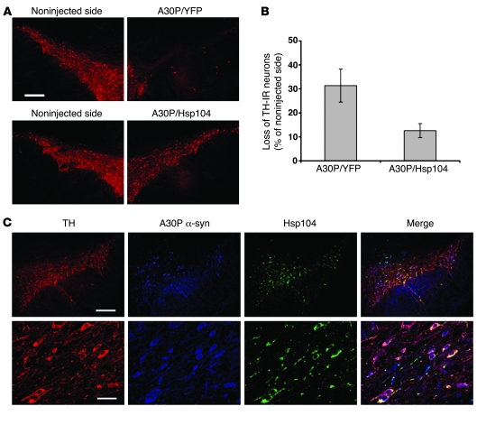
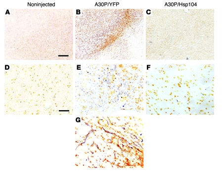
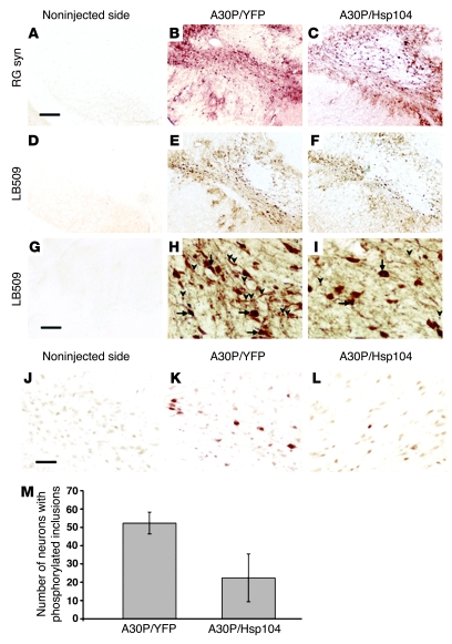
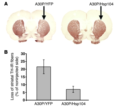
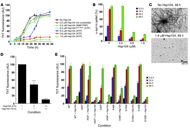
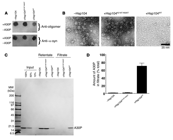
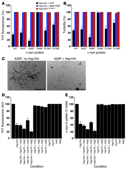
Parkinson disease (PD) is characterized by dopaminergic neurodegeneration and intracellular inclusions of alpha-synuclein amyloid fibers, which are stable and difficult to dissolve. Whether inclusions are neuroprotective or pathological remains controversial, because prefibrillar oligomers may be more toxic than amyloid inclusions. Thus, whether therapies should target inclusions, preamyloid oligomers, or both is a critically important issue. In yeast, the protein-remodeling factor Hsp104 cooperates with Hsp70 and Hsp40 to dissolve and reactivate aggregated proteins. Metazoans, however, have no Hsp104 ortholog. Here we introduced Hsp104 into a rat PD model. Remarkably, Hsp104 reduced formation of phosphorylated alpha-synuclein inclusions and prevented nigrostriatal dopaminergic neurodegeneration induced by PD-linked alpha-synuclein (A30P). An in vitro assay employing pure proteins revealed that Hsp104 prevented fibrillization of alpha-synuclein and PD-linked variants (A30P, A53T, E46K). Hsp104 coupled ATP hydrolysis to the disassembly of preamyloid oligomers and amyloid fibers composed of alpha-synuclein. Furthermore, the mammalian Hsp70 and Hsp40 chaperones, Hsc70 and Hdj2, enhanced alpha-synuclein fiber disassembly by Hsp104. Hsp104 likely protects dopaminergic neurons by antagonizing toxic alpha-synuclein assemblies and might have therapeutic potential for PD and other neurodegenerative amyloidoses.


Figures

Figure 1
Figure 1. Hsp104 reduces dopaminergic cell loss in the substantia nigra of rats expressing human α-syn A30P.
(A) Staining for TH at 6 weeks in the substantia nigra of rats injected with either lenti-A30P/lenti-YFP or lenti-A30P/lenti-Hsp104. (B) Quantitation of TH-IR nigral neurons at 6 weeks relative to the contralateral side in rats unilaterally injected with the different lentiviral constructs. Values represent mean ± SEM; n = 7 animals for both A30P/YFP and A30P/Hsp104 groups; P < 0.05. (C) Confocal imaging of triple labeling for TH (red), A30P human α-syn (blue), and Hsp104 (green) in the rat substantia nigra injected with lenti-A30P/lenti-Hsp104. Higher magnification (bottom row) reveals numerous TH neurons positive for Hsp104 and A30P human α-syn at 6 weeks after injection. Scale bar: 350 μm (A and C, top row); 60 μm (C, bottom row).
Figure 3
Figure 3. Hsp104 prevents A30P α-syn–induced neurodegeneration.
(AG) Degenerating nigral neurons were detected by silver staining. (A and D) Noninjected sides did not show any specific silver staining. (DG) Higher magnification reveals numerous silver-positive dark structures in the substantia nigra of animals expressing A30P/YFP (E). (G) Abundant neuritic pathology clearly showed the presence of degenerating neurites. (C and F) Coexpression of Hsp104 with A30P α-syn prevents the appearance of silver-positive degenerating neurons. Scale bar: 300 μm (AC); 100 μm (DG).
Figure 4
Figure 4. Hsp104 reduces the number of phosphorylated α-syn inclusions.
(AC) Robust expression of human α-syn was detected with both RG syn and (DI) the human specific LB509 Abs. (GI) Higher magnification revealed the accumulation and aggregation of human α-syn in both the cell bodies (arrows) and neurites (arrowheads). (H and I) Note that there are fewer dense puncta in neurites in the A30P/Hsp104 condition. (J) Sections of the substantia nigra from the noninjected side and (K) from rats expressing A30P/YFP or (L) A30P/Hsp104 were immunostained with Ab specific for phosphorylated Ser 129 of α-syn. The substantia nigra of animals overexpressing either A30P/YFP or A30P/Hsp104 reveals the presence of hyperphosphorylated inclusions reminiscent of Lewy bodies. Scale bar: 300 μm (AF); 40 μm (GI); 50 μm (JL). (M) Quantitation of neurons containing Pser129-positive aggregates in the substantia nigra of rats expressing A30P/YFP or A30P/Hsp104. Values represent mean ± SEM; n = 6 animals per group; P < 0.05.
Figure 2
Figure 2. Hsp104 reduces α-syn–induced dopaminergic fiber loss in the striatum of rats.
(A) Striatal sections stained for TH from rats after intranigral injections of either lenti-A30P/lenti-YFP or lenti-A30P/lenti-Hsp104 (arrows indicate the injected sides). Original magnification, ×3. (B) Quantitation of the loss of TH fibers at 6 weeks, relative to the contralateral side in rats unilaterally injected with the different lentiviral vectors. Values represent mean ± SEM; n = 7 animals per group; P < 0.05.
Figure 5
Figure 5. Hsp104 inhibits α-syn fibrillization.
(A and B) Kinetics of rotated (80 rpm) α-syn (80 μM) fibrillization at 37°C without or with either Hsp104 (0.2–1.6 μM) plus ATP and regeneration system (20 mM creatine phosphate and 0.1 mg/ml creatine kinase), Hsp104 (0.2 μM) with no nucleotide, Hsp104 (0.2 μM) plus nonhydrolyzable ATP analogue (AMP-PNP), or Hsp104K218T:K620T (0.2 μM) plus ATP and regeneration system. Fibrillization was measured by ThT fluorescence (A) or sedimentation analysis (B). Values represent mean ± SD; n = 3. (C) EM of α-syn (80 μM) fibrillization at 37°C for 48 hours without or with Hsp104 (1.6 μM) plus ATP and regeneration system. Scale bar: 200 nm. (D) α-syn (80 μM) was incubated for 96 hours with rotation at 37°C in the presence or absence of Hsp104 (1.6 μM) plus ATP and regeneration system. After 72 hours, reactions were either supplemented with buffer or fresh Hsp104 (1.6 μM). Fibrillization was measured by ThT fluorescence. Values represent mean ± SD; n = 3. (E) Fibrillization kinetics of wild-type, A53T, A30P, E46K, S129A, and S129E α-syn (80 μM) in the presence or absence of Hsp104 (2 μM) plus ATP and regeneration system. Another reaction contained A53T (80 μM) plus Hsp104 (4 μM) with ATP and regeneration system. Reactions were rotated (80 rpm) at 37°C. Fibrillization was measured by ThT fluorescence. Values represent mean ± SD; n = 4.
Figure 6
Figure 6. Hsp104 remodels α-syn A30P preamyloid oligomers.
(AD) Purified α-syn A30P preamyloid oligomers (0.5 μM) were incubated without or with Hsp104 or Hsp104K218T:K620T (10 μM) plus ATP and regeneration system for 1 hour at 37°C. Reactions were spotted on nitrocellulose and probed with anti-oligomer Ab or anti–α-syn Ab (A). (B) Alternatively, reactions were processed for EM (scale bar: 25 nm) or (C) depleted of Hsp104 and passed through a 100-kDa molecular weight filter. (C and D) Retentate and filtrate fractions were collected and analyzed by SDS-PAGE, and the amount of A30P in the filtrate was determined by densitometry. Values represent mean ± SD; n = 4 (D).
Figure 7
Figure 7. Hsp104 remodels α-syn fibers.
(A and B) Fibers composed of wild-type, A53T, A30P, E46K, S129A, or S129E α-syn (0.5 mM monomer) were incubated with Hsp104K218T:K620T, Hsp104 (10 μM) plus ATP and regeneration system, or AMP-PNP system for 1 hour at 37°C. Fiber integrity was determined by ThT fluorescence (A) or turbidity (B). For each fiber, 100% assembly reflects untreated fibers. Values represent mean ± SD; n = 3. (C) EM of A30P fibers incubated without or with Hsp104 with ATP and regeneration system as in A. Scale bar: 0.5 μm. (D and E) Fibers composed of wild-type α-syn (0.5 μM monomer) were incubated for 1 hour at 37°C without or with the indicated combinations of Hsp104 (10 μM), Hsp70 (10 μM), Hsc70 (10 μM), Hdj1 (10 μM), or Hdj2 (10 μM) plus ATP and regeneration system. Fiber integrity was determined by ThT fluorescence (D) or sedimentation analysis (E). Values represent mean ± SD; n = 3.

Similar articles

Cited by

References

    1. Skovronsky D.M., Lee V.M., Trojanowski J.Q. Neurodegenerative diseases: new concepts of pathogenesis and their therapeutic implications. Annu. Rev. Pathol. 2006;1:151–170. doi: 10.1146/annurev.pathol.1.110304.100113. - DOI - PubMed
    1. Dorsey E.R., et al. Projected number of people with Parkinson’s disease in the most populous nations, 2005 through 2030. Neurology. 2007;68:384–386. doi: 10.1212/01.wnl.0000247740.47667.03. - DOI - PubMed
    1. Braak H., et al. Staging of brain pathology related to sporadic Parkinson’s disease. Neurobiol. Aging. 2003;24:197–211. doi: 10.1016/S0197-4580(02)00065-9. - DOI - PubMed
    1. Spillantini M.G., et al. Alpha-synuclein in Lewy bodies. Nature. 1997;388:839–840. doi: 10.1038/42166. - DOI - PubMed
    1. Moore D.J., West A.B., Dawson V.L., Dawson T.M. Molecular pathophysiology of Parkinson’s disease. Annu. Rev. Neurosci. 2005;28:57–87. doi: 10.1146/annurev.neuro.28.061604.135718. - DOI - PubMed

Publication types