. 2008 Sep;118(9):3123-31.
doi: 10.1172/JCI35147.

Drosophila are protected from Pseudomonas aeruginosa lethality by transgenic expression of paraoxonase-1

Affiliations

Drosophila are protected from Pseudomonas aeruginosa lethality by transgenic expression of paraoxonase-1

David A Stoltz et al. J Clin Invest. 2008 Sep.

Abstract

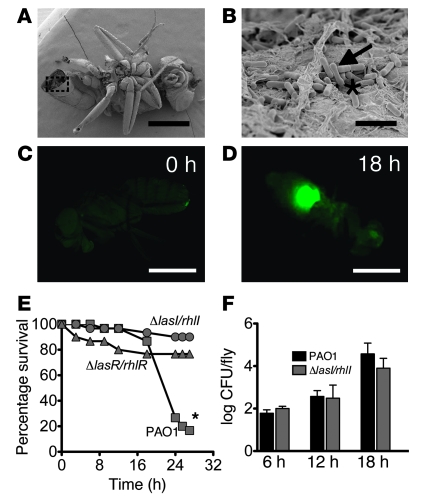
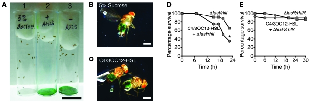
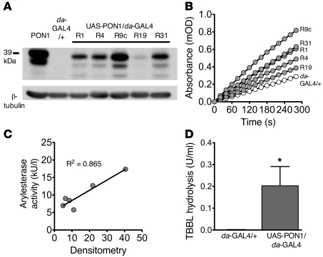
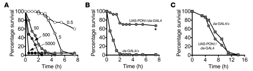
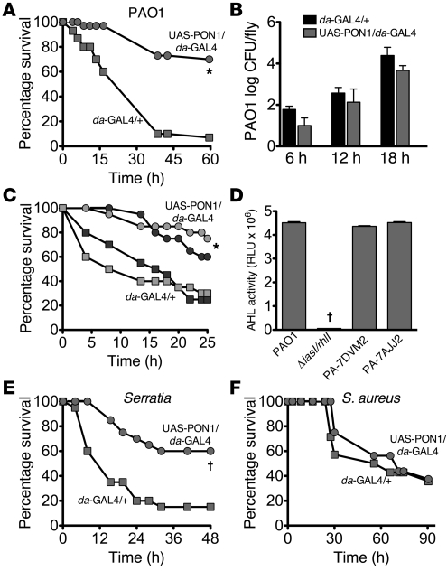
Pseudomonas aeruginosa uses quorum sensing, an interbacterial communication system, to regulate gene expression. The signaling molecule N-3-oxododecanoyl homoserine lactone (3OC12-HSL) is thought to play a central role in quorum sensing. Since 3OC12-HSL can be degraded by paraoxonase (PON) family members, we hypothesized that PONs regulate P. aeruginosa virulence in vivo. We chose Drosophila melanogaster as our model organism because it has been shown to be a tractable model for investigating host-pathogen interactions and lacks PONs. By using quorum-sensing-deficient P. aeruginosa, synthetic acyl-HSLs, and transgenic expression of human PON1, we investigated the role of 3OC12-HSL and PON1 on P. aeruginosa virulence. We found that P. aeruginosa virulence in flies was dependent upon 3OC12-HSL. PON1 transgenic flies expressed enzymatically active PON1 and thereby exhibited arylesterase activity and resistance to organophosphate toxicity. Moreover, PON1 flies were protected from P. aeruginosa lethality, and protection was dependent on the lactonase activity of PON1. Our findings show that PON1 can interfere with quorum sensing in vivo and provide insight into what we believe is a novel role for PON1 in the innate immune response to quorum-sensing-dependent pathogens. These results raise intriguing possibilities about human-pathogen interactions, including potential roles for PON1 as a modifier gene and for PON1 protein as a regulator of normal bacterial florae, a link between infection/inflammation and cardiovascular disease, and a potential therapeutic modality.


Figures

Figure 1
Figure 1. Quorum-sensing–dependent P. aeruginosa infection of Drosophila.
(A and B) Scanning electron micrographs at low and high magnification of Drosophila at 24 hours following infection with wild-type P. aeruginosa (PAO1). (A) Low-magnification image shows a whole fly with an opening in the distal abdomen. Scale bar: 0.5 mm. (B) High-magnification image of boxed area on whole fly. Arrow indicates bacteria and asterisk denotes extracellular matrix. Scale bar: 2.5 μm. (C and D) GFP signal (quorum-sensing activity) after infection of Drosophila with a quorum-sensing reporter strain of P. aeruginosa (under control of the lasB promoter). GFP signal at 0 hours (C) and 18 hours (D) following abdominal inoculation with PAO1. Scale bars: 0.5 mm. (E) Fly survival after infection with PAO1 and quorum-sensing–deficient strains of P. aeruginosa. da-GAL4/+ flies were infected with PAO1 (squares), ΔlasI/rhlI (circles), or ΔlasR/rhlR (triangles) strains. n = 30 flies per group for each experiment. *P < 0.001, comparing PAO1 versus ΔlasI/rhlI and PAO1 versus ΔlasR/rhlR fly survival; log-rank test. (F) P. aeruginosa bacterial counts after infection with PAO1 or ΔlasI/rhlI. At 6, 12, and 18 hours following infection, flies were anesthetized, surface sterilized, and homogenized for performance of quantitative bacterial counts. Data are displayed as log10 CFU/fly and represent the mean ± SEM. n = 3–5 per time point with 10–20 flies per group per experiment.
Figure 2
Figure 2. acyl-HSL feeding enhances virulence of quorum-sensing–deficient P. aeruginosa mutants.
(A) Photomicrograph of fly-feeding vials with filter paper at the base containing either 5% sucrose alone (tubes 1 and 3) or 5% sucrose with C4- and 3OC12-HSL (tube 2). Filter paper in tubes 2 and 3 contained green food dye to confirm similar rates of food consumption between feeding groups and also consumption of acyl-HSLs. Scale bar: 2.5 cm. (B and C) Representative low-power images of Drosophila after 24 hours feeding on either 5% sucrose alone (B) or 5% sucrose containing C4- and 3OC12-HSL (containing green dye) (C). Note similar levels of green dye in the abdomen of flies consuming sucrose alone or sucrose containing acyl-HSLs. Scale bars: 0.25 mm. (D and E) Flies were fed 5% sucrose (squares) or 5% sucrose containing C4-HSL (5 μM) and 3OC12-HSL (60 μM) (circles). 48 hours later, flies were infected with ΔlasI/rhlI (D) or ΔlasR/rhlR (E) strains of P. aeruginosa. Fly survival was monitored over time. n = 30 flies per group for each experiment. *P < 0.01, comparing survival between ΔlasI/rhlI versus ΔlasI/rhlI + C4- and 3OC12-HSL groups; log-rank test.
Figure 3
Figure 3. PON1 expression and activity in PON1 transgenic Drosophila.
(A) Western blot analysis for PON1 in da-GAL4/+ (control) and UAS-PON1/da-GAL4 flies. PON1 flies were constructed using the GAL4-UAS binary system under control of the da promoter driving ubiquitous PON1 expression. Fly heads were obtained from 1- to 3-day-old flies, homogenized, and used for immunoblotting with PON1 antibody. Lysates from CHO cells infected with an adenovirus-expressing human PON1 served as the positive control. All PON1 fly lines obtained were tested for PON1 protein. β-tubulin was used as a loading control. (B) Arylesterase activity was tested in fly homogenates from da-GAL4/+ and UAS-PON1/da-GAL4 transgenic fly lines by measuring phenylacetate degradation. mOD, optical density × 10–3. (C) Correlation of arylesterase activity and PON1 protein expression in control and PON1 transgenic flies. Densitometry analysis of PON1 and β-tubulin immunoreactive bands was performed, and data are expressed in relative units. (D) Lactonase activity in control and PON1 transgenic flies. Whole-fly lysates were combined with the synthetic lactone TBBL (0.25 mM), and lactonase activity (thiol moiety release) was monitored with DTNB (0.5 mM) at an absorbance of 412 nm. Data represent the mean ± SEM with n = 3–4 per group. *P < 0.01, lactonase activity between da-GAL4/+ and UAS-PON1/da-GAL4 flies; Student’s t test.
Figure 4
Figure 4. PON1 flies are protected from chlorpyrifos toxicity.
(A) da-GAL4/+ flies were fed 0.5, 5, 50, 500, or 5000 ppm chlorpyrifos in 5% sucrose. Fly survival was monitored over time. (B and C) da-GAL4/+ (squares) or UAS-PON1/da-GAL4 (circles) flies were fed chlorpyrifos (50 ppm in 5% sucrose) (B) or DSM (150 ppm in 5% sucrose) (C), and survival was followed over time. n = 30 flies per group for each experiment. *P < 0.001, comparing survival between da-GAL4/+ and UAS-PON1/da-GAL4 flies; log-rank test.
Figure 5
Figure 5. PON1 flies are protected from P. aeruginosa lethality.
(A) da-GAL4/+ (squares) and UAS-PON1/da-GAL4 (circles) flies were infected with P. aeruginosa, and fly survival was monitored over time. n = 30 flies per group for each experiment. *P < 0.01, comparing da-GAL4/+ and UAS-PON1/da-GAL4 survival; log-rank test. (B) P. aeruginosa bacterial counts following infection in da-GAL4/+ and UAS-PON1/da-GAL4 flies. Bacterial quantification was performed as described for Figure 1. Data are displayed as log 10 CFU/fly and represent the mean ± SEM. n = 3–5 per time point with 10–20 flies per group per experiment. (C) PON1 transgenic flies are protected from lethality following infection with clinical isolates of P. aeruginosa. Light-shaded symbols, PA-7JJA2; and dark-shaded symbols, PA-7DVM2. *P < 0.01, comparing da-GAL4/+ and UAS-PON1/da-GAL4 fly survival following either PA-7JJA2 or PA-7DVM2 infection; log-rank test. (D) acyl-HSL quorum-sensing activity by P. aeruginosa isolates. Supernatants from overnight bacterial cultures were incubated with the acyl-HSL detector strain A. tumefaciens NTL4, and β-galactosidase activity was determined. Data are mean ± SEM and n = 3 per bacterial strain. P < 0.001, compared with PAO1. (EF) da-GAL4/+ and UAS-PON1/da-GAL4 flies were infected with either (E) S. marcescens or (F) S. aureus (SH1000), and survival was followed over time. n = 30 flies per group for each experiment. P < 0.001, comparing survival between da-GAL4/+ and UAS-PON1/da-GAL4 flies following S. marcescens infection; log-rank test.
Figure 6
Figure 6. 3OC12-HSL fails to enhance P. aeruginosa virulence in PON1 flies.
(A) da-GAL4/+ or (B) UAS-PON1/da-GAL4 flies were fed 5% sucrose (squares) or 5% sucrose containing 3OC12-HSL (60 μM) (circles). 48 hours later, flies were infected with the ΔlasI mutant strain of P. aeruginosa (deficient in 3OC12-HSL production). n = 30 flies per group for each experiment. Fly survival was monitored over time. *P < 0.05, comparing ΔlasI and 3OC12-HSL + ΔlasI survival in da-GAL4/+ flies; log-rank test.
Figure 7
Figure 7. Quorum-sensing–dependent virulence gene activation is suppressed in PON1 flies.
(A) da-GAL4/+ and UAS-PON1/da-GAL4 flies were infected with a quorum-sensing reporter strain of P. aeruginosa, PAO-1qsc102-lacZ. 18 hours later, X-gal staining was performed. Shown are representative images of da-GAL4/+ and UAS-PON1/da-GAL4 flies following infection. Scale bars: 0.5 mm. (B) Quorum-sensing–regulated (QS-regulated) and nonregulated genes. da-GAL4/+ and UAS-PON1/da-GAL4 flies were infected with P. aeruginosa, and 18 hours later, flies were sacrificed for mRNA analysis with RT-PCR. Data are expressed as percentage change (± SEM) in mRNA levels between daGAL4/+ and UAS-PON1/da-GAL4 flies. *P < 0.05 for mRNA expression between da-GAL4/+ and UAS-PON1/da-GAL4 flies following P. aeruginosa infection.

Similar articles

Cited by

References

    1. Gan K.N., Smolen A., Eckerson H.W., La Du B.N. Purification of human serum paraoxonase/arylesterase. Evidence for one esterase catalyzing both activities. Drug Metab. Dispos. 1991;19:100–106. - PubMed
    1. Furlong C.E., et al. Human and rabbit paraoxonases: purification, cloning, sequencing, mapping and role of polymorphism in organophosphate detoxification. Chem. Biol. Interact. 1993;87:35–48. doi: 10.1016/0009-2797(93)90023-R. - DOI - PubMed
    1. Furlong C.E., Richter R.J., Chapline C., Crabb J.W. Purification of rabbit and human serum paraoxonase. Biochemistry. 1991;30:10133–10140. doi: 10.1021/bi00106a009. - DOI - PubMed
    1. Durrington P.N., Mackness B., Mackness M.I. Paraoxonase and atherosclerosis. Arterioscler. Thromb. Vasc. Biol. 2001;21:473–480. - PubMed
    1. Shih D.M., et al. Mice lacking serum paraoxonase are susceptible to organophosphate toxicity and atherosclerosis. Nature. 1998;394:284–287. doi: 10.1038/28406. - DOI - PubMed

Publication types

Substances