

Sodium current properties of primary skeletal myocytes and cardiomyocytes derived from different mouse strains
- PMID: 18704489
- PMCID: PMC3016614
- DOI: 10.1007/s00424-008-0570-x
Sodium current properties of primary skeletal myocytes and cardiomyocytes derived from different mouse strains
Abstract
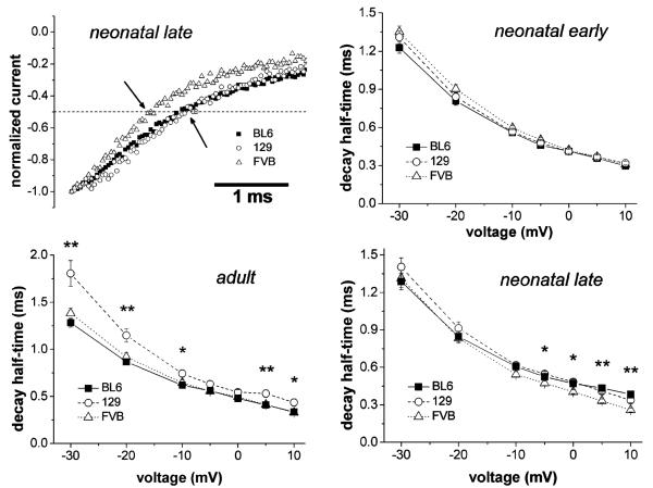
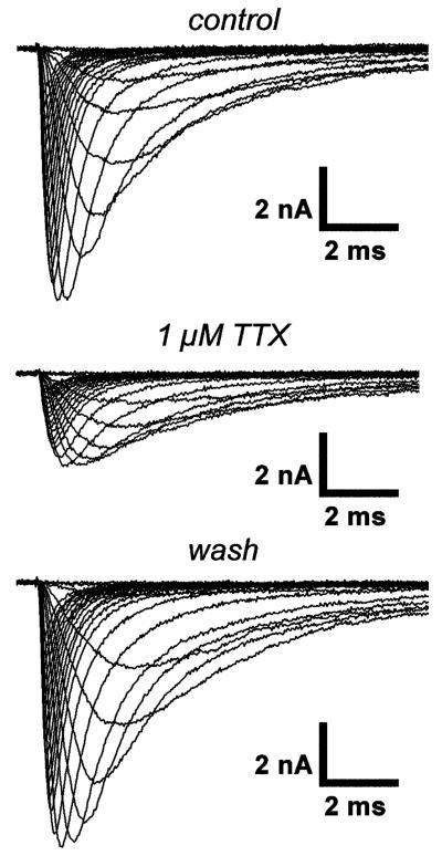
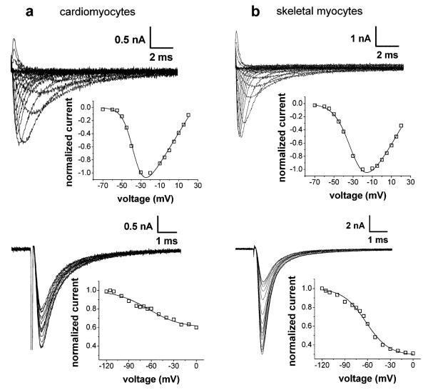
The mouse has become the preferred animal for genetic manipulations. Because of the diverse genetic backgrounds of various mouse strains, these can manifest strikingly different characteristics. Here, we studied the functional properties of currents through voltage-gated sodium channels in primary cultures of skeletal myocytes and cardiomyocytes derived from the three commonly used mouse strains BL6, 129/Sv, and FVB, by using the whole-cell patch-clamp technique. We found strain-specific sodium current function in skeletal myocytes, which could partly be explained by differences in sodium channel isoform expression. In addition, we found significant effects of cell source (neonatal or adult animal-derived) and variation of the differentiation time period. In contrast to skeletal myocytes, sodium current function in cardiomyocytes was similar in all strains. Our findings are relevant for the design and proper interpretation of electrophysiological studies, which use excitable cells in primary culture as a model system.
Figures
Similar articles
-
Voltage-gated ion channel dysfunction precedes cardiomyopathy development in the dystrophic heart.PLoS One. 2011;6(5):e20300. doi: 10.1371/journal.pone.0020300. Epub 2011 May 23. PLoS One. 2011. PMID: 21677768 Free PMC article.
-
C2C12 skeletal muscle cells adopt cardiac-like sodium current properties in a cardiac cell environment.Am J Physiol Heart Circ Physiol. 2007 Jan;292(1):H439-50. doi: 10.1152/ajpheart.00119.2006. Epub 2006 Sep 15. Am J Physiol Heart Circ Physiol. 2007. PMID: 16980339
-
Differential sialylation modulates voltage-gated Na+ channel gating throughout the developing myocardium.J Gen Physiol. 2006 Mar;127(3):253-65. doi: 10.1085/jgp.200509423. Epub 2006 Feb 13. J Gen Physiol. 2006. PMID: 16476705 Free PMC article.
-
Ca2+-dependent modulation of voltage-gated myocyte sodium channels.Biochem Soc Trans. 2021 Nov 1;49(5):1941-1961. doi: 10.1042/BST20200604. Biochem Soc Trans. 2021. PMID: 34643236 Free PMC article. Review.
-
Autonomic regulation of voltage-gated cardiac ion channels.J Cardiovasc Electrophysiol. 2006 May;17 Suppl 1:S34-S42. doi: 10.1111/j.1540-8167.2006.00387.x. J Cardiovasc Electrophysiol. 2006. PMID: 16686680 Review.
Cited by
-
Anti-addiction drug ibogaine inhibits voltage-gated ionic currents: a study to assess the drug's cardiac ion channel profile.Toxicol Appl Pharmacol. 2013 Dec 1;273(2):259-68. doi: 10.1016/j.taap.2013.05.012. Epub 2013 May 22. Toxicol Appl Pharmacol. 2013. PMID: 23707769 Free PMC article.
-
Mylk3 null C57BL/6N mice develop cardiomyopathy, whereas Nnt null C57BL/6J mice do not.Life Sci Alliance. 2020 Mar 25;3(4):e201900593. doi: 10.26508/lsa.201900593. Print 2020 Apr. Life Sci Alliance. 2020. PMID: 32213617 Free PMC article.
-
Characterisation of connexin expression and electrophysiological properties in stable clones of the HL-1 myocyte cell line.PLoS One. 2014 Feb 28;9(2):e90266. doi: 10.1371/journal.pone.0090266. eCollection 2014. PLoS One. 2014. PMID: 24587307 Free PMC article.
-
Gating behaviour of sodium currents in adult mouse muscle recorded with an improved two-electrode voltage clamp.J Physiol. 2011 Feb 1;589(Pt 3):525-46. doi: 10.1113/jphysiol.2010.199430. Epub 2010 Dec 6. J Physiol. 2011. PMID: 21135045 Free PMC article.
-
Voltage-gated ion channel dysfunction precedes cardiomyopathy development in the dystrophic heart.PLoS One. 2011;6(5):e20300. doi: 10.1371/journal.pone.0020300. Epub 2011 May 23. PLoS One. 2011. PMID: 21677768 Free PMC article.
References
-
- Burton KA, Johnson BD, Hausken ZE, Westenbroek RE, Idzerda RL, Scheuer T, Scott JD, Catterall WA, McKnight GS. Type II regulatory subunits are not required for the anchoring-dependent modulation of Ca2+ channel activity by cAMP-dependent protein kinase. Proc Natl Acad Sci U S A. 1997;94:11067–11072. - PMC - PubMed
-
- Menasche P. Skeletal myoblasts as a therapeutic agent. Prog Cardiovasc Dis. 2007;50:7–17. - PubMed
Publication types
MeSH terms
Substances
Grants and funding
LinkOut - more resources
Full Text Sources
