

Shape priors for segmentation of the cervix region within uterine cervix images
- PMID: 18704582
- PMCID: PMC3043693
- DOI: 10.1007/s10278-008-9134-z
Shape priors for segmentation of the cervix region within uterine cervix images
Abstract
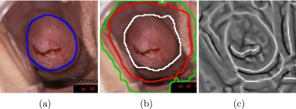
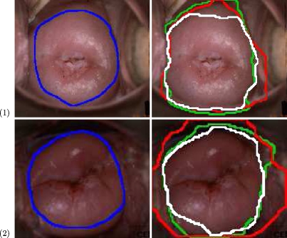
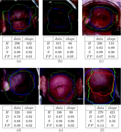
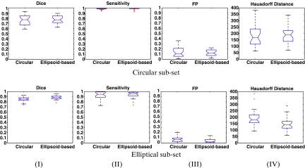
The work focuses on a unique medical repository of digital uterine cervix images ("cervigrams") collected by the National Cancer Institute (NCI), National Institute of Health, in longitudinal multiyear studies. NCI together with the National Library of Medicine is developing a unique web-based database of the digitized cervix images to study the evolution of lesions related to cervical cancer. Tools are needed for the automated analysis of the cervigram content to support the cancer research. In recent works, a multistage automated system for segmenting and labeling regions of medical and anatomical interest within the cervigrams was developed. The current paper concentrates on incorporating prior-shape information in the cervix region segmentation task. In accordance with the fact that human experts mark the cervix region as circular or elliptical, two shape models (and corresponding methods) are suggested. The shape models are embedded within an active contour framework that relies on image features. Experiments indicate that incorporation of the prior shape information augments previous results.
Figures


References
-
- Chen Y, Tagare HD, Thiruvenkadam S, Huang F, Wilson D, Gopinath KS, Briggs RW, Geiser EA. Using prior shapes in geometric active contours in a variational framework. Int J Comput Vis. 2002;50(3):315–328. doi: 10.1023/A:1020878408985. - DOI
-
- Cootes TF, Taylor CJ, Cooper DH, Graham J. Active shape models—their training and application. Comput Vis Image Underst. 1995;61(1):38–59. doi: 10.1006/cviu.1995.1004. - DOI
-
- Gerig G, Jomier M, Chakos M. Valmet: a new validation tool for assessing and improving 3D object segmentation. Proc. of Medical Image Computing and Computer-Assisted Intervention (MICCAI’01) 2001;2208:516–523.
-
- Gordon S, Zimmerman G, Long R, Antani S, Jeronimo J, Greenspan H. Content analysis of uterine cervix images: initial steps towards content based indexing and retrieval of cervigrams. Proc. of SPIE Medical Imaging. 2006;6144:1549–1556.
