. 2008 Oct;48(4):1242-50.
doi: 10.1002/hep.22439.

Hepatocyte nuclear factor 4alpha is implicated in endoplasmic reticulum stress-induced acute phase response by regulating expression of cyclic adenosine monophosphate responsive element binding protein H

Affiliations

Hepatocyte nuclear factor 4alpha is implicated in endoplasmic reticulum stress-induced acute phase response by regulating expression of cyclic adenosine monophosphate responsive element binding protein H

Jennifer Luebke-Wheeler et al. Hepatology. 2008 Oct.

Abstract

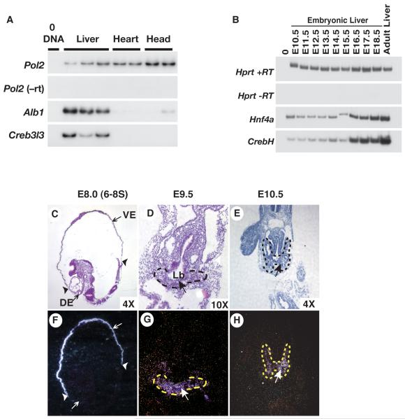
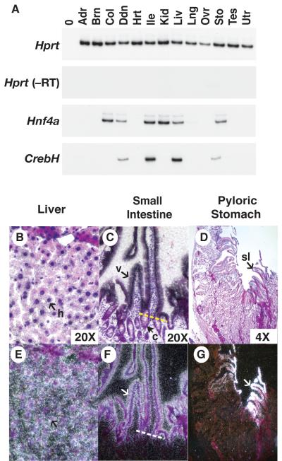
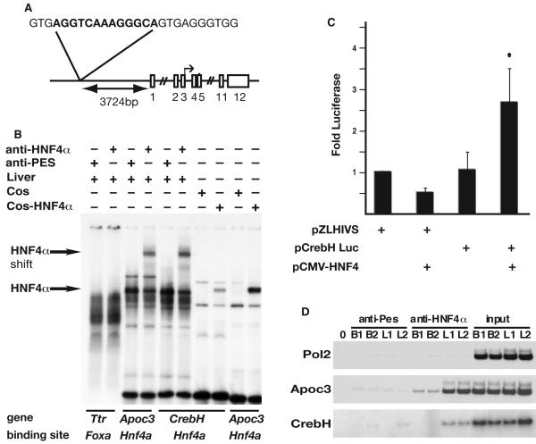
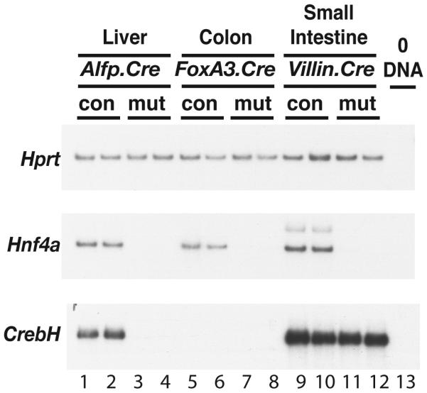
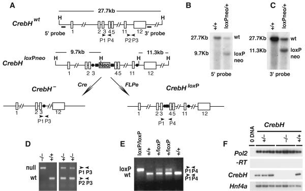
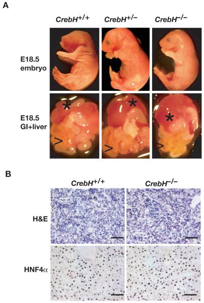
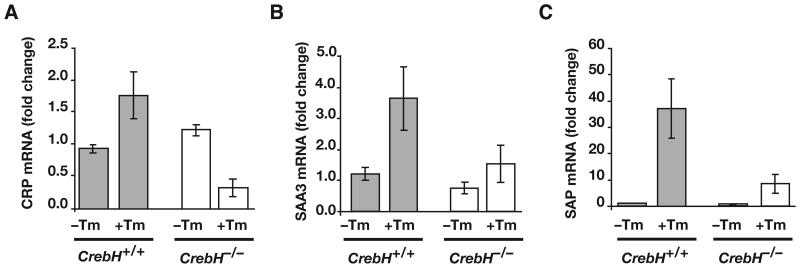
Loss of the nuclear hormone receptor hepatocyte nuclear factor 4alpha (HNF4alpha) in hepatocytes results in a complex pleiotropic phenotype that includes a block in hepatocyte differentiation and a severe disruption to liver function. Recent analyses have shown that hepatic gene expression is severely affected by the absence of HNF4alpha, with expression of 567 genes reduced by > or =2.5-fold (P < or = 0.05) in Hnf4alpha(-/-) fetal livers. Although many of these genes are direct targets, HNF4alpha has also been shown to regulate expression of other liver transcription factors, and this raises the possibility that the dependence on HNF4alpha for normal expression of some genes may be indirect. We postulated that the identification of transcription factors whose expression is regulated by HNF4alpha might reveal roles for HNF4alpha in controlling hepatic functions that were not previously appreciated. Here we identify cyclic adenosine monophosphate responsive element binding protein H (CrebH) as a transcription factor whose messenger RNA can be identified in both the embryonic mouse liver and adult mouse liver and whose expression is dependent on HNF4alpha. Analyses of genomic DNA revealed an HNF4alpha binding site upstream of the CrebH coding sequence that was occupied by HNF4alpha in fetal livers and facilitated transcriptional activation of a reporter gene in transient transfection analyses. Although CrebH is highly expressed during hepatogenesis, CrebH(-/-) mice were viable and healthy and displayed no overt defects in liver formation. However, upon treatment with tunicamycin, which induces an endoplasmic reticulum (ER)-stress response, CrebH(-/-) mice displayed reduced expression of acute phase response proteins.

Conclusion: These data implicate HNF4alpha in having a role in controlling the acute phase response of the liver induced by ER stress by regulating expression of CrebH.


Figures

Fig. 1
Fig. 1. Fetal expression of CrebH initiates in the primary liver bud and continues throughout hepatogenesis
A) RT-PCR analyses revealed the presence of CrebH and Albumin (Alb1) mRNAs in livers isolated from E10.5 embryos. Amplification of Rna pol2 (Pol2) was used as a loading control while reactions lacking reverse transcriptase (-RT) and DNA template (0DNA) confirmed the absence of contaminating DNA. B) RT-PCR analyses uncovered CrebH and Hnf4α mRNA in livers isolated mouse embryos at daily intervals ranging from E10.5 through E18.5, as well as in adult livers. Amplification of Hprt was used as loading control while reactions lacking reverse transcriptase (-RT) and DNA template (0DNA) confirmed the absence of contaminating DNA. C-H) Radioactive in situ hybridization analyses revealed the presence of CrebH mRNA (arrows; silver grains) during early development. Sagittal sections through an E8.5 (6-8 somite stage) embryo (C, F) identified CrebH mRNA in the extraembryonic visceral endoderm (VE) but not in the definitive endoderm (DE); extraembryonic/embryonic boundary indicated by arrowheads. CrebH mRNA was also found to be present in the primary liver bud (Lb; outlined with dashes) in transverse sections through an E9.5 embryo (D, G), and in the expanding clusters of hepatoblasts (outlined with dashes) in transverse sections through an E10.5 embryo (E, H). H&E-stained bright field images (C, D, E) and corresponding dark field images (F, G, H) are presented.
Fig. 2
Fig. 2. CrebH is expressed in the adult liver and gastrointestinal tract
A) RT-PCR analyses of CrebH and Hnf4α was performed on mRNA extracts from the adrenal gland (Adr), brain (Brn), colon (Col), duodenum (Ddn), heart (Hrt) ileum (Ile), kidney (Kid), liver (Liv), lung (Lng), ovary (Ovr), stomach (Sto), testis (Tes) and uterus (Utr). B-G) The distribution of CrebH mRNA in the liver (B, E), small intestine (C, F), and stomach (D, G), was identified (white silver grains) using radioactive in situ hybridization analysis. CrebH mRNA was present in the hepatocytes (h, arrow) of the liver, the epithelial cells of the villi (v,arrow), but not the crypts (c, white arrows; a yellow dashed line demarcates the villi/crypt border) of the small intestine, and in the surface lining cells (sl) of the stomach. H&E-stained bright field images (B, C, D) and corresponding dark field images (E, F, G) are presented.
Fig. 3
Fig. 3. CrebH is a direct target of HNF4α transcriptional activity
(A) Schematic showing the genomic location and sequence of the identified HNF4α binding site relative to CrebH (exons shown as boxes). (B) The ability of HNF4α protein to bind the putative HNF4α-binding site was confirmed by EMSA. Radiolabeled oligonucleotides representing binding sites were incubated with liver nuclear extracts in the presence of anti-HNF4α antibody, which resulted in a retarded migration of HNF4α-bound complexes (arrows), or anti-Pes1 antibody (negative control). Alternatively, EMSAs were performed using nuclear extracts from COS-7 cells or COS-7 cells expressing HNF4α. A previously described HNF4α binding site in the Apolipoprotein c3 (Apoc3) promoter served as a positive control and a Foxa (Hnf3) binding site within the Transthyretin (Ttr) promoter served as a negative control. (C) 293T cells were transfected with plasmids in which expression of luciferase was driven by the HIV basal promoter (pZLHIVS) or in addition a 207bp fragment from the CrebH gene that contained the HNF4α binding site (pCrebH-Luc1) in the presence or absence of exogenously expressed HNF4α. Luciferase levels from five independent experiments are presented as fold difference relative to cells transfected with pZLHIVS alone. Significance was determined by Student’s t-test (p<0.05). D) ChIP analyses were performed on chromatin extracted from two independent E18.5 livers (L1, L2) or brains (B1, B2), which acted as a negative control tissue that does not express HNF4α. Chromatin was precipitated using anti-HNF4α or anti-Pes1 (negative control), and specific primers were used to amplify input chromatin or chromatin precipitated from the Pol2 promoter (negative control), Apoc3 promoter (positive control), or CrebH.
Fig. 4
Fig. 4. HNF4α is essential for expression of CrebH in the liver, but is dispensable for expression in the small intestine
RT-PCR analyses of Hnf4α and CrebH mRNA were performed on RNA isolated from liver (lanes 1-4), colon (lanes 5-8), or small intestine (lanes 9-13), that had been collected from Hnf4αloxP/+ (con) or Hnf4αloxP/loxP (mut) mice that expressed Cre recombinase either in the hepatocytes (Alfp.Cre; lanes 1-4)), colonic epithelial cells (foxa3.Cre; lanes 5-8), or small intestinal epithelial cells (Villin.Cre; lanes 9-13), respectively. Amplification of Hprt was used as a loading control and omitting DNA template from the reaction (0DNA) served as a negative control.
Fig. 5
Fig. 5. Generation of mice harboring conditional and null alleles of CrebH
A) Diagram showing the targeting strategy used to generate a CrebHloxPneo allele as well as the alleles CrebH-, in response to Cre recombinase activity, and CrebHloxP, in response to Flpe recombinase activity. The position of HindIII restriction endonuclease recognition sequences (H), a neomycin phosphotransferase cassette (Neo), loxP (solid circles) and Frt (solid rectangles) sites, oligonucleotide PCR primers, and Southern blot probes are shown relative to exons (numbered open rectangles). B) Autoradiograph of a Southern blot of HindIII-digested ES cell genomic DNA hybridized to 5′ (left) and 3′ (right) probes, which identify a 27.7kb wild type CrebH (wt) fragment and 9.7kb and 11.3kb CrebHloxPneo (loxPneo) fragments, respectively. D, E) PCR analyses of ear-punch genomic DNA from CrebH+/+, CrebH+/-, CrebH-/-, CrebH+/loxP, and CrebHloxP/loxP mice using primers denoted in A). Sizes of CrebH+, (wt), CrebH- (null), and CrebHloxP (loxP) amplicons are indicated in base pairs. F) RT-PCR analyses of RNA extracted from the individual livers of CrebH+/+(+/+), CrebH+/- (+/-), and CrebH-/- (-/-) mice using primers that identify CrebH, Pol2 (input RNA control), and Hnf4α (hepatocyte control) mRNA. The omission of reverse transcriptase (-RT) and template (0 DNA) ensured the absence of contaminating DNA.
Fig. 6
Fig. 6. CrebH is not essential for development of the liver
A) Micrographs of viscera (lower panels) showing liver (*) and G.I. tract (>) dissected from E18.5 CrebH+/+, CrebH+/-, and CrebH-/- embryos (upper panels). B) Micrographs of sections through CrebH+/+ and CrebH-/- E18.5 livers stained with hematoxylin and eosin (H&E) or for the presence of HNF4α using immunohistochemistry. Scale bar=100μM.
Fig. 7
Fig. 7. Response to tunicamycin-induced endoplasmic reticulum stress is reduced by loss of CrebH
CrebH+/+ (grey bars) and CrebH-/- (white bars) mice were given 2μg/gram body weight tunicamycin by intraperitoneal injection. Livers were isolated at 24 hours and processed for qRT-PCR of mRNAs encoding the acute phase proteins CRP, SAA3, and SAP.

References

    1. Li J, Ning G, Duncan SA. Mammalian hepatocyte differentiation requires the transcription factor HNF-4alpha. Genes Dev. 2000;14:464–474. - PMC - PubMed
    1. Hayhurst GP, Lee YH, Lambert G, Ward JM, Gonzalez FJ. Hepatocyte nuclear factor 4alpha (nuclear receptor 2A1) is essential for maintenance of hepatic gene expression and lipid homeostasis. Mol Cell Biol. 2001;21:1393–1403. - PMC - PubMed
    1. Parviz F, Matullo C, Garrison WD, Savatski L, Adamson JW, Ning G, Kaestner KH, et al. Hepatocyte nuclear factor 4alpha controls the development of a hepatic epithelium and liver morphogenesis. Nat Genet. 2003;34:292–296. - PubMed
    1. Battle MA, Konopka G, Parviz F, Gaggl AL, Yang C, Sladek FM, Duncan SA. Hepatocyte nuclear factor 4alpha orchestrates expression of cell adhesion proteins during the epithelial transformation of the developing liver. Proc Natl Acad Sci U S A. 2006;103:8419–8424. - PMC - PubMed
    1. Odom DT, Zizlsperger N, Gordon DB, Bell GW, Rinaldi NJ, Murray HL, Volkert TL, et al. Control of pancreas and liver gene expression by HNF transcription factors. Science. 2004;303:1378–1381. - PMC - PubMed

Publication types

MeSH terms