. 2008 Jul;9(4):447-61.
doi: 10.1111/j.1364-3703.2008.00474.x.

Distribution and pathway for phloem-dependent movement of Melon necrotic spot virus in melon plants

Affiliations

Distribution and pathway for phloem-dependent movement of Melon necrotic spot virus in melon plants

Blanca Gosalvez-Bernal et al. Mol Plant Pathol. 2008 Jul.

Abstract

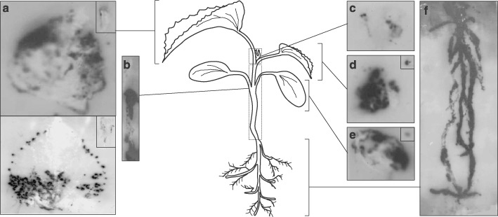
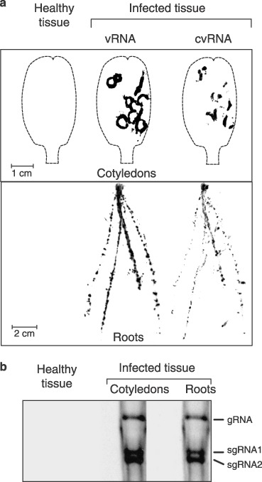
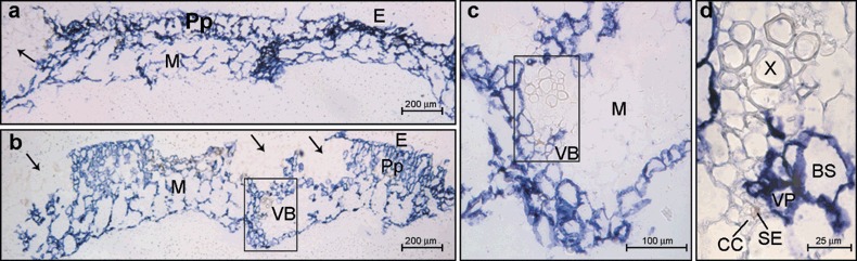
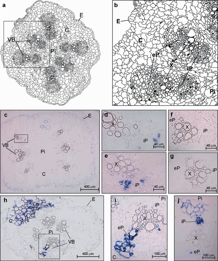
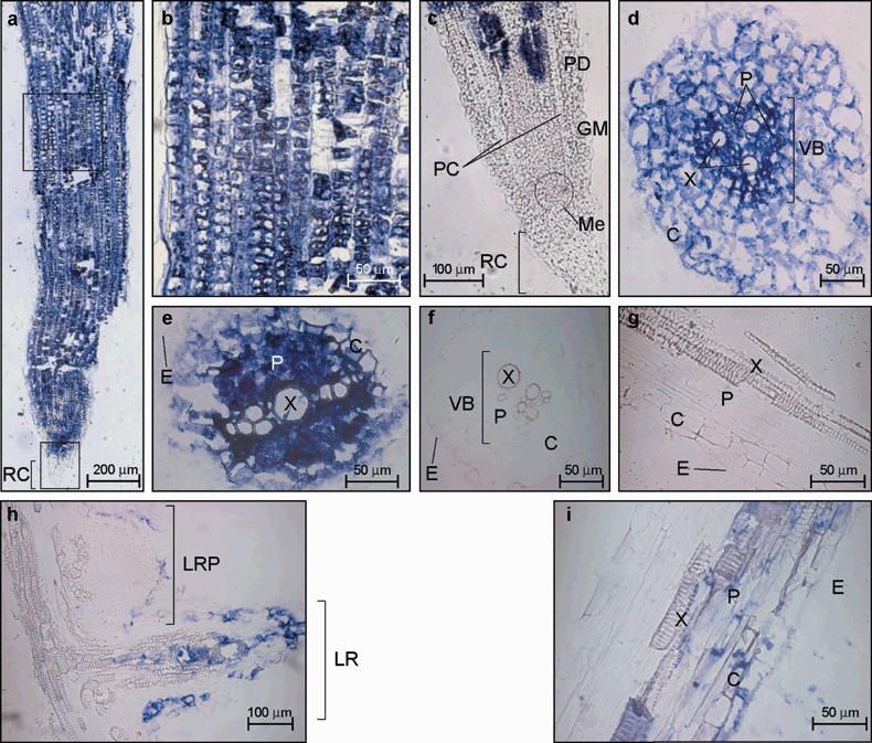
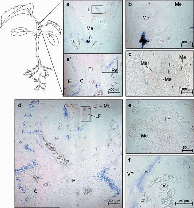
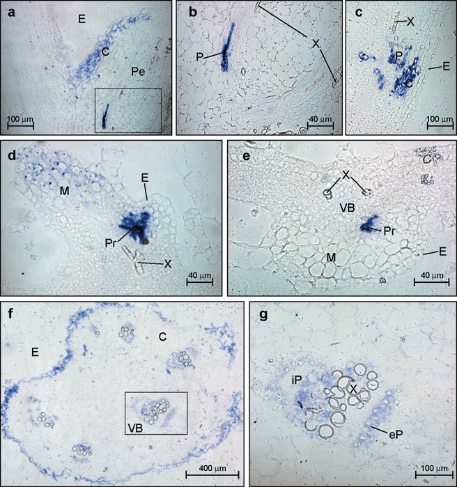
The translocation of Melon necrotic spot virus (MNSV) within tissues of inoculated and systemically infected Cucumis melo L. 'Galia' was studied by tissue-printing and in situ hybridization techniques. The results were compatible with the phloem vascular components being used to spread MNSV systemically by the same assimilate transport route that runs from source to sink organs. Virus RNAs were shown to move from the inoculated cotyledon toward the hypocotyl and root system via the external phloem, whereas the upward spread through the stem to the young tissues took place via the internal phloem. Virus infection was absent from non-inoculated source tissues as well as from both shoot and root apical meristems, but active sink tissues such as the young leaves and root system were highly infected. Finally, our results suggest that the MNSV invasion of roots is due to virus replication although a destination-selective process is probably necessary to explain the high levels of virus accumulation in roots. This efficient invasion of the root system is discussed in terms of natural transmission of MNSV by the soil-borne fungal vector.


Figures

Figure 1
Figure 1
Analysis of the spatial distribution of MNSV in infected melon plants (8 dpi) by tissue printing hybridization of (a) first systemic leaves (developed leaves), (b) hypocotyl and main stem, (c) shoot tip, (d) second systemic leaf (young leaf), (e) MNSV‐inoculated cotyledon and (f) roots. Either longitudinal or cross‐section impressions of the petioles from both developed or young leaves and cotyledons, respectively, are displayed in the insets located in the top right of the corresponding panel. A diagram of the melon plant in the developmental stage when the samples were taken is displayed at the centre of the figure to better understand the origin of each panel. Tissue‐printing hybridization was performed using a digoxigenin‐labelled RNA probe for detecting MNSV vRNA as described in the Experimental Procedures. Five different plants were analysed but only those results clearly representative are shown.
Figure 2
Figure 2
(a) Detection of either vRNA or cvRNA in both cotyledon and roots from MNSV‐infected melon plants (10 dpi) by tissue printing hybridization. Impressions of healthy tissues hybridized using the vRNA complementary riboprobe are shown. The contour of the cotyledons is shown by a broken line. (b) Northern‐blot analysis of the dsRNAs isolated either from the cotyledons or from the roots of MNSV‐infected melon plants and hybridized against an MNSV vRNA complementary riboprobe. A negative control from healthy tissue (cotyledons and roots mix) is shown. The position of the MNSV gRNA and both sgRNAs is indicated on the right.
Figure 3
Figure 3
Microscopic imaging of the cellular localization of Melon necrotic virus (MNSV) RNAs by in situ RNA–RNA hybridization with a cvRNA riboprobe in cotyledons of systemically infected melon plants (6 dpi). The blue colour indicates the presence of target viral RNAs. (a,b) Cross‐sections of symptomatic and asymptomatic areas from two different cotyledons showing the viral‐specific blue signal in: epidermal cells, E; palisade parenchyma, Pp; and mesophyll cells, M. Black arrows point to the uninfected areas of the adaxial face of the cotyledon. The area inside the black box in panel (b) is enlarged in panel (c) to show a closer view of the cotyledon vascular bundle (VB). Panel (d) is a higher magnification of the same vascular bundle from panel (c) showing that vascular parenchyma (VP) and bundle sheath (BS) cells were infected but not the companion cells–sieve elements (CC–SE) complex.
Figure 4
Figure 4
(a) Schematic representation of a cross‐section of the melon stem showing six vascular bundles (VB), the pith (Pi), the cortex (C) and the epidermis (E). (b) Diagram from the area enclosed by the black box in (a) showing an enlarged view of the vascular anatomy. The components of VB are designated as external, eP, and internal, iP, fascicular phloem, respectively and X, xylem. (c–j) Microscopic imaging of the cellular localization of the Melon necrotic spot virus (MNSV) RNAs by in situ RNA–RNA hybridization with a cvRNA riboprobe in transport tissues of systemically infected melon plants (6 dpi). The blue colour indicates the presence of target viral RNAs. (c) Internodal cross‐section of the stem region above the cotyledonary node showing MNSV RNA‐specific signals in cortical (C), and vascular bundle (VB) cells. The VB enclosed in the black box in panel (c) is enlarged in panel (d). (e) A closer view of another vascular bundle from a different cross‐section of the stem. (f,g) Two vascular bundles from different stem cross‐sections of healthy melon plant. (h) Hypocotyl cross‐section with only four vascular bundles showing the MNSV RNA‐specific signals in cortex (C) and vascular bundle (VB) cells. Panel (i) is an enlarged image of the vascular bundle enclosed by the black box in panel (h). (j) Vascular bundle from a hypocotyl cross‐section at 10 dpi where iP shows high hybridization levels.
Figure 5
Figure 5
Microscopic imaging of the cellular localization of the Melon necrotic spot virus (MNSV) RNAs by in situ RNA–RNA hybridization with a cvRNA riboprobe in both the primary root tip (panels a–e) and lateral roots (panels h and i) of systemically infected melon plants. The blue colour indicates the presence of target viral RNAs. (a) Longitudinal section of a primary root tip. RC, root cap. (b) Enlarged image of the distal root‐tip sector enclosed by the upper black box from the longitudinal section of panel (a) showing discrete uninfected cells. (c) Longitudinal section through the central axis of a primary root tip (lower black box in panel (a)). MNSV invasion was not allowed at RC and primary meristem including protoderm (PD), ground meristem (GM), procambium (PC) and apical meristem (Me). (d) Cross‐section of the zone of cell elongation of primary root tip. (e) Cross‐section of the zone of cell division of primary root tip. (f,g) Cross‐section and longitudinal section of a primary root tip from healthy tissue. (h) Longitudinal section of both a lateral root primordial (LRP) and an elongated lateral root (LR). (i) Enlarged image of a longitudinal section of a lateral root. VB, vascular cylinder; E, epidermis; C, cortex; X, xylem; P, phloem.
Figure 6
Figure 6
Microscopic imaging of the cellular localization of the Melon necrotic spot virus (MNSV) RNAs by in situ RNA–RNA hybridization with a cvRNA riboprobe in the shoot tip of systemically infected melon plants. The blue colour indicates the presence of target viral RNAs. (a,a′) Longitudinal section of the apical shoot tip showing the cortex (C), the stem pith (Pi), the insertion point of the young leaf petiole (Pe), an immature leaf (IL) and an apical meristem (Me) at 6 dpi. The meristem in panel (a) is enlarged in panel (b). The areas enclosed by both the lower and upper black boxes are enlarged in Fig. 7 in panels (a) and (d), respectively. (c) Longitudinal section of the multi‐meristematic apical shoot tip from a healthy melon plant. (d) Longitudinal section of the apical shoot tip at 10 dpi. Enlarged images of the apical meristem and the leaf primordia (LP) (black box) as well as a vascular bundle showing the MNSV infection into phloem (P) and vascular parenchyma (VP) are displayed on panels (e) and (f), respectively.
Figure 7
Figure 7
Microscopic imaging of the cellular localization of the Melon necrotic spot virus (MNSV) RNAs by in situ RNA–RNA hybridization with a cvRNA riboprobe in both the petiole of a young leaf (panels a–c), an immature leaf (panels d and e) and a petiole of a developed leaf (panels f and g) of systemically infected melon plants. The blue colour indicates the presence of target viral RNAs. (a) Longitudinal section of the young leaf petiole (Pe) at the stem insertion point showing MNSV infection (blue signal) at the epidermis (E) and adjacent cortical (C) cells. (b) A closer view of a vascular bundle (VB) showing MNSV infection on phloem (P) but not in xylem (X) components. (c) Longitudinal section of the young leaf petiole (Pe) at distal region from their stem insertion point. (d,e) Longitudinal and cross‐sections, respectively, of an immature leaf showing MNSV infection on immature epidermis and mesophyll cells (M) as well as in the protophloem (Pr). X, xylem. (f) Cross‐section of a developed leaf petiole taken close to the leaf insertion point. (g) A closer view of the vascular bundle inside the box in panel (f). The components of VB are designated as external, eP, and internal, iP, fascicular phloem, respectively.

References

    1. Al‐Kaff, N.S. and Covey, S.N. (1996) Unusual accumulations of Cauliflower mosaic virus in local lesions, dark green leaf tissue, and roots of infected plants. Mol. Plant–Microbe Interact., 9, 357–363.
    1. Andrianifahanana, M. , Lovins, K. , Dute, R. , Sikora, E. and Murphy, J.F. (1997) Pathway for phloem‐dependent movement of Pepper mottle potyvirus in the stem of Capsicum annuum . Phytopathology, 87, 892–898. - PubMed
    1. Aoki, K. , Suzui, N. , Fujimaki, S. , Dohmae, N. , Yonekura‐Sakakibara, K. , Fujiwara, T. , Hayashi, H. , Yamaya, T. and Sakakibara, H. (2005) Destination‐selective long‐distance movement of phloem proteins. Plant Cell, 17, 1801–1814. - PMC - PubMed
    1. Appiano, A. and D’Agostino, G. (1983) Distribution of Tomato bushy stunt virus in root tips of systemically infected Gomphrena globosa . J. Ultrastruct. Res. 85, 239–248. - PubMed
    1. Astruc, N. , Marcos, J.F. , Macquaire, G. , Candresse, T. and Pallas, V. (1996) Studies on the diagnosis of hop stunt viroid in fruit trees: identification of new hosts and application of a nucleic acid extraction procedure based on non‐organic solvents. Eur. J. Plant Pathol. 102, 837–846

Publication types

LinkOut - more resources