. 2008 Jul;9(4):463-78.
doi: 10.1111/j.1364-3703.2008.00479.x.

Histological and molecular analysis of Rdg2a barley resistance to leaf stripe

Affiliations

Histological and molecular analysis of Rdg2a barley resistance to leaf stripe

Anita Haegi et al. Mol Plant Pathol. 2008 Jul.

Abstract

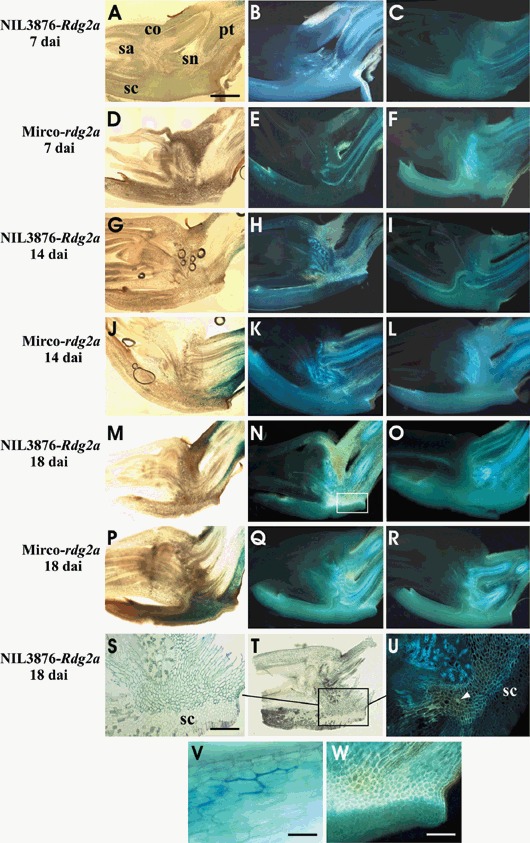
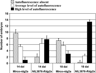
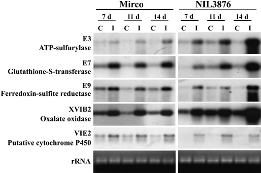
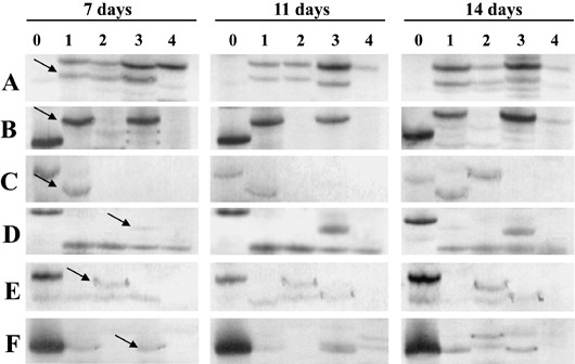
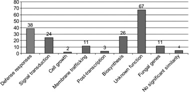
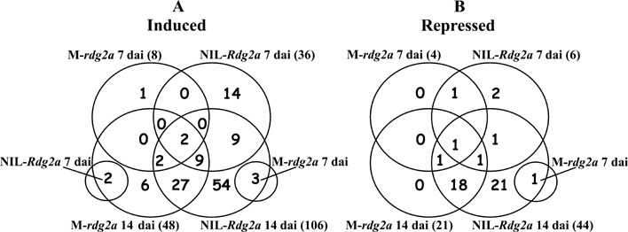
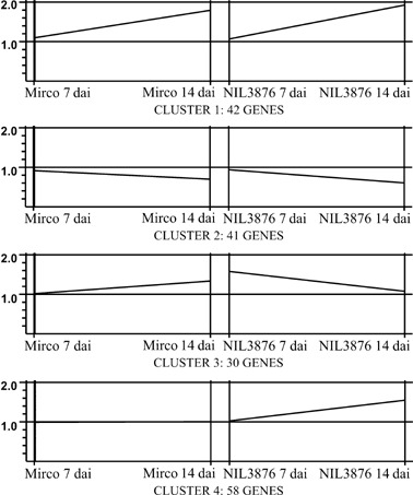
Barley (Hordeum vulgare L.) leaf stripe is caused by the seed-borne fungus Pyrenophora graminea. We investigated microscopically and molecularly the reaction of barley embryos to leaf stripe inoculation. In the resistant genotype NIL3876-Rdg2a, fungal growth ceased at the scutellar node of the embryo, while in the susceptible near-isogenic line (NIL) Mirco-rdg2a fungal growth continued past the scutellar node and into the embryo. Pathogen-challenged embryos of resistant and susceptible NILs showed different levels of UV autofluorescence and toluidine blue staining, indicating differential accumulation of phenolic compounds. Suppression subtractive hybridization and cDNA amplified fragment-length polymorphism (AFLP) analyses of embryos identified P. graminea-induced and P. graminea-repressed barley genes. In addition, cDNA-AFLP analysis identified six pathogenicity-associated fungal genes expressed during barley infection but at low to undetectable levels during growth on artificial media. Microarrays representing the entire set of differentially expressed cDNA-AFLP fragments and 100 barley homologues of previously described defence-related genes were used to study gene expression changes at 7 and 14 days after inoculation in the resistant and susceptible NILs. A total of 171 significantly modulated barley genes were identified and assigned to four groups based on timing and genotype dependence of expression. Analysis of the changes in gene expression during the barley resistance response to leaf stripe suggests that the Rdg2a-mediated response includes cell-wall reinforcement, signal transduction, generation of reactive oxygen species, cell protection, jasmonate signalling and expression of plant effector genes. The identification of genes showing leaf stripe inoculation or resistance-dependent expression sets the stage for further dissection of the resistance response of barley embryo cells to leaf stripe.


Figures

Figure 1
Figure 1
Barley embryo sections indicating P. graminea growth and defence reactions. The transgenic P. graminea Dg2‐GUS isolate containing the β‐glucuronidase transgene was used, allowing detection of fungal colonization by (blue) GUS staining. A, D, G, J, M and P are bright‐field views of GUS‐stained sections from NIL3876‐Rdg2a and from Mirco‐rdg2a at the time points of inoculation indicated. A close‐up view of the Mirco‐rdg2a embryo at 14 dai (V) shows blue GUS staining of fungal mycelium. The same sections were also observed by UV epifluorescence microscopy (B, E, H, K, N, and Q) for NIL3876‐Rdg2a and for Mirco‐rdg2a at the time points of inoculation indicated. C, F, I, L, O and R show epifluorescence views of sections of non‐inoculated control embryos of NIL3876‐Rdg2a and Mirco‐rdg2a harvested at the same time points as the inoculated samples. S and T show high‐ and low‐magnification views of an inoculated NIL3876‐Rdg2a embryo treated with Toluidine Blue O, which stains phenolic compounds blue‐green. U is the same view as in S except observed under UV light, illustrating co‐localization of pathogen‐induced UV autofluorescence with Toluidine Blue O staining. W is a magnified view of the boxed area in N, illustrating cell‐wall localization of autofluorescence. Scale bars represent 1 mm (A), 300 µm (S), 50 µm (V) and 200 µm (W). Arrowhead indicates a cell showing whole‐cell autofluorescence. co = coleoptile; pt = provascular tissue; sa = shoot apex; sc = scutellum; sn = scutellar node.
Figure 2
Figure 2
Quantification of UV autofluorescence of resistant and susceptible embryos inoculated with P. graminea isolate Dg2. Embryos of Mirco‐rdg2a and NIL3876‐Rdg2a were observed by epifluorescence microscopy at 14 and 18 dai and assigned to categories according to their level of autofluorescence. Shown are mean and SD obtained from three biological replicates comprising 16 embryos each per genotype and time point.
Figure 3
Figure 3
Northern blot analysis of genes induced in barley embryos by P. graminea inoculation. The genes used as probes had been identified by SSH and were those subsequently confirmed by Northern analysis to be induced. Blots contain mRNAs from inoculated (I) or non‐inoculated control (C) embryos of susceptible Mirco‐rdg2a or resistant NIL3876‐Rdg2a, harvested after 7, 11 or 14 days. Ethidium bromide‐stained gel images (bottom panels) show RNA loading.
Figure 4
Figure 4
cDNA‐AFLP profiles illustrating expression patterns influenced by barley inoculation with P. graminea. Samples were collected from fungal mycelium grown on PDA (0) or from embryos at different time‐points after inoculation (7, 11 and 14 days), from NIL3876‐Rdg2a inoculated (1) or non inoculated (2) with leaf stripe or from Mirco‐rdg2a inoculated (3) or non‐inoculated (4) with leaf stripe. The six primer combinations (A, B, C, D, E and F) provide examples of different expression patterns as explained in the text. Arrows indicate cDNA‐AFLP bands eluted from the gels.
Figure 7
Figure 7
Functional characterization of significantly modulated genes. Genes showing significant (P < 0.05) induction/repression on microarrays based on three biological replicates and a ratio of at least 1.3 relative to untreated controls were assigned to functional classes based on homology (e value > 10e‐6) to characterized sequences in the GenBank (nr) and EST (dbEST) databases.
Figure 5
Figure 5
Venn diagrams showing numbers of barley genes shown by the microarray analysis to be significantly induced (A) or repressed (B) in barley embryos after inoculation with P. graminea for various combinations of genotypes and time‐points. The genotypes are Mirco‐rdg2a (M‐rdg2a) and NIL3876‐Rdg2a (NIL‐Rdg2a), and the time points are 7 and 14 dai. For each individual genotype/time‐point combination, the total number of induced/repressed genes is shown in parentheses.
Figure 6
Figure 6
Cluster analysis. The 171 genes significantly regulated (> 1.3‐fold induced relative to untreated controls; P < 0.05) in barley embryos were classified into four different expression profiles. The x‐axes show the genotypes and the infection time‐points. The y‐axes show the normalized signal intensity ratios (global mean fold change) between infected and control samples.
Figure 8
Figure 8
RT‐PCR expression analysis of fungal genes. mRNA was extracted from P. graminea Dg2 fungus that was grown on PDA medium (F) and from Mirco‐rdg2a and NIL3876‐Rdg2a barley embryos inoculated with Dg2 and collected at 7, 11 and 14 dai. The fungal gene CAT1 (ubiquitin) was used to adjust input template amounts so that templates used for each TGG10:2, CAC15, CTC14, TGC57, CCC81 and TCC38 RT‐PCR reaction contained equal amounts of CAT1 transcript (see Experimental procedures).

Similar articles

Cited by

References

    1. Altschul, S.F. , Madden, T.L. , Schäffer, A.A. , Zhang, J. , Zhang, Z. , Miller, W. and Lipmann, D.J. (1997) Gapped BLAST and PSI‐BLAST: a new generation of protein database search programs. Nucleic Acids Res. 25, 3389–3402. - PMC - PubMed
    1. Anthony, J.R. , Warczak, K.L. and Donohue, T.J. (2005) A transcriptional response to singlet oxygen, a toxic byproduct of photosynthesis. Proc. Natl Acad. Sci. USA, 102, 6502–6507. - PMC - PubMed
    1. Aragona, M. and Porta‐Puglia, A. (1999) Identification of resistance to leaf stripe using a Pyrenophora graminea transformant expressing β‐glucuronidase. Eur. J. Plant Pathol. 105, 831–834.
    1. Arru, L. , Francia, E. and Pecchioni, N. (2003) Isolate‐specific QTLs of resistance to leaf stripe (Pyrenophora graminea) in the Steptoe × Morex spring barley cross. Theor. Appl. Genet. 106, 668–675. - PubMed
    1. Arru, L. , Nicks, R.E. , Lindhout, P. , Valè, G. , Francia, E. and Pecchioni, N. (2002) Genomic regions determining resistance to leaf stripe (Pyrenophora graminea) in barley. Genome, 45, 460–466. - PubMed

Publication types

MeSH terms