. 2008;10(4):R91.
doi: 10.1186/ar2475. Epub 2008 Aug 15.

Interferon-induced protein IFIT4 is associated with systemic lupus erythematosus and promotes differentiation of monocytes into dendritic cell-like cells

Affiliations

Interferon-induced protein IFIT4 is associated with systemic lupus erythematosus and promotes differentiation of monocytes into dendritic cell-like cells

Xiangyang Huang et al. Arthritis Res Ther. 2008.

Abstract

Introduction: Using oligonucleotide microarray, many IFN-inducible genes have been found to be highly expressed in peripheral blood mononuclear cells (PBMCs) from most patients with systemic lupus erythematosus (SLE). Among these IFN-inducible genes, IFN-induced protein with tetratricopeptide repeats 4 (IFIT4) is a novel gene whose function is unknown.

Methods: In this study we examined the role played by IFIT4 in monocyte differentiation and the correlation between IFIT4 expression and the clinical manifestation of SLE. To this end, we used plasmid transfection, flow cytometry, mixed leucocyte responses, ELISA, quantitative RT-PCR and Western blotting.

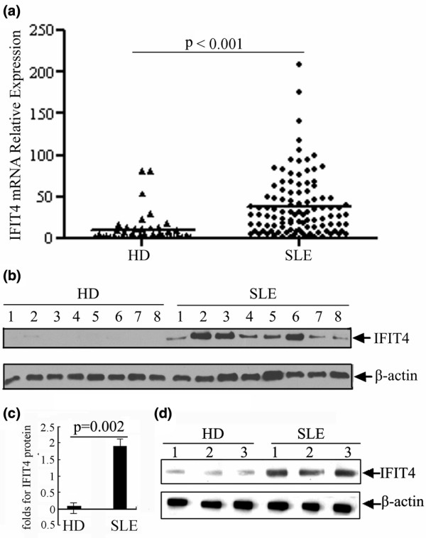
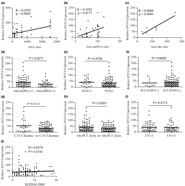
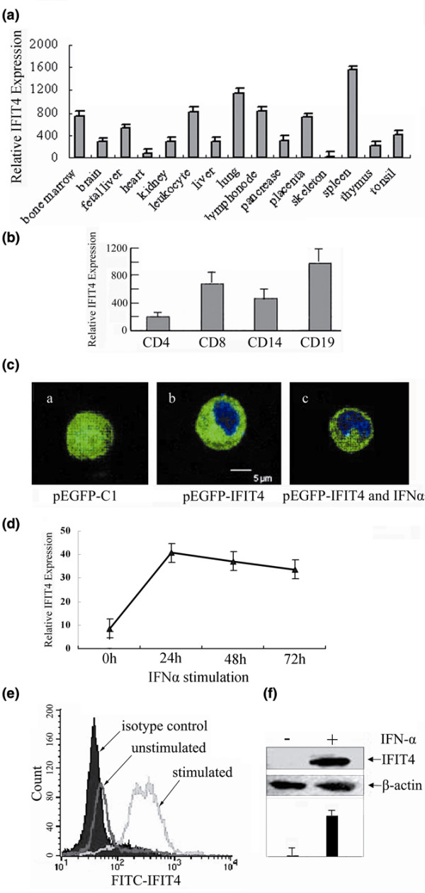
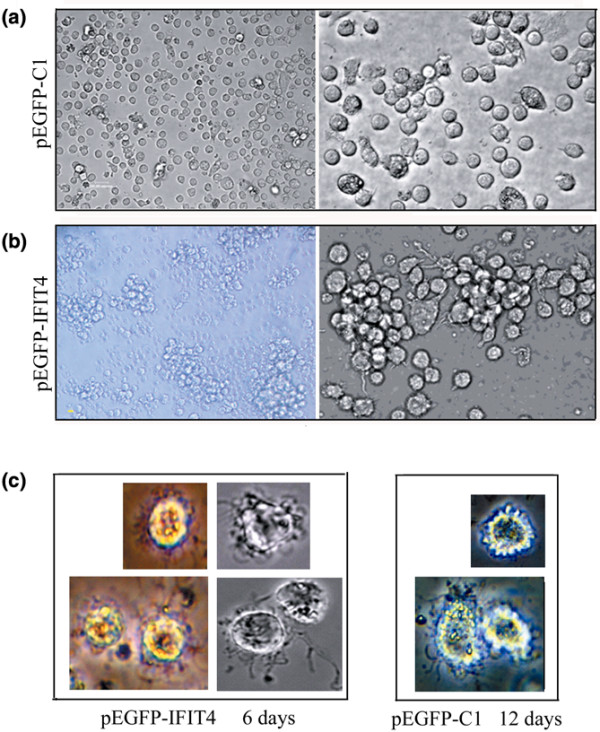
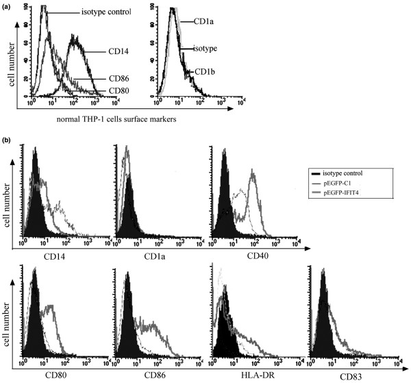
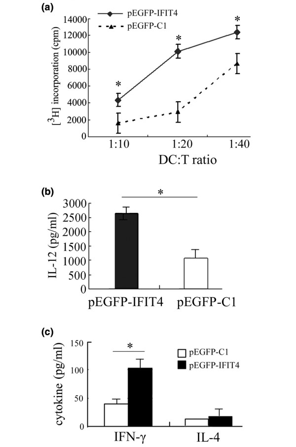
Results: We found that both IFIT4 mRNA and protein expression levels were significantly higher in PBMCs and monocytes from SLE patients than in those from healthy control individuals. IFIT4 expression was positively correlated with antinuclear antibodies, anti-double-stranded DNA, and anti-Sm auto-immune antibodies in SLE. Patients with SLE exhibiting higher expression of IFIT4 had a higher prevalence of leucopenia, thrombocytopenia and C3/C4 decrease. IFIT4 protein was localized exclusively to the cytoplasm, and it was significantly upregulated by IFN-alpha in normal PBMCs. To determine the role played by IFIT4 in monocyte differentiation, the monocytic cell line THP-1 was transfected with pEGFP-IFIT4 expression plasmid and stimulated with granulocyte-macrophage colony-stimulating factor/IL-4 to generate IFIT4-primed dendritic cell-like cells (DCLCs). IFIT4-primed DCLCs acquired morphological characteristics of dendritic cells more quickly, with greater resemblance to dendritic cells, as compared with DCLCs primed with pEGFP-C1 control plasmid trasfection. Furthermore, they exhibited higher expressions of CD40, CD86, CD80, HLA-DR and CD83, along with lower expression of CD14; increased IL-12 secretion; and an increased ability to stimulate T-cell proliferation. In addition, IFIT4-primed DCLCs enhanced IFN-gamma secretion (about 2.4-fold) by T cells compared with controls.

Conclusion: Our findings suggest that IFIT4 might play roles in promoting monocyte differentiation into DCLCs and in directing DCLCs to modulate T-helper-1 cell differentiation; these actions might contribute to the autoimmunity and pathogenesis of SLE.


Figures

Figure 1
Figure 1
Expression levels of IFIT4 in patients with SLE and healthy control individuals. (a) Total RNA from the peripheral blood mononuclear cells (PBMCs) of 108 systemic lupus erythematosus (SLE) patients and 46 healthy donor (HDs) was isolated, and the relative expression level of IFIT4 mRNA was determined by real-time quantitative RT-PCR. Glyceraldehyde-3-phosphate dehydrogenase (GAPDH) was co-amplified as an internal control to normalize the amount of IFIT4 mRNA. All experiments were repeated three times with similar results. Horizontal lines indicate the mean (P < 0.001, Mann-Whitney test). (b,c) Total protein was extracted from the PBMCs of 24 SLE patients and 24 HD control individuals. IFIT4 protein expression levels were detected using Western blotting. β-Actin was used as a protein loading control. A set of random data from eight SLE patients and eight HD controls is presented (P = 0.002, Mann-Whitney test). (d) Monocytes from three SLE patients and three HDs were isolated using magnetic beads. The IFIT4 protein expression level in these monocytes was determined by Western blotting. IFIT4, interferon-induced protein with tetratricopeptide repeats 4.
Figure 2
Figure 2
Correlation analysis between IFIT4 expression and clinical assessments in SLE patients. Total RNA from the peripheral blood mononuclear cells (PBMCs) of 108 systemic lupus erythematosus (SLE) patients and 46 healthy donors (HDs) was isolated, and the relative expression levels of IFIT4 mRNA were determined using real-time quantitative RT-PCR. Glyceraldehyde-3-phosphate dehydrogenase (GAPDH) was coamplified as an internal control to normalize the amount of RNA. In the total SLE population, the relative expression of IFIT4 was plotted against the following: (a) the titre of antinuclear antibody (ANA) in a group of 108 SLE patients; (b) the titre of anti-double-stranded DNA antibody (anti-dsDNA) in 36 SLE patients; and (c) the titre of anti-Smith antibody (anti-Sm) in 7 SLE patients. Spearman's correlation test was used to analyze these data. In the SLE population as a whole, the relative expression of IFIT4 was determined using real-time quantitative RT-PCR in patients who were positive or negative for the following: (d) anti-dsDNA antibody; (e) anti-SSA antibody; (f) anti-cardiolipid antibody or β-GP1 (ACL/b-GP1); (g) hypocomplementaemia (C3/C4 decrease); (h) haematocytopenia (wbc/PLT decrease); and (i) lupus nephritis. Mann-Whitney test was used to analyze these data. (j) The relative expression of IFIT4 was plotted against Systemic Lupus Erythematosus Disease Activity Index-2,000 (SLEDAI-2000) and analyzed usingy Spearman's test. IFIT4, interferon-induced protein with tetratricopeptide repeats 4.
Figure 3
Figure 3
Distribution of IFIT4 in tissues and cells, and the effect of IFN-α on IFIT4 expression. Using real-time quantitative RT-PCR, IFIT4 mRNA relative expression was determined among (a) 14 normal human tissues and (b) four kinds of immune cells. To determine the subcellular location of IFIT4 protein, THP-1 cells were transfected with (c) (subpanel a) pEGFP-C1 control or (subpanel b) pEGFP-IFIT4 plasmid. Forty-eight hours later, cells were stained with DAPI for nuclear staining and examined by confocal microscopy. The effect of IFN-α2a on the localization of IFIT4 protein was also observed. (c) (subpanel c) THP-1 cells transfected with pEGFP-IFIT4 were further stimulated with 3,000 μ/ml IFN-α2a for 72 hours. Blue colour shows the location of the nucleus, whereas green colour shows the sublocalization of green fluorescent protein alone or fused with IFIT4 protein. (d) To analyze the effect of IFN-α on the expression level of IFIT4, peripheral blood mononuclear cells (PBMCs) from healthy donors were treated with 3,000 μ/ml IFN-α2a for 24 hours, 48 hours or 72 hours, and then IFIT4 mRNA in the PBMCs was detected by real-time quatitative RT-PCR; all experiments were repeated three times with similar results. IFIT4 protein levels from normal PBMCs treated with IFN-α2a for 72 hours was examined by (e) flow cytometry with intracellular staining or (f) Western blotting with β-actin as a protein loading control. The experiments were performed three times and a set of representative histograms and data is presented. IFIT4, interferon-induced protein with tetratricopeptide repeats 4.
Figure 4
Figure 4
Morphological comparison of DCLCs primed with or without IFIT4 over-expression by inverted microscope. Cell morphology changes were observed to evaluate the effects of IFIT4 on dendritic cell (DC) differentiation upon treatment with granulocyte-macrophage colony-stimulating factor (GM-CSF; 50 ng/ml) or IL-4 (20 ng/ml). (a) THP-1 cells were transfected with pEGFP-C1; 36 hours later cells were further stimulated with GM-CSF/IL-4 for 86 hours (left lane: ×20; right lane: ×40). (b) THP-1 cells were transfected with pEGFP-IFIT4 fusion plasmid; 36 hours later cells were further stimulated with GM-CSF/IL-4 for 48 hours (left lane: ×20; right lane: ×40). (c) Effect of IFIT4 on morphological changes that occured in monocytes differentiation into mature and activated DC-like cells (DCLCs). THP-1 cells were transfected with pEGFP-IFIT4 (left lane) or pEGFP-C1 (right lane) plasmid; 36 hours later cells were stimulated with GM-CSF/IL-4 for 6 days (left lane) or 12 days (right lane) followed by lipopolysaccharide (LPS; 100 ng/ml) for another 2 days. All the cells above were observed with an inverted microscope. More than three fields of view (containing ≥100 cells/field) per sample were examined. IFIT4, interferon-induced protein with tetratricopeptide repeats 4.
Figure 5
Figure 5
Comparison of the phenotypic profiles of DCLCs primed with or without IFIT4 over-expression. (a) Surface antigens of normal THP-1 cells were examined by flow cytometry. (b) To analyze the effect of IFIT4 on phenotypic changes of dendritic cell-like cells (DCLCs) during the process of differentiation, THP-1 cells were transfected with pEGFP-IFIT4 or pEGFP-C1; 36 hours later, cells were stimulated with granulocyte-macrophage colony-stimulating factor (GM-CSF; 50 ng/ml) and IL-4 (20 ng/ml) for 90 hours to generate DCLCs. These DCLCs primed with pEGFP-IFIT4 or pEGFP-C1 transfection were incubated with fluorochrome-conjugated monoclonal antibodies (mAbs) and the antigens of CD40, CD80, CD86, CD83, HLA-DR, CD14 and CD1a on the surface of those DCLCs were analyzed by flow cytometry. Appropriate fluorochrome or isotype control mAbs of each antibody species were used as negative controls. Shaded histograms represent isotype control antibodies. The thick line represents DCLCs primed with pEGFP-IFIT4 transfection, whereas the slender lines represent DCLCs primed with pEGFP-C1 transfection. All experiments were performed three times and a set of representative histograms was presented. IFIT4, interferon-induced protein with tetratricopeptide repeats 4.
Figure 6
Figure 6
Functional analysis of IFIT4-primed DCLCs. Mature and activated dendritic cell-like cells (DCLCs) were generated from THP-1 cells transfected with pEGFP-IFIT4 or pEGFP-C1 and harvested after 6 days of culture with granulocyte-macrophage colony-stimulating factor (GM-CSF)/IL-4 plus 2 days of stimulation with lipopolysaccharide (LPS; 100 ng/ml). (a) Effect of IFIT4 on modulating the ability of DCLCs to present antigens to T cells was examined by allogeneic mixed leukocyte responses (MLRs). Allogeneic CD4+ T cells were cultured for 3 days with γ-irradiated mature DCLCs primed with pEGFP-C1 or pEGFP-ITFIT4 transfection at different ratios of DCLCs to T cells (1:10, 1:20 and 1:40). The cells were harvested and the incorporated radioactivity was measured using a beta counter. Responses are reported as the mean of triplicate samples (counts/minute [cpm] ± standard deviation). (b) Effect of IFIT4 on IL-12 production by DCLCs. Mature and activated DCLCs were generated as described above. Supernatant from the DCLCs (1 × 10⁵ cells/ml) primed with pEGFP-C1 or pEGFP-ITFIT4 transfection was analyzed for IL-12p70 by ELISA. (c) Analysis of the cytokine-production in T cells primed with co-stimulation by DCLCs. Mature and activated DCLCs primed with pEGFP-C1 or pEGFP-ITFIT4 transfection were generated as described above. Then these mature and activated DCLCs were γ-irradiated and cultured with T cells at a ratio of DCLCs to T cells of 1:10 for 6 days. Phorbol-12-myristate-13-acetate (PMA) (10 ng/ml) was added for another day. Finally, the culture medium was measured to assess IFN-γ and IL-4 produced by T cells by ELISA (T cells: 2 × 10⁵ cells/ml; DCLC to T cell ratio = 1:10). DCLCs primed with pEGFP-IFIT4 (shaded histogram, test group) or pEGFP-C1 (open histogram, control group) were compared with each other. The results are expressed as the mean ± standard deviation. Data represent the mean of triplicate experiments. The asterisk indicates a highly significant difference between the test group and the control (P < 0.05). IFIT4, interferon-induced protein with tetratricopeptide repeats 4.

Similar articles

Cited by

References

    1. Vakharia DD, Szebenyi SE, Gutterman JU, Rich SA. Interferon-alpha-induced human lupus inclusions and p36 protein in cancer and AIDS. J Interferon Cytokine Res. 1996;16:709–715. - PubMed
    1. Blanco P, Palucka AK, Gill M, Pascual V, Banchereau J. Induction of dendritic cell differentiation by IFN-alpha in systemic lupus erythematosus. Science. 2001;294:1540–1543. doi: 10.1126/science.1064890. - DOI - PubMed
    1. Baechler EC, Batliwalla FM, Karypis G, Gaffney PM, Ortmann WA, Espe KJ, Shark KB, Grande WJ, Hughes KM, Kapur V, Gregersen PK, Behrens TW. Interferon-inducible gene expression signature in peripheral blood cells of patients with severe lupus. Proc Natl Acad Sci USA. 2003;100:2610–2615. doi: 10.1073/pnas.0337679100. - DOI - PMC - PubMed
    1. Feng X, Wu H, Grossman JM, Hanvivadhanakul P, FitzGerald JD, Park GS, Dong X, Chen W, Kim MH, Weng HH, Furst DE, Gorn A, McMahon M, Taylor M, Brahn E, Hahn BH, Tsao BP. Association of increased interferon-inducible gene expression with disease activity and lupus nephritis in patients with systemic lupus erythematosus. Arthritis Rheum. 2006;54:2951–2962. doi: 10.1002/art.22044. - DOI - PubMed
    1. Crow MK. Interferon pathway activation in systemic lupus erythematosus. Curr Rheumatol Rep. 2005;7:463–468. doi: 10.1007/s11926-005-0053-4. - DOI - PubMed

Publication types

MeSH terms