

doi: 10.1186/1757-1626-1-97.
Ultrasound and MR-imaging in preoperative evaluation of two rare cases of scar endometriosis
Affiliations
- PMID: 18706122
- PMCID: PMC2533002
- DOI: 10.1186/1757-1626-1-97
Item in Clipboard
Ultrasound and MR-imaging in preoperative evaluation of two rare cases of scar endometriosis
Cases J.
.
Abstract
Scar or incisional endometriosis is a rare, often misdiagnosed, pathologic condition of the abdominal wall. Two cases of incisional endometriosis are presented. Both patients presented with atypical cyclic pain and palpable nodules on scars of previous cesarean sections. In both cases, the mass was totally excised, after accurate preoperative evaluation with 2-D ultrasound, power Doppler and MRI. Microscopic examination confirmed the preoperatively presumed diagnosis of cutaneous endometriosis. In cases of suspected scar endometriosis, preoperative diagnostic imaging is valuable in determining the extent of disease, thus enhancing accurate and total excision.
Figures
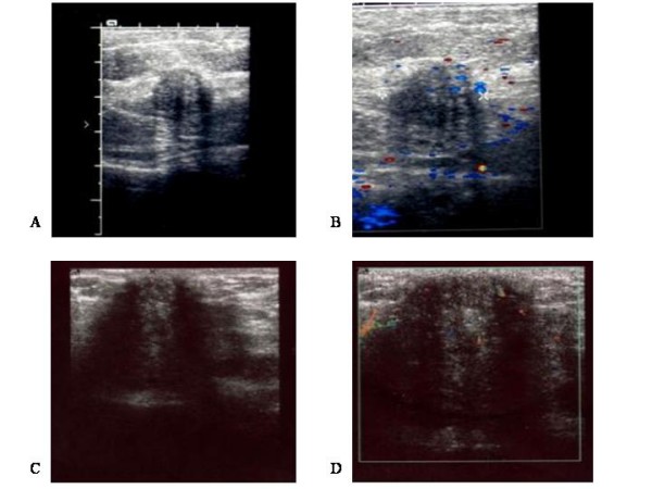
2-D and power Doppler sonographic views of scar endometriosis nodules. Using 7.5 MHz transducers, scar endometriotic lesions appeared hypoechoic, with internal echoes on 2-D ultrasound (A, C), and with internal vascularity on power Doppler (B, D). A and B: Case 1; C and D: Case 2.
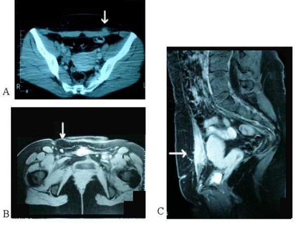
MRI of scar endometriosis nodules. Endometriotic lesions (arrows) appear isodense to muscle on transverse (A, B) and axial (C) T1-weighted spin-echo MRI. A: Case 1; B and C: Case 2.
References
-
- Zafrakas M, Tarlatzis BC, Streichert T, Pournaropoulos F, Wölfle U, Smeets SJ, Wittek B, Grimbizis G, Brakenhoff RH, Pantel K, Bontis J, Günes C. Genome-wide microarray gene expression, array-CGH analysis, and telomerase activity in advanced ovarian endometriosis: A high degree of differentiation rather than malignant potential. Int J Mol Med. 2008;21:335–344. - PubMed
LinkOut - more resources
Full Text Sources
