Clinical Trial
. 2009 Apr;12(3):305-16.
doi: 10.1017/S146114570800922X. Epub 2008 Aug 18.

Brain nicotinic acetylcholine receptor occupancy: effect of smoking a denicotinized cigarette

Affiliations
Clinical Trial

Brain nicotinic acetylcholine receptor occupancy: effect of smoking a denicotinized cigarette

Arthur L Brody et al. Int J Neuropsychopharmacol. 2009 Apr.

Abstract

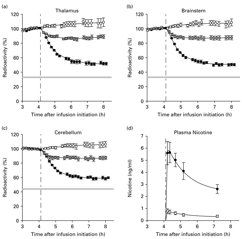
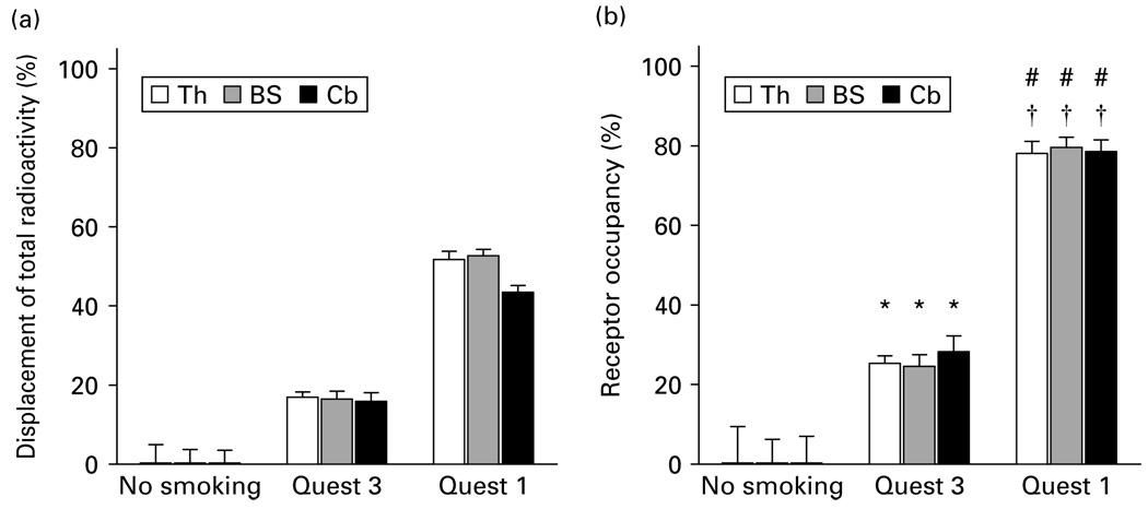
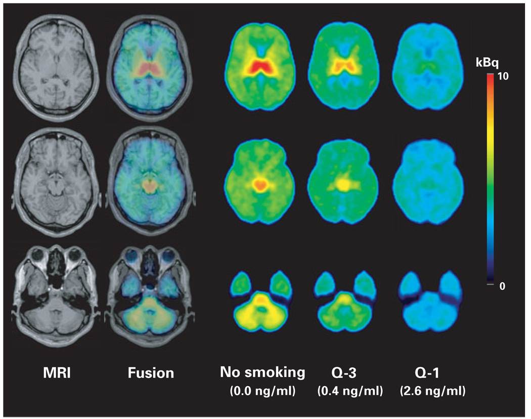
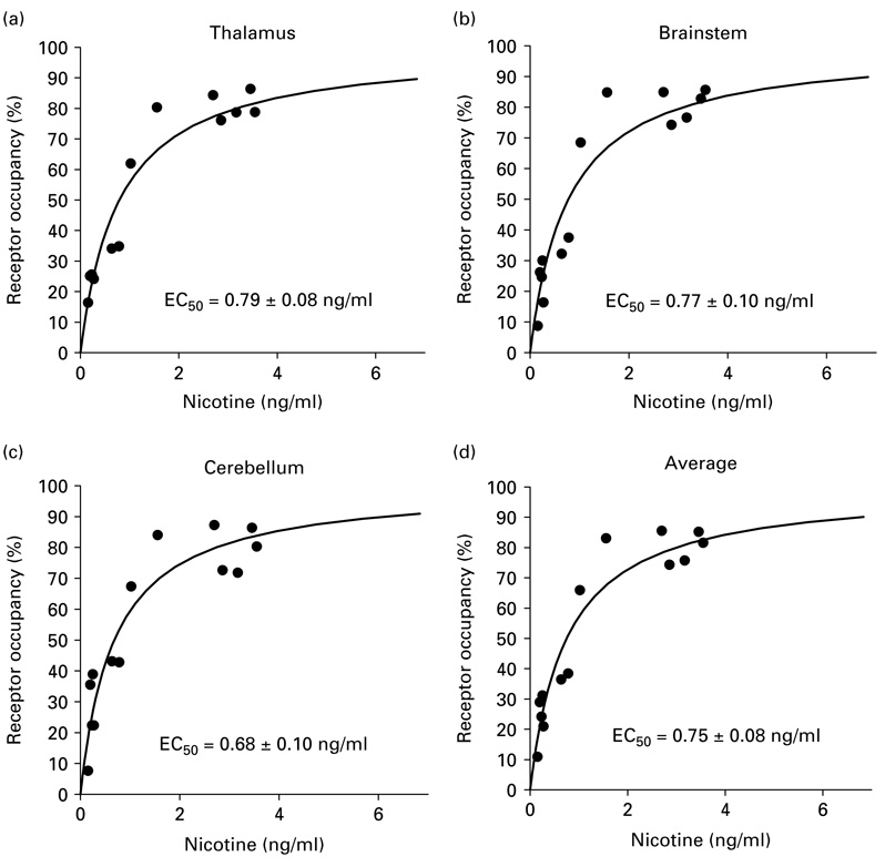
Our group recently reported that smoking a regular cigarette (1.2-1.4 mg nicotine) resulted in 88% occupancy of brain alpha4beta2* nicotinic acetylcholine receptors (nAChRs). However, this study did not determine whether nicotine inhalation or the many other pharmacological and behavioural factors that occur during smoking resulted in this receptor occupancy. If nicotine is solely responsible for alpha4beta2* nAChR occupancy from smoking, then (as estimated from our previous data) smoking a denicotinized (0.05 mg nicotine) or a low-nicotine (0.6 mg nicotine) cigarette (commonly used for research and clinical purposes) would result in substantial 23% and 78% alpha4beta2* nAChR occupancies, respectively, and a plasma nicotine concentration of 0.87 ng/ml would result in 50% alpha4beta2* nAChR occupancy (EC50). Twenty-four positron emission tomography sessions were performed on tobacco-dependent smokers, using 2-[F-18]fluoro-A-85380 (2-FA), a radiotracer that binds to alpha4beta2* nAChRs. 2-FA displacement was determined from before to 3.1 hours after either: no smoking, smoking a denicotinized cigarette, or smoking a low-nicotine cigarette. Analysis of this PET data revealed that smoking a denicotinized and a low-nicotine cigarette resulted in 26% and 79% alpha4beta2* nAChR occupancies, respectively, across three regions of interest. The EC50 determined from this dataset was 0.75 ng/ml. Given the consistency of findings between our previous study with regular cigarettes and the present study, nicotine inhalation during smoking appears to be solely responsible for alpha4beta2* nAChR occupancy, with other factors (if present at all) having either short-lived or very minor effects. Furthermore, smoking a denicotinized cigarette resulted in substantial nAChR occupancy.


Figures

Figure 1
Figure 1
(a)–(c) Time–activity curves demonstrating changes in percent radioactivity for the three regions of interest (thalamus, brainstem, and cerebellum) over time following the initiation of bolus + infusion of 2-FA. The white, grey, and black circles represent the mean values (±S.E.M.) for the scans with no smoking (n = 7), smoking of a denicotinized cigarette (0.05 mg nicotine yield) (n = 10), and smoking of a low-nicotine cigarette (0.6 mg nicotine yield) (n = 7), respectively. The horizontal grey lines in panels (a)–(c) represent the percent of maximum fractional displaceable radioactivity for each region, which were obtained from the asymptotic portions of the curves in figure 4 of our previous study in which subjects smoked regular cigarettes to satiety (Brody et al., 2006). (d) Changes in mean plasma nicotine levels (ng/ml) with time in subjects who smoked a low-nicotine (●) or denicotinized (○) cigarette.
Figure 2
Figure 2
(a) The mean (±S.E.M.) percent displacements of total radioactivity for the three smoking conditions [no smoking, Quest 3 (denicotinized cigarette), Quest 1 (low-nicotine cigarette)] for the three regions of interest (Th, thalamus, BS, brainstem, Cb, cerebellum) from before to 3.1 h after smoking. (b) The mean (±S.E.M.) receptor occupancies for the three smoking conditions for the three regions of interest (ROIs). The * symbol indicates a significance level of p < 0.05 for Student’s t test comparisons (following Bonferroni correction for the three ROIs) between the denicotinized cigarette and the no-smoking conditions. The # and † symbols indicate significance levels of p < 0.0001 for Student’s t test comparisons (following Bonferroni correction for the three ROIs) between the low-nicotine cigarette condition and the no-smoking and denicotinized cigarette conditions.
Figure 3
Figure 3
Radioactivity levels on mean positron emission tomography (PET) images 3.1 h after the smoke break in scanning. Column 1 shows a magnetic resonance imaging (MRI) scan with representative slices from three levels of interest for this study (row 1 at the level of the thalamus, row 2 at the level of the brainstem, and row 3 at the level of the cerebellum). Column 2 demonstrates a PET/MRI fusion used for transfer of drawn regions of interest from MRI to PET. Columns 3–5 show mean PET images from the three study groups [no smoking, Q-3 (denicotinized cigarette), Q-1 (low-nicotine cigarette)] 3.1 h after the smoking break in scanning, demonstrating radiotracer displacement for the two active smoking conditions, but not for the no-smoking condition. Mean plasma nicotine levels at the 3.1 h time-point are shown below columns 3–5.
Figure 4
Figure 4
(a)–(c) Graphs showing percent α4β2* nicotinic acetylcholine receptor occupancy as a function of plasma nicotine concentration (ng/ml) for the three regions of interest and (d) for the mean of these three regions. From these graphs, the effective plasma concentration of nicotine needed to occupy 50% of available receptors (EC50) was determined for the three regions separately and for the mean of the regions.

Similar articles

Cited by

References

    1. Benowitz NL, Porchet H, Jacob PD. Pharmacokinetics, metabolism, and pharmacodynamics of nicotine. In: Wonnacott S, Russell MAH, Stolerman IP, editors. Nicotine Psychopharmacology: Molecular, Cellular and Behavioral Effects. Oxford, UK: Oxford University Press; 1990.
    1. Brody AL, Mandelkern MA, London ED, Childress AR, Bota RG, Ho ML, Lee GS, Saxena S, Baxter LR, Madsen D, Jarvik ME. Brain metabolic changes during cigarette craving. Archives of General Psychiatry. 2002;59:1162–1172. - PubMed
    1. Brody AL, Mandelkern MA, London ED, Olmstead RE, Farahi J, Scheibal D, Jou J, Allen V, Tiongson E, Chefer SI, Koren AO, Mukhin AG. Cigarette smoking saturates brain alpha4beta2 nicotinic acetylcholine receptors. Archives of General Psychiatry. 2006;63:907–915. - PMC - PubMed
    1. Buchhalter AR, Schrinel L, Eissenberg T. Withdrawal-suppressing effects of a novel smoking system: comparison with own brand, not own brand, and de-nicotinized cigarettes. Nicotine & Tobacco Research. 2001;3:111–118. - PubMed
    1. Butschky MF, Bailey D, Henningfield JE, Pickworth WB. Smoking without nicotine delivery decreases withdrawal in 12-hour abstinent smokers. Pharmacology Biochemistry & Behavior. 1995;50:91–96. - PubMed

Publication types

MeSH terms