

Expression and localization of the novel and highly conserved gametocyte-specific factor 1 during oogenesis and spermatogenesis
- PMID: 18706558
- PMCID: PMC4852867
- DOI: 10.1016/j.fertnstert.2008.05.042
Expression and localization of the novel and highly conserved gametocyte-specific factor 1 during oogenesis and spermatogenesis
Abstract
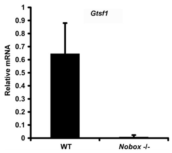
Objective: To determine the onset of gametocyte-specific factor 1 (Gtsf1) expression in embryogenesis and its relation to Nobox; and to determine its localization during gonadal development and gametocyte maturation.
Design: Developmental animal study.
Setting: University reproductive biology laboratory.
Animal(s): Mice ranging in age from embryonic day 12.5 to 8 weeks.
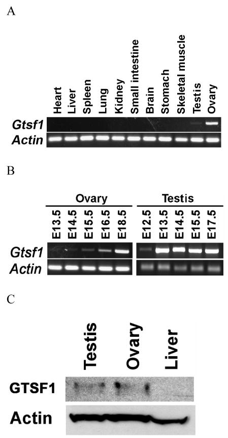
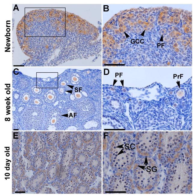
Intervention(s): Polymerase chain reaction and quantitative polymerase chain reaction were performed to determine the onset of and relative messenger RNA expression. Western blot was performed to confirm protein expression and antibody specificity. In situ hybridization and immunohistochemistry were used determine localization of expression.
Main outcome measure(s): Gtsf1 messenger RNA expression levels during embryogenesis through adulthood in wild-type mice and in newborn Nobox knockout mice; GTSF1 expression and localization in postnatal mice.
Result(s): Gtsf1 functions downstream of Nobox and is highly expressed in embryonic male and female gonads, localizing to germ cells throughout development. GTSF1 expression is confined to the cytoplasm in all stages of postnatal oocyte maturation and to prespermatogonia during early postnatal testicular development.
Conclusion(s): The expression pattern of Gtsf1 and its high conservation suggests that it may play an important role in germ cell development. Further characterization of Gtsf1 may elucidate mechanisms involved in premature ovarian failure.
Conflict of interest statement
None of the above authors has any potential conflict of interest, financial or otherwise.
Figures
Similar articles
-
Gtsf1/Cue110, a gene encoding a protein with two copies of a CHHC Zn-finger motif, is involved in spermatogenesis and retrotransposon suppression in murine testes.Dev Biol. 2009 Nov 1;335(1):216-27. doi: 10.1016/j.ydbio.2009.09.003. Epub 2009 Sep 6. Dev Biol. 2009. PMID: 19735653
-
Molecular characterization and expression profiles of cyclin B1, B2 and Cdc2 kinase during oogenesis and spermatogenesis in rainbow trout (Oncorhynchus mykiss).Anim Reprod Sci. 2008 May;105(3-4):209-25. doi: 10.1016/j.anireprosci.2007.03.005. Epub 2007 Mar 7. Anim Reprod Sci. 2008. PMID: 17399922
-
TOPAZ1, a novel germ cell-specific expressed gene conserved during evolution across vertebrates.PLoS One. 2011;6(11):e26950. doi: 10.1371/journal.pone.0026950. Epub 2011 Nov 1. PLoS One. 2011. PMID: 22069478 Free PMC article.
-
Expression analysis and evolutionary conservation of the mouse germ cell-specific D6Mm5e gene.Dev Dyn. 2006 Sep;235(9):2613-9. doi: 10.1002/dvdy.20907. Dev Dyn. 2006. PMID: 16881047
-
Transcriptional regulation of early oogenesis: in search of masters.Hum Reprod Update. 2006 Jan-Feb;12(1):65-76. doi: 10.1093/humupd/dmi033. Epub 2005 Sep 2. Hum Reprod Update. 2006. PMID: 16143663 Review.
Cited by
-
Sex differences in gene expression and alternative splicing in the Chinese horseshoe bat.PeerJ. 2023 Apr 24;11:e15231. doi: 10.7717/peerj.15231. eCollection 2023. PeerJ. 2023. PMID: 37123006 Free PMC article.
-
Asterix/Gtsf1 links tRNAs and piRNA silencing of retrotransposons.Cell Rep. 2021 Mar 30;34(13):108914. doi: 10.1016/j.celrep.2021.108914. Cell Rep. 2021. PMID: 33789107 Free PMC article.
-
GTSF1 gene may serve as a novel potential diagnostic biomarker for liver cancer.Oncol Lett. 2018 Mar;15(3):3133-3140. doi: 10.3892/ol.2017.7695. Epub 2017 Dec 27. Oncol Lett. 2018. PMID: 29435047 Free PMC article.
-
Postovulatory aging affects dynamics of mRNA, expression and localization of maternal effect proteins, spindle integrity and pericentromeric proteins in mouse oocytes.Hum Reprod. 2016 Jan;31(1):133-49. doi: 10.1093/humrep/dev279. Epub 2015 Nov 17. Hum Reprod. 2016. PMID: 26577303 Free PMC article.
-
Developmental roles and molecular mechanisms of Asterix/GTSF1.Wiley Interdiscip Rev RNA. 2022 Sep;13(5):e1716. doi: 10.1002/wrna.1716. Epub 2022 Feb 2. Wiley Interdiscip Rev RNA. 2022. PMID: 35108755 Free PMC article. Review.
References
-
- Lawson KA, Hage WJ. Clonal analysis of the origin of primordial germ cells in the mouse. Ciba Found Symp. 1994;182:68–84. discussion 84–91. - PubMed
-
- McClellan KA, Gosden R, Taketo T. Continuous loss of oocytes throughout meiotic prophase in the normal mouse ovary. Dev Biol. 2003;258:334–348. - PubMed
-
- Elvin JA, Yan C, Matzuk MM. Oocyte-expressed TGF-beta superfamily members in female fertility. Mol Cell Endocrinol. 2000;159:1–5. - PubMed
-
- Pepling ME, Spradling AC. Mouse ovarian germ cell cysts undergo programmed breakdown to form primordial follicles. Dev Biol. 2001;234:339–51. - PubMed
Publication types
MeSH terms
Substances
Grants and funding
LinkOut - more resources
Full Text Sources
Molecular Biology Databases
