Review
. 2008 Sep;18(9):430-42.
doi: 10.1016/j.tcb.2008.06.006. Epub 2008 Aug 15.

Decoding the quantitative nature of TGF-beta/Smad signaling

Affiliations
Review

Decoding the quantitative nature of TGF-beta/Smad signaling

David C Clarke et al. Trends Cell Biol. 2008 Sep.

Abstract

How transforming growth factor-beta (TGF-beta) signaling elicits diverse cell responses remains elusive, despite the major molecular components of the pathway being known. We contend that understanding TGF-beta biology requires mathematical models to decipher the quantitative nature of TGF-beta/Smad signaling and to account for its complexity. Here, we review mathematical models of TGF-beta superfamily signaling that predict how robustness is achieved in bone-morphogenetic-protein signaling in the Drosophila embryo, how changes in receptor-trafficking dynamics can be exploited by cancer cells and how the basic mechanisms of TGF-beta/Smad signaling conspire to promote Smad accumulation in the nucleus. These studies demonstrate the power of mathematical modeling for understanding TGF-beta biology.


Figures

Figure I
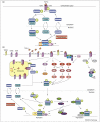
Figure I
A dynamic view of canonical TGF-β/Smad signaling. The abbreviations associated with the rate constants for the depicted reactions identify the process and in most cases, the species involved, separated by an underscore. Process abbreviations: a, association; deg, degradation; d, dissociation; exp, nuclear export; imp, nuclear import; int, internalization; kcat, enzyme turnover number; KM, Michaelis-Menten constant; prod, production; rec, recycling. Species abbreviations: TGF-β, transforming growth factor-β ligand; TβRI, TGF-β type I receptor; TβRII, TGF-β type II receptor; L-TβRII, ligand-TβRII complex; LRC, ligand-TβRII-TβRI heterotetrameric complex; M2, Smad2 or Smad3; M4, Smad4; Phosphatases, nuclear phosphatases (e.g. PPM1A/PP2C [1] and possibly others).
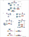
Figure 1
Figure 1
TGF-β/Smad signaling. (a) An overview of canonical TGF-β/Smad signaling. The TGF-β signal is delivered intracellularly by the TGF-β receptors and Smad transcription factors. (b) TGF-β activates non-canonical pathways and engages in crosstalk. Depicted examples include additional proteins that are putatively involved in canonical signaling, such as the TGF-β type III receptor (TβRIII) and SARA. In addition, TGF-β signaling interacts with other major cell-signaling pathways such as MAPK signaling (i.e. p38, Jnk and ERK), whereby TGF-β is able to activate MAPK signaling but also be modulated by it through phosphorylation of the Smad linker domain. TGF-β can also regulate non-Smad signaling such as RhoA GTPase degradation via activation of Par6. In the nucleus, multiple interactions occur, including repression of Smad signaling by SnoN, which is alleviated by the E3 ubiquitin ligase Arkadia, and additional signaling mediated by interactions between the phospho-R-Smads with other transcriptional regulators, such as ectodermin (Ecto), which is also known as transcriptional intermediary factor 1-γ (TIF1γ).
Figure 2
Figure 2
BMP signaling in Drosophila dorsal-ventral patterning. (a) Molecular-level events in BMP signaling at the dorsal surface of the Drosophila embryo. BMP ligands consist of homodimers of either Dpp or Scw or a heterodimer of Dpp and Scw. These ligands (generically represented with gray coloring) form ternary complexes with the proteins Sog and Tsg, which prevents binding of these ligands to their receptors. Tolloid (Tld), a metalloprotease, degrades Sog, the loss of which promotes dissociation of the complex and liberation of the BMP ligand and Tsg. Free Dpp can bind the type II receptor, Punt, and its type I receptor, Tkv, whereas Scw binds to Punt and another type I receptor, Sax. The active type I receptors within the receptor complex phosphorylate the Drosophila Smad1 homolog, Mad. Phospho-Mad forms a complex with the Smad4 homolog, Medea. The phospho-Mad-Medea complex shuttles into the nucleus and regulates the transcription of target genes. (b) Sites of production of the BMP ligands and putative extracellular modulators of BMP signaling. Represented is a transverse cross-section of the Drosophila embryo. Dpp, Scw, Tld, and Tsg are broadly expressed in the dorsal region whereas Sog is expressed ventro-laterally [53]. In the dorsal region, BMP receptors and Mad are expressed uniformly [53]. (c). A simplified view of the dynamics of the extracellular events in BMP signaling (for simplicity, only Dpp is only shown). Solid arrows indicate biochemical reactions, broken arrows represent transport by diffusion. The basic mechanism operates as follows: Dpp diffuses down its concentration gradient ventro-laterally (i); Sog counteracts the movement of Dpp by binding to it and diffusing down its own concentration gradient (ii and iii) transporting Dpp dorsally (iv). Tsg facilitates complex formation between Sog and Dpp. Tld cleaves Sog, which liberates Dpp and Tsg from the complex (v) so that Dpp is free to bind its receptors to initiate signaling (vi). Degradation of Sog by Tld also provides a ‘sink’ for Sog that maintains its concentration gradient. The net result is a sharp phospho-Mad (P-Mad) gradient and downstream signaling in cells located near the dorsal midline.
Figure 3
Figure 3
A potential mechanism by which TGF-β changes from a tumor suppressor to tumor promoter. TGF-β superfamily members signal through common receptors in a combinatorial manner [54]. In this example, TGF-β and BMP-7 signal through different type II receptors but through the same type I receptor, Alk2 (a). The degree of signal coupling (i.e. the degree to which the signals interact) depends on the ratio of the rates of constitutive degradation and ligand-induced degradation of the receptors. If constitutive degradation dominates, then the signals are independent of each other - compare the number of active receptor complexes for BMP-7 signaling in (b) and (c) - presumably, because the number of receptors is kept constant through balanced production and degradation independently of signaling. Conversely, if ligand-induced degradation is the dominant mechanism for negatively regulating receptor activity, then one signal can limit the other and vice versa because the common receptor becomes depleted during signaling (d). The potential for TGF-β signaling to deplete a shared receptor could underlie its switch from tumor suppressor to tumor promoter, because, in the presence of TGF-β, Alk2 is degraded, which limits the ability of BMP-7 to exert its effects on the cell. The tumor cell would therefore develop resistance to any ligand that shares a common receptor with TGF-β.
Figure 4
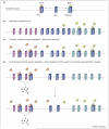
Figure 4
Mechanisms of Smad nuclear accumulation. (a). Summary of the four postulated mechanisms for Smad nuclear accumulation in the context of canonical TGF-β/Smad signaling. (b). Smad nuclear accumulation results from several mechanisms. Red arrows indicate the rate-limiting reactions that could promote Smad nuclear accumulation. The thickness of the arrows indicates the relative reaction rates. Species that would exist at very low abundance under the assumed conditions are colored gray. In the absence of TGF-β, the Smads localize predominantly in the cytoplasm because the rate of nuclear export exceeds the rate of nuclear import [39] (1). In the presence of TGF-β signaling, enhanced nuclear import of Smad complexes compared to monomeric Smads might occur [47] (1), but most evidence to date indicates that it does not [39,40,55,56]. Furthermore, the nuclear-export machinery neither recognizes phosphorylated R-Smads strongly [57] nor Smad4 contained within Smad complexes [47,55], thereby contributing to Smad complex nuclear accumulation (1). Therefore, dissociation of Smad complexes and dephosphorylation of phospho-R-Smad are probably prerequisites for nuclear export, such that these two mechanisms represent potential causes of Smad nuclear accumulation during signaling. If dephosphorylation by the nuclear phosphatase(s) is rate-limiting, then Smad nuclear accumulation occurs if the rate of R-Smad phosphorylation is higher than that of dephosphorylation [43] (2). If dephosphorylation is not rate-limiting, then Smad oligomerization in the cytoplasm, for example with Smad4, could protect the phospho-R-Smads from the phosphatase upon nuclear translocation [43] (3). In this case, the rate at which the Smad complex dissociates in the nucleus would primarily determine the degree of Smad nuclear accumulation. A third possibility is that both mechanisms contribute substantially to Smad nuclear accumulation, which seems to be the case in vivo [47]. Finally, Smad nuclear accumulation caused by either (2) or (3) promotes the reversible binding of nuclear Smads to transcriptional cofactors, coactivators and corepressors, and DNA. Binding to such nuclear-retention factors could further sequester the phospho-R-Smads from dephosphorylation and Smad4 from the nuclear-export machinery [39,41] (4). As signaling ends, the rate of R-Smad phosphorylation decreases, which decreases the driving force for Smad complex formation. Specifically, continual phospho-R-Smad dephosphorylation depletes the concentration of monomeric phospho-R-Smads such that reversible binding reactions are driven towards dissociation, thus reducing R-Smad oligomer abundance and the abundance of Smads bound to nuclear-retention factors. The rates at which the Smad complexes dissociate probably contribute to determining the observed rate of Smad nuclear export.

Similar articles

Cited by

References

    1. Lin X, et al. PPM1A functions as a Smad phosphatase to terminate TGFβ signaling. Cell. 2006;125:915–928. - PMC - PubMed
    1. Inman GJ, et al. Nucleocytoplasmic shuttling of Smads 2, 3 and 4 permits sensing of TGF-β receptor activity. Mol. Cell. 2002;10:283–294. - PubMed
    1. Karlsson G, et al. Gene expression profiling demonstrates that TGF-β1 signals exclusively through receptor complexes involving Alk5 and identifies targets of TGF-β signaling. Physiol. Genomics. 2005;21:396–403. - PubMed
    1. Levy L, Hill CS. Smad4 dependency defines two classes of transforming growth factor (TGF-β) target genes and distinguishes TGF-β-induced epithelial-mesenchymal transition from its antiproliferative and migratory responses. Mol. Cell. Biol. 2005;25:8108–8125. - PMC - PubMed
    1. Yang YC, et al. Hierarchical model of gene regulation by transforming growth factor β. Proc. Natl. Acad. Sci. U. S. A. 2003;100:10269–10274. - PMC - PubMed

Publication types

LinkOut - more resources