. 2008 Oct 24;375(3):425-9.
doi: 10.1016/j.bbrc.2008.08.022. Epub 2008 Aug 14.

Combined sphingosine, S1P and ischemic postconditioning rescue the heart after protracted ischemia

Affiliations

Combined sphingosine, S1P and ischemic postconditioning rescue the heart after protracted ischemia

Donald A Vessey et al. Biochem Biophys Res Commun. .

Abstract

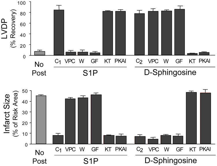
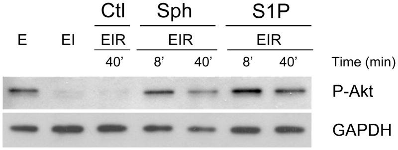
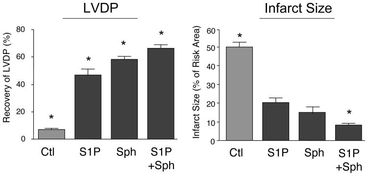
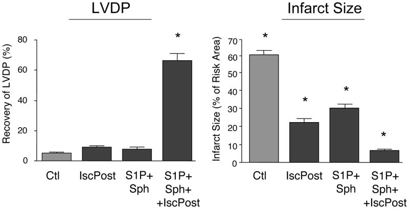
Both sphingosine and sphingosine-1-phosphate (S1P) were able to protect the ex vivo rat heart from ischemia reperfusion injury when added to the perfusion medium at the time of reperfusion after a 40min ischemia (postconditioning). Inhibitor studies revealed distinct mechanisms of protection, with S1P employing a G-protein coupled receptor pathway and sphingosine a cyclic nucleotide dependent protein kinase pathway. However, both restored ischemia-induced depletion of phospho-AKT. Extending the ischemia to 75min reduced protection by both S1P and sphingosine, but protection could be enhanced by employing them in combination. Extending the time of ischemia further to 90min almost eliminated cardioprotection by S1P or sphingosine; and their combination gave only modest protection. However, when S1P plus sphingosine was combined with a novel ramped ischemic postconditioning regimen, left ventricle developed pressure recovered by 66% and there was only a 6% infarct size. The data indicate that detrimental changes are accumulating during protracted ischemia but for up to 90min this damage is not irreversible and hearts can still recover with proper treatment.


Figures

Figure 1
Figure 1. The Effect of Postconditioning with either S1P or Sphingosine on the Recovery of LVDP and on Infarct Size in Hearts Reperfused after 40 Min of Ischemia, and the Effect of Inhibitors
Ex vivo hearts were equilibrated for 20 minutes and then exposed to 40 min of global ischemia. This was followed by 40 min of reperfusion in the presence of either 0.4 μM S1P or 0.4 μM sphingosine in the presence or absence of inhibitors. Recovery of LVDP (A) is expressed as a percentage of the pre-index ischemia value. The infarct size (B) is expressed as a percentage of the area at risk determined at the end of the 40 min reperfusion. The data represent the mean and the error bars reflect the standard deviation (n≥4). The vehicle control (No Post) consisted of reperfusion in the absence of any addition. Reperfusion was done with S1P alone (C1) and sphingosine alone (C2), or with the further addition of the inhibitors: 1 μM VPC23019 (VPC), 50 nm GF109203X (GF), 0.1 μM wortmanin (W), 0.2 μM KT5823 (KT), or 0.1 μM PKA-I 14–22 amide-myristoylated (PKAI).
Figure 2
Figure 2. Effect of Postconditioning with S1P and Sphingosine on the Phosphorylation of Akt
Ex vivo hearts were either equilibrated for 20’ (E), equilibrated followed by 40 min of ischemia (EI), or equilibrated followed by 40 min of ischemia followed by reperfusion (EIR). For the vehicle control (Ctl), reperfusion was for 40 min with medium containing vehicle only (Ctl EIR-40’). For postconditioning, after 40 min of global ischemia (EI), hearts were reperfused with medium containing either 0.4 μM S1P or 0.4 μM sphingosine for either 8 min or 40 min (EIR-8’and EIR-40’). After 40 min of reperfusion, the hearts were collected, homogenized and separated into cytosolic and particulate fractions. The Figure shows the western analysis for the cytosolic fraction using an antibody to phospho-AKT (ser473). Each lane was loaded with 10 μg of protein. The lanes were loaded as follows, lane 1- equilibrated; lane 2-equilibrated plus ischemia; lane 3-equilibrated plus ischemia plus vehicle reperfusion for 40’: lane 4- 8’ of reperfusion in the presence of sphingosine, lane 5– 40’ of reperfusion in the presence of sphingosine,, lane 6– 8’ of reperfusion in the presence of S1P, lane 7– 40’ of reperfusion in the presence of S1P.
Figure 3
Figure 3. Effect of Combined S1P and Sphingosine Postconditioning on Recovery from 75 Min of Ischemia
Ex vivo hearts were equilibrated for 20 minutes and then exposed to 75 min of global ischemia. This was followed by 40 min of reperfusion in the presence of either vehicle (Ctl), 0.4 μM S1P, 0.4 μM sphingosine, or 0.2 μM S1P plus 0.2 μM sphingosine. Recovery of LVDP (A) is expressed as a percentage of the pre-index ischemia value. The infarct size (B) is expressed as a percentage of the area at risk determined at the end of the 40 min reperfusion. The data represent the mean and the error bars reflect the standard deviation (n≥4). Statistical significance (p<0.05) is indicated by (*)
Figure 4
Figure 4. Effect of Combined S1P and Sphingosine and Ischemic Postconditioning on Recovery from 90 Min of Ischemia
Ex vivo hearts were equilibrated for 20 minutes and then exposed to 90 min of global ischemia. This was followed by 40 min of reperfusion in the presence of 0.2 μM S1P plus 0.2 μM sphingosine (S1P+Sph) either alone or with the addition of an ischemic postconditioning regimen of 4 cycles of 15” reperfusion/15” ischemia (S1P+Sph+IscPost). The control (Ctl) received no postconditioning and ischemic postconditioning alone (IscPost) received only ischemic postconditioning in the absence of S1P or sphingosine. Recovery of LVDP (A) is expressed as a percentage of the pre-index ischemia value. The infarct size (B) is expressed as a percentage of the area at risk determined at the end of the 40 min reperfusion. The data represent the mean and the error bars reflect the standard deviation (n≥4). Statistical significance (p<0.05) is indicated by (*)

References

    1. Jennings RB, Reimer KA. The Cell Biology of Acute Myocardial Ischemia. Ann Revs Med. 1991;42:225–246. - PubMed
    1. Buja LM, Entman ML. Modes of myocardial cell injury and cell death in ischemic heart disease. Circulation. 1998;98:1355–1357. - PubMed
    1. Hansen PR. Myocardial reperfusion injury: experimental evidence and clinical relevance. Eur Heart J. 1995;16:734–740. - PubMed
    1. Maxwell SR, Lip GY. Reperfusion injury: a review of the pathophysiology, clinical manifestations and therapeutic options. Int J Cardiol. 1997;58:95–117. - PubMed
    1. Zhao ZQ, Vinten-Johansen J. Myocardial apoptosis and ischemic preconditioning. Cardiovasc Res. 2002;55:438–455. - PubMed

Publication types

MeSH terms

LinkOut - more resources