. 2009 Jan;23(1):64-74.
doi: 10.1016/j.bbi.2008.07.007. Epub 2008 Jul 30.

Detection of autoantibodies to neural cells of the cerebellum in the plasma of subjects with autism spectrum disorders

Affiliations

Detection of autoantibodies to neural cells of the cerebellum in the plasma of subjects with autism spectrum disorders

Sharifia Wills et al. Brain Behav Immun. 2009 Jan.

Abstract

Autism spectrum disorders (ASD) are a group of heterogeneous, behaviorally defined disorders characterized by disturbances in social interaction and communication, often with repetitive and stereotyped behavior. Previous studies have described the presence of antibodies to various neural proteins in autistic individuals as well as post-mortem evidence of neuropathology in the cerebellum. We examined plasma from children with ASD, as well as age-matched typically developing controls, for antibodies directed against human cerebellar protein extracts using Western blot analysis. In addition, the presence of cerebellar specific antibodies was assessed by immunohistochemical staining of sections from Macaca fascicularis monkey cerebellum. Western blot analysis revealed that 13/63 (21%) of subjects with ASD possessed antibodies that demonstrated specific reactivity to a cerebellar protein with an apparent molecular weight of approximately 52 kDa compared with only 1/63 (2%) of the typically developing controls (p=0.0010). Intense immunoreactivity, to what was determined morphologically to be the Golgi cell of the cerebellum, was noted for 7/34 (21%) of subjects with ASD, compared with 0/23 of the typically developing controls. Furthermore, there was a strong association between the presence of antibodies reactive to the 52 kDa protein by Western blot with positive immunohistochemical staining of cerebellar Golgi cells in the ASD group (r=0.76; p=0.001) but not controls. These studies suggest that when compared with age-matched typically developing controls, children with ASD exhibit a differential antibody response to specific cells located in the cerebellum and this response may be associated with a protein of approximately 52 kDa.


Figures

Figure 1
Figure 1
a. Western blot of human cerebellum proteins. Representative blots, shown from left to right, include plasma reactivity of two subjects with ASD, a typically developing control (TD), and a subject with developmental delay (DD), along with the molecular weight markers. The arrow denotes a protein with an apparent molecular weight of approximately 52 kDa that is specifically recognized by the autoantibodies from subjects with ASD. b. Western blot comparing reactivity to human and monkey cerebellum proteins. Cerebellum protein extracts from human and Rhesus macaque were run together in alternate lanes to determine the presence of the 52 kDa band in monkey cerebellum. The left lane of each duplex contains the human cerebellum preparation (H), while the right lane contains adult monkey cerebellum (M). The left blot was reacted with plasma from a child with autism (AU) while the right was reacted with plasma from a typically developing control (TD).
Figure 1
Figure 1
a. Western blot of human cerebellum proteins. Representative blots, shown from left to right, include plasma reactivity of two subjects with ASD, a typically developing control (TD), and a subject with developmental delay (DD), along with the molecular weight markers. The arrow denotes a protein with an apparent molecular weight of approximately 52 kDa that is specifically recognized by the autoantibodies from subjects with ASD. b. Western blot comparing reactivity to human and monkey cerebellum proteins. Cerebellum protein extracts from human and Rhesus macaque were run together in alternate lanes to determine the presence of the 52 kDa band in monkey cerebellum. The left lane of each duplex contains the human cerebellum preparation (H), while the right lane contains adult monkey cerebellum (M). The left blot was reacted with plasma from a child with autism (AU) while the right was reacted with plasma from a typically developing control (TD).
Figure 2
Figure 2
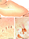
Immunohistochemical staining of macaque monkey cerebellum with plasma from a subject with intense (++) reactivity. A) Low magnification (4X objective) photomicrograph of one folia in a coronal section of the cerebellum. This image illustrates the consistent staining of Golgi cells lying just subjacent to the molecular layer. B) Higher magnification (40X objective) phase-contrast photomicrograph of a subset of the cells from panel A. Note that the labeled neurons have both apical dendrites that extend into the molecular layer and basal dendrites that remain in the granular layer. C) High magnification (100X objective) photomicrograph of immunostained Golgi cells. Panel C illustrates that the immunoreactivity has a particulate appearance and is distributed within the somal cytoplasm and extends into the proximal dendrites. Calibration bars: A) 250 µm, B) 50µm, C) 20 µm. Abbreviations: GL: granular layer. ML: molecular layer.
Figure 3
Figure 3
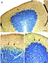
Nissl counterstained (blue) immunohistochemically processed coronal sections through the macaque monkey cerebellum illustrating an additional (++) case not illustrated in preceding figures. A: low power (10X objective) photomicrograph illustrates the Golgi cells (dark brown) at the interface between the molecular and granule cell layers. Panels B and C are higher magnification (20 X) photomicrographs and illustrate Golgi cells with dark brown reaction product and lightly Nissl-stained Purkinje cells (arrows) which are devoid of immunohistochemical reaction product. Calibration bars equal 100µm. Bar in panel C also applies to panel B.
Figure 4
Figure 4
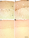
This figure illustrates the range of immunohistochemical staining intensities and patterns observed in this study. Panel A illustrates strong and selective staining of Golgi cells (++). Panel B illustrates less intense and less consistent staining of Golgi cells (+). Panel C depicts intermittent positive staining (−/+) and panel D illustrates a lack of specific staining in the referenced cell type (−). Calibration bar equals 100µm and applies to all panels.
Figure 5
Figure 5
Immunohistochemical staining of macaque monkey cerebellum with plasma from six individual subjects. Despite slight differences in the nature of the staining, each of these cases was rated as (++) for Golgi cell staining. Panels A and B both represent strong and selective staining of Golgi cells similar to the subject presented in Figure 2. Of particular note in both panels A and B is the particulate dendritic staining in the molecular layer. In panel B, there is also some light staining of basket cell somata in the deep portion of the molecular layer. Panels C and D illustrates staining resulting from plasma from subjects whose antibodies produced both positive staining of the Golgi cells as well as more intense staining of the deep molecular layer basket cells. As illustrated in Figure 4, there is occasionally light staining of these basket cells in negative control sections. E illustrates a case in which the staining was lighter using the same concentration of plasma and procedures as in cases shown in panels A and B, nonetheless, the staining is selective for Golgi neurons. In F, the staining is also light but selective for Golgi neurons. More prominent dendritic labeling is observed in the superficial portion of the molecular layer. Calibration bar equals 100µm and applies to all panels.

References

    1. Prevalence of autism spectrum disorders--autism and developmental disabilities monitoring network, 14 sites, United States, 2002. MMWR Surveill Summ. 2007;56:12–28. - PubMed
    1. Alarcon-Segovia D, Ruiz-Arguelles A, Fishbein E. Antibody to nuclear ribonucleoprotein penetrates live human mononuclear cells through Fc receptors. Nature. 1978;271:67–69. - PubMed
    1. Amaral DG, Schumann CM, Nordahl CW. Neuroanatomy of autism. Trends Neurosci. 2008;31:137–145. - PubMed
    1. Ashwood P, Wills S, Van de Water J. The immune response in autism: a new frontier for autism research. J Leukoc Biol. 2006;80:1–15. - PubMed
    1. Association AP. Diagnostic and Statistical Manual of Mental Disorders. Fourth Edition. Washington, DC: American Psychiatric Association; 1994.

Publication types

MeSH terms