. 2008 Sep 9;47(36):9380-93.
doi: 10.1021/bi800677k. Epub 2008 Aug 16.

Molecular basis for the structural instability of human DJ-1 induced by the L166P mutation associated with Parkinson's disease

Affiliations

Molecular basis for the structural instability of human DJ-1 induced by the L166P mutation associated with Parkinson's disease

Peter C Anderson et al. Biochemistry. .

Abstract

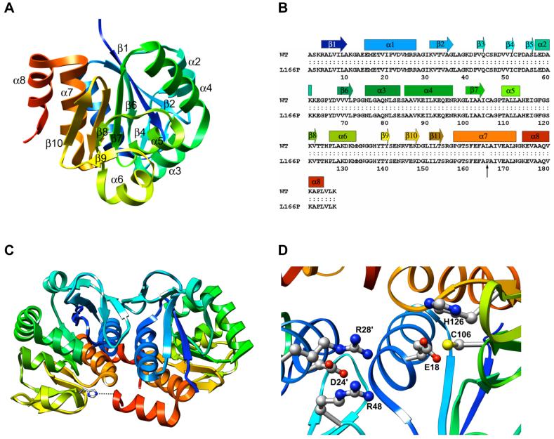
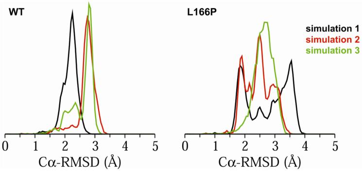
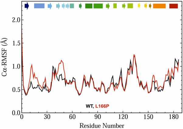
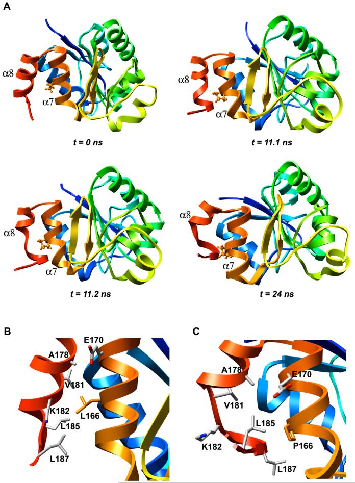
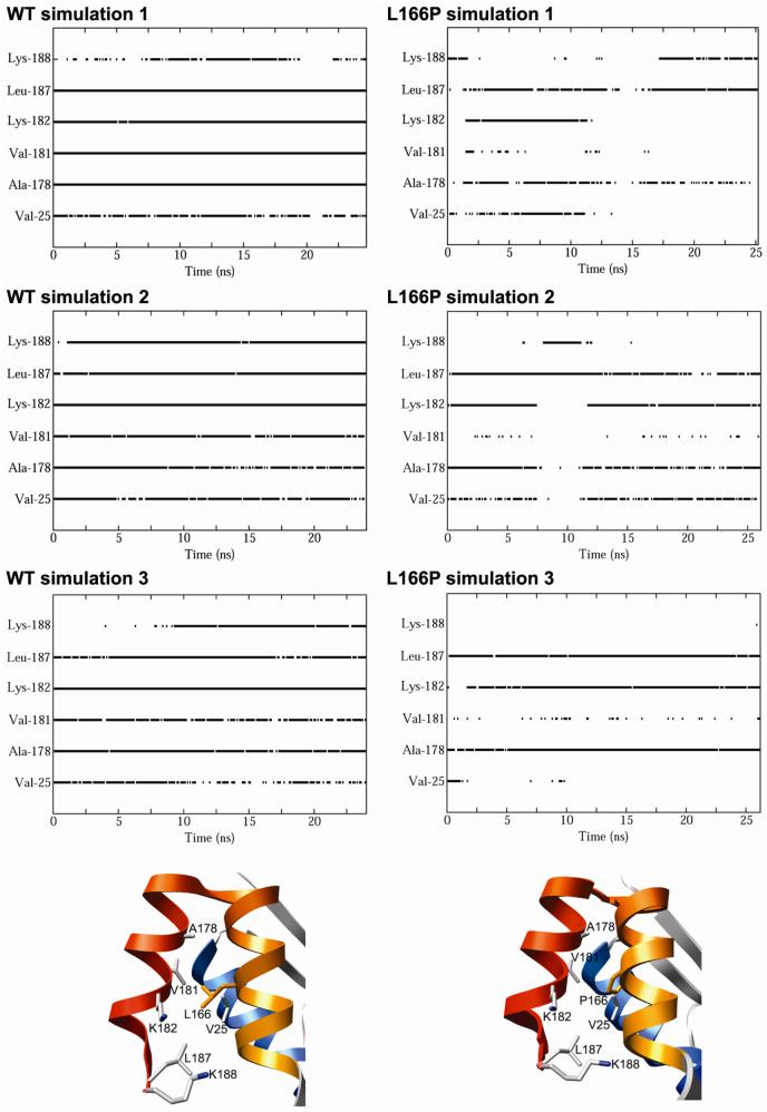
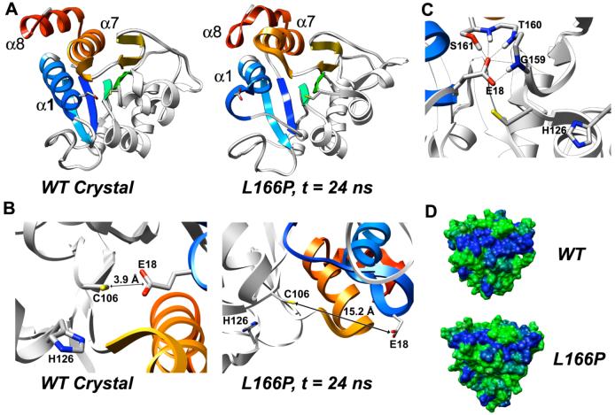
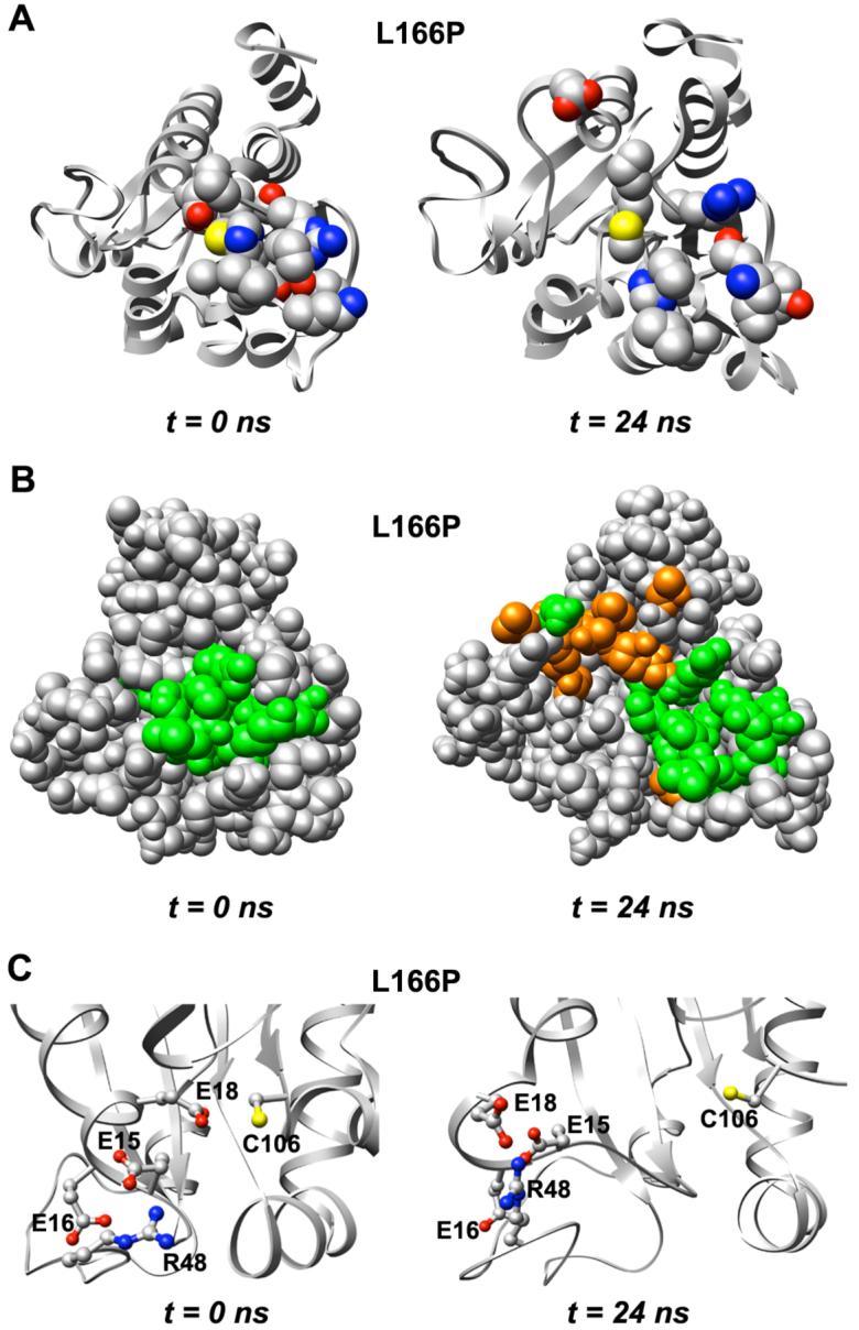
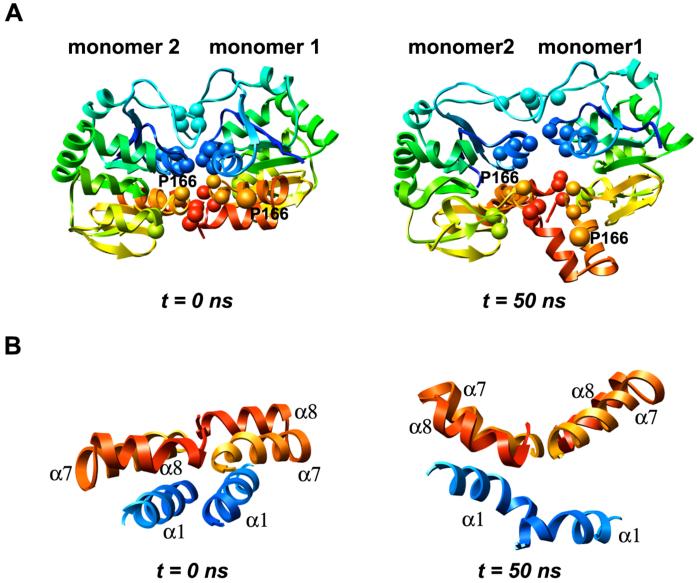
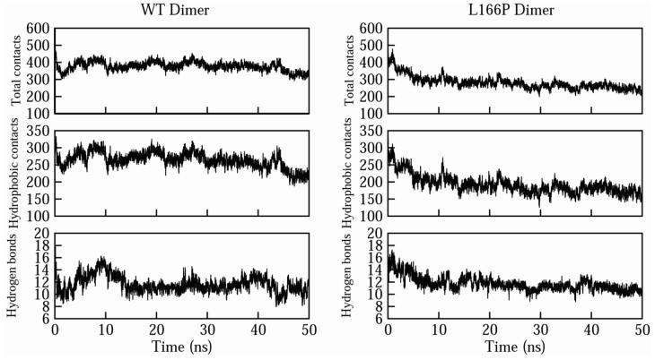
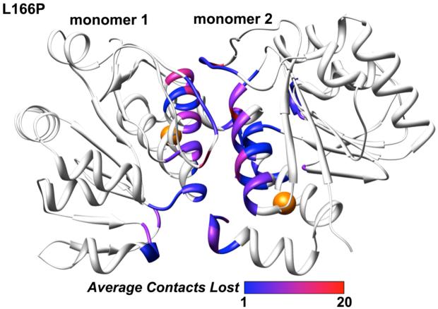
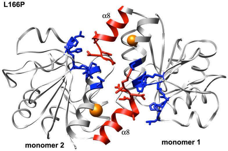
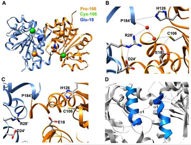
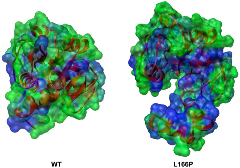
DJ-1 is a dimeric protein of unknown function in vivo. A mutation in the human DJ-1 gene causing substitution of proline for leucine at residue 166 (L166P) has been linked to early onset Parkinson's disease. Lack of structural stability has precluded experimental determination of atomic-resolution structures of the L166P DJ-1 polymorph. We have performed multiple molecular dynamics (MD) simulations ( approximately 1/3 mus) of the wild-type and L166P DJ-1 polymorph at physiological temperature to predict specific structural effects of the L166P substitution. L166P disrupted helices alpha1, alpha5, alpha6 and alpha8 with alpha8 undergoing particularly severe disruption. Secondary structural elements critical for protein stability and dimerization were significantly disrupted across the entire dimer interface, as were extended hydrophobic surfaces involved in dimer formation. Relative to wild-type DJ-1, L166P DJ-1 populated a broader ensemble of structures, many of which corresponded to distorted conformations. In a L166P dimer model the substitution significantly destabilized the dimer interface, interrupting >100 intermolecular contacts that are important for dimer formation. The L166P substitution also led to major perturbations in the region of a highly conserved cysteine residue (Cys-106) that participates in dimerization and that is critical for a proposed chaperone function of DJ-1. Cys-106 is located approximately 16 A from the substitution site, demonstrating that structural disruptions propagate throughout the whole protein. Furthermore, L166P DJ-1 showed a significant increase in hydrophobic surface area relative to wild-type protein, possibly explaining the tendency of the mutant protein to aggregate. These simulations provide details about specific structural disturbances throughout L166P DJ-1 that previous studies have not revealed.


Figures

Figure 1
Figure 1
DJ-1 structure. (A) Ribbon diagram of human DJ-1 (1PDV, ref. 26). The structure is colored from blue (N-terminus) to red (C-terminus). (B) Sequence of human wt DJ-1 and L166P polymorph. α-helices and α-strands are indicated by bars and arrows, respectively, and are colored to match the ribbon representation of DJ-1. The site of the L166P substitution is indicated by a vertical arrow. (C) Ribbon diagram of human DJ-1 dimer (1UCF, ref. 25). A hydrogen bond critical for dimer formation is formed by His-126 and the backbone carbonyl oxygen atom of Pro-184 of the opposite subunit (represented as a dotted line at the bottom of the figure). (D) The side chain of highly conserved Cys-106 is located near the side chains of His-126 and buried Glu-18. Residues Asp-24′ and Arg-28′ from the opposite subunit contribute to stabilizing interactions across the subunit-subunit interface in the dimer.
Figure 2
Figure 2
Distributions of Cα-RMS deviations (in Å) during the last 10 ns of DJ-1 wt and L166P monomer simulations. Simulations 1, 2 and 3 are indicated by black, red and green, respectively.
Figure 3
Figure 3
Average Cα-RMS fluctuations (in Å) per residue for the three wt (black) and three L166P (red) simulations of DJ-1 monomer. C α-RMSFs were calculated relative to the average structure over the last 10 ns of all three wt and all three L166P simulations. Secondary structural elements are indicated at the top of the plot and colored to match the ribbon representations of DJ-1 in Figure 1. Key: box, α-helix; arrow, β-strand.
Figure 4
Figure 4
L166P DJ-1 simulations show significant loss of helical content in α8. (A) Time sequence of loss of helical structure of L166P DJ-1 residues 181-184 at C-terminal end of helix α8. Pro-166 is displayed in ball-and-stick format. Structures shown are taken from the indicated time points of L166P simulation 1. (B) In the wt DJ-1 simulations Leu-166 retains 67 of the 69 interresidue atomic contacts with helices α7 and α8 that are observed in the wt DJ-1 monomer crystal structure. The side chains of the residues contacted by Leu-166 are displayed in stick format and colored by atom type. The Leu-166 side chain is shown in orange. (C) The L166P DJ-1 simulations show an average of only 52 interresidue atomic contacts between Pro-166 and α7 and α8. The side chains of the residues that are contacted by Leu-166 in the wt simulations are displayed in stick format and colored by atom type. The Pro-166 residue is shown in orange.
Figure 5
Figure 5
(A) Contacts formed by residue 166 as a function of time for DJ-1 wt (left) and L166P (right) monomer simulations. The residues with which residue 166 forms atomic contacts are specified on the vertical axis. A solid line indicates that at least one intermolecular atomic contact occurs between the specific residue on the vertical axis and residue 166 at the given time. Gaps in lines signify that no intermolecular contacts with the specific residue occur at the given time. (B) Contacts with Leu-166 and (C) Pro-166 monitored in part A. Relevant amino acid side chains are represented by sticks. The structures depicted in (A) and (B) are the starting structures of the wt and L166P simulations, respectively.
Figure 6
Figure 6
Structural distortion of region around conserved Cys-106 in L166P DJ-1 and disruption of dimer interface. (A) The N-terminal end of α1 loses its helicity in the L166P simulation and the helix moves away from the central β-sheet. (B) In the wt crystal structure the side chain of Glu-18 is buried in the protein structure. The distance between atom Sγ of the Cys-106 side chain and the Cα atom of the Glu-18 side chain is 3.9 Å. In the L166P simulations the side chain of Glu-18 loses its buried position and swings outward into solution. At the end of L166P simulation 1 (24 ns), the distance between atom Sγ of the Cys-106 side chain and the Cδ atom of the Glu-18 side chain is 15.2 Å. (C) In L166P simulations 2 and 3, the displaced Glu-18 side chain is stabilized by forming hydrogen bonds (indicated by black lines) with the side chain of Ser-161 and with backbone amide hydrogen atoms of Gly-159, Thr-160 and Ser-161. The side chain of Thr-160 is not shown. (D) Surface representation of DJ-1 wt and L166P monomers colored according to hydrophobicity. Each subunit of the DJ-1 dimer contains a large hydrophobic surface (extended blue patch). In L166P simulations this surface is divided in two by repositioning of the hydrophilic Asp-24 and Arg-27 residues. Colors range continuously from green (most hydrophilic) to blue (most hydrophobic). Hydrophobicity values were calculated by Chimera using the algorithm of Kyte and Doolittle (47).
Figure 7
Figure 7
Expansion of structure around conserved Cys-106 in L166P DJ-1. (A) Residues that have atoms located within 4 Å of Cys-106 (shown with yellow sulfur atom) or His-126 in the starting structure (left) are displayed in CPK format. (B) Space-filling view of L166P DJ-1 and expansion of structure around Cys-106. Residues displayed in CPK format in part A are colored green (left). Residues other than those depicted in CPK format in part A and that experience increased solvent exposure as a result of the structural expansion are shown in orange for the structure at 24 ns (right). (C) Two acidic residues near the Glu-18 side chain, Glu-15 and Glu-16, as well as the basic residue Arg-48, move away from Cys-106.
Figure 8
Figure 8
Perturbations in the DJ-1 dimer interface following L166P substitution in each subunit. (A) Loss of intermolecular contacts that mediate DJ-1 dimerization after 50 ns. Key hydrophobic interactions across the interface are formed by Met-17, Val-20, Ile-21, Val-23, Val-50, Ile-52, His-126, Pro-127, Pro-158, Phe-162, Pro-184, Leu-185 and Val-186, which are represented by spheres in both subunits. Residue 166 positions are indicated by orange spheres. (B) Helices α7 and α8 from both subunits are roughly coplanar in the wt simulations and in the starting structure for L166P simulations (left). During the L166P simulations the α7-turn-α8 motif in subunit 1 loses packing with α1 and moves out of the plane of the opposite α7-turn-α8 motif, undergoing a dynamic swinging motion that creates a V-shape with the opposite motif (right).
Figure 9
Figure 9
Effects of L166P substitution on intermolecular contacts across the DJ-1 dimer interface. The average numbers of total intermolecular contacts (top), hydrophobic contacts (center), and hydrogen bonds (bottom) are plotted for the wt simulations (left) and L166P simulations (right).
Figure 10
Figure 10
Loss of atomic intermolecular contacts across L166P DJ-1 dimer interface. The color for each residue corresponds to the average number of total contacts lost by that residue across the interface during the course of the three L166P simulations. The color scale spans the range from 1 (blue) to 20 (red). Residues that do not constitute the interface are not colored. Positions of residue 166 are indicated by orange spheres.
Figure 11
Figure 11
Intermolecular contacts formed by the C-terminal ends of helices α8 in each subunit of L166P DJ-1 dimer structure. Side chains of amino acid residues in α8 (red) and in the opposite subunit (blue) that form contacts across the dimer interface are shown in stick format. Positions of residue 166 are indicated by orange spheres.
Figure 12
Figure 12
Disruption of structure around conserved Cys-106 in L166P DJ-1 dimer simulations. (A) L166P DJ-1 dimer starting structure showing positions of Pro-166 (orange spheres), Cys-106 (green spheres) and Glu-18 (blue spheres). Subunits 1 and 2 are colored orange and blue, respectively. (B) Hydrogen bonding network involving conserved Cys-106 side chain in DJ-1 wt dimer crystal structure. Oxygen atoms of waters participating in hydrogen bonds are represented as red spheres. Hydrogen bonds are denoted by dotted lines. (C) Disruption of hydrogen bonding network shown in part B caused by L166P substitution in DJ-1 dimer. (D) Distortion of α-helical structure of N-terminal end of α1.
Figure 13
Figure 13
Increase in hydrophobic SASA of L166P DJ-1 (right) relative to wt (left). Molecular surface is colored by residue from least hydrophobic (green) to most hydrophobic (blue). Ribbons are colored red for entire protein structure. Both structures have been aligned by C α-RMSD.

References

    1. Farrer MJ. Genetics of Parkinson disease: paradigm shifts and future prospects. Nat. Rev. Genet. 2006;7:306–318. - PubMed
    1. Spillantini MG, Schmidt ML, Lee VM, Trojanowski JQ, Jakes R, Goedert M. Alpha-synuclein in Lewy bodies. Nature. 1997;388:839–840. - PubMed
    1. Kitada T, Asakawa S, Hattori N, Matsumine H, Yamamura Y, Minoshima S, Yokochi M, Mizuno Y, Shimizu N. Mutations in the parkin gene cause autosomal recessive juvenile parkinsonism. Nature. 1998;392:605–608. - PubMed
    1. Shimura H, Hattori N, Kubo S, Mizuno Y, Asakawa S, Minoshima S, Shimizu N, Iwai K, Chiba T, Tanaka K, Suzuki T. Familial Parkinson disease gene product, parkin, is a ubiquitin-protein ligase. Nat. Genet. 2000;25:302–305. - PubMed
    1. Leroy E, Boyer R, Auburger G, Leube B, Ulm G, Mezey E, Harta G, Brownstein MJ, Jonnalagada S, Chernova T, Dehejia A, Lavedan C, Gasser T, Steinbach PJ, Wilkinson KD, Polymeropoulos MH. The ubiquitin pathway in Parkinson’s disease. Nature. 1998;395:451–452. - PubMed

Publication types

MeSH terms

Substances